Weekly Digests
‹ Back to December

Why CSF1R inhibition doesn’t slow tumor growth - and how it could

December 6, 2017

CSF1R inhibition for the depletion of tumor-associated macrophages (TAMs) seemed like a promising therapeutic strategy, but while TAM depletion occurred as expected, antitumor efficacy has been incredibly limited. To understand why, Kumar et al., in a study published in Cancer Cell, revealed the additional immunosuppressive mechanism activated by CSF1R inhibition and exploited this information to create an effective anti-tumor strategy.

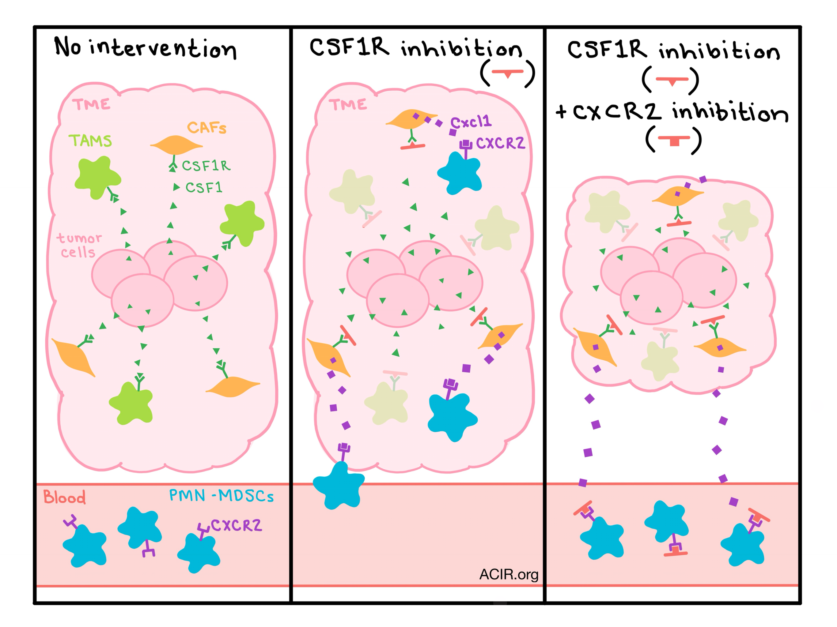
One way that tumors support immunosuppressive TAMs is by producing CSF1, which is recognized by TAMs via the receptor CSF1R. This pathway supports differentiation and survival of TAMs, so researchers hypothesized that inhibiting CSF1R would deplete TAMs and delay tumor growth. In preclinical models, various small molecule inhibitors have been tested, and while they appear to successfully deplete TAMs, little to no anti-tumor effects have been observed. Kumar et al. tested the CSF1R inhibitor JNJ-40346527 on a variety of tumor models; in addition to depletion of TAMs, the researchers also observed accumulation of granulocytes with potent immunosuppressive activity, which were identified as polymorphonuclear myeloid-derived suppressor cells (PMN-MDSCs). Further, the team was able to prove that the increase in PMN-MDSCs was due to enhanced recruitment to the tumor site, rather than proliferation of precursor cells.

To understand how this second pro-tumor immune cell type was being recruited to the tumor, Kumar et al. evaluated chemokines in tumor cell lysates following CSF1R inhibition and noticed an upregulation of chemokines, most notably Cxcl1, that are known to recruit granulocytes via the CXCR2 receptor. Carcinoma-associated fibroblasts (CAFs) were identified as the major source of Cxcl1 and the researchers unexpectedly found that CAFs also express the CSF1R protein on their cell surface and in the cytoplasm. A series of mechanistic experiments showed that CSF1 activates the CSF1/CSF1R pathway on CAFs, causing the histone deacetylase HDAC2 to migrate to the Cxcl1 promoter and silence the production of Cxcl1. Blocking this pathway with a CSF1R inhibitor reinstates Cxcl1 expression and increases the recruitment of tumorigenic PMN-MDSCs to the tumor site, abrogating any effect of TAM depletion.

In humans, CXCL1 is not as potent a recruiting cytokine as Cxcl1 in mice, however, evaluation of human tumors following surgical resection showed that CAFs produced high levels of CXCL8, which is known for recruiting neutrophils. The researchers went on to show that in addition to recruiting neutrophils, CXCL8 also plays a role in recruiting PMN-MDSCs. Gene silencing and neutralizing antibody experiments demonstrated that CXCL8 production is controlled by the CSF1/CSF1R pathway.

In an effort to prevent the recruitment of tumor-promoting PMN-MDSCs following CSF1R inhibition, Kumar et al. tested a CSF1R inhibitor in combination with a CXCR2 inhibitor. Neither single agent showed antitumor efficacy, but together they resulted in a highly significant delay in tumor progression in two mouse models. Evaluation of the tumors showed that the combination successfully decreased the presence of TAMs and abrogated the CSF1R inhibitor-induced accumulation PMN-MDSCs. The combination therapy also enhanced the therapeutic effect of anti-PD-1 therapy, resulting in a dramatic antitumor effect.

Speculating on why tumors would contribute to a mechanism that inherently prevents the recruitment of pro-tumor PMN-MDSCs, Kumar et al. suggest that early in tumor development, infiltration of highly inflammatory neutrophil populations might be damaging to the nascent tumor; thus, early-stage tumors might utilize CSF1 production to limit this population until the tumor is more advanced.

Kumar et al. show that the use of CSF1R inhibitors to deplete immunosuppressive TAMs and slow the growth of tumors is not a lost cause as long as the recruitment of PMN-MDSCs to the tumor site is addressed. Further, they show that this therapy may enhance the efficacy of anti-PD-1 therapy, which could one day lead to improved clinical efficacy.

by Lauren Hitchings

References:

Kumar V., Donthireddy L., Marvel D., Condamine T., Wang F., Lavilla-Alonso S., Hashimoto A., Vonteddu P., Behera R., Goins M.A., Mulligan C., Nam B., Hockstein N., Denstman F., Shakamuri S., Speicher D.W., Weeraratna A.T., Chao T., Vonderheide R.H., Languino L.R., Ordentlich P., Liu Q., Xu X., Lo A., Puré E., Zhang C., Loboda A., Sepulveda M.A., Snyder L.A., Gabrilovich D.I. Cancer-Associated Fibroblasts Neutralize the Anti-tumor Effect of CSF1 Receptor Blockade by Inducing PMN-MDSC Infiltration of Tumors. Cancer Cell. 2017 Nov 13.

In the Spotlight...

Interferon-γ signaling in melanocytes and melanoma cells regulates expression of CTLA-4

Mo et al. discovered that CTLA-4 is expressed not only in activated T cells but also in most human melanoma cell lines and normal human melanocytes, and its expression is controlled by IFNγ signaling via the JAK/STAT pathway and by the IFNγ-independent MAPK pathway. A subset of melanoma patients receiving anti-CTLA-4 therapy had upregulated IFNγ and CTLA-4 gene expression in the tumor, which correlated with durable response, suggesting that the success of anti-CTLA-4 therapy may be due in part to targeting melanoma cells themselves.

T cell receptor sequencing of early-stage breast cancer tumors identifies altered clonal structure of the T cell repertoire

Beausang et al. performed TCR β-chain deep sequencing on lymphocytes from tumor tissue, adjacent normal tissue, and peripheral blood from 16 patients with early-stage breast cancer. While CDR3 repertoires overlapped within patients, tumor tissue had more lymphocyte infiltration, but less clonal diversity than normal breast tissue; tumor and normal tissue were also individually distinct from blood. Most abundant tumor-enriched clonotypes could be found in adjacent normal tissue and tissue from healthy volunteers, suggesting lack of tumor specificity.

Targeting the T cell receptor beta-chain constant region for immunotherapy of T cell malignancies

Since the β-chain constant region is encoded by either the TRBC1 or TRBC2 gene, normal T cell populations have a mix of the two genotypes, while T cell malignancies tend to skew toward one or the other. Maciocia et al. confirmed this for many T cell malignancies and took advantage of a TRBC1-specific monoclonal antibody to generate and test proof-of-concept CAR-T cells to demonstrate that such therapy selectively eliminates TRBC1+ malignant and healthy T cells, while leaving the TRBC2+ T cell repertoire intact, preserving cellular immunity.

CRISPR-mediated TCR replacement generates superior anticancer transgenic T-cells

Transducing T cells with αβ or γδ TCRs specific for target cancer antigens while simultaneously knocking out the endogenous TCR-β locus using CRISPR/Cas9 increased the surface expression of transgenic TCRs, led to higher sensitivity to cognate antigen, and demonstrated stronger, polyfunctional response in vitro and ex vivo to various blood cancers compared to standard TCR transfer. γδ TCRs recognizing pan-cancer targets are attractive for adoptive cell therapy, as they do not exhibit MHC restriction.

Precision Targeting of Tumor Macrophages with a CD206 Binding Peptide

Scodeller et al. performed in vivo phage display on cells from tumor-bearing mice and identified a peptide (UNO) that homes to M2-like tumor-associated macrophages (MEMs) in tumors and sentinel lymph nodes, interacts with CD206, and gets internalized. Binding to MEMs required linearization of the cyclic peptide, which is facilitated by the TME. Coating nanoparticles with UNO allows for specific drug delivery to MEMs in solid tumors; UNO could also be used to guide contrast agents to the sentinel lymph node for live-imaging or diagnostic testing.

KIR downregulation by IL-12/15/18 unleashes human NK cells from KIR/HLA-I inhibition and enhances killing of tumor cells

Ewen et al. demonstrated that stimulating human NK cells with IL-12/15/18 downregulated the surface expression and mRNA levels of inhibitory KIRs, resulting in increased cytotoxicity against tumor cells expressing cognate HLA-I molecules. KIR expression was re-inducible by IL-2 culture, suggesting that the reversible nature of KIR downregulation could help prevent autoimmunity upon treatment with autologous NK cells.

CDK4/6 Inhibition Augments Anti-Tumor Immunity by Enhancing T Cell Activation

In a screen for small molecules that could increase T cell activity (measured by IL-2 production), Deng et al. found that CDK4/6 inhibitors increase IL-2 even in the presence of suppressive PD-1 signaling. Dosing for short-term exposure to CDK4/6 inhibitors decreased T cell proliferation, but increased effector T cell activation (in part due to de-repression of NFAT family proteins) and tumor infiltration and decreased expression of coinhibitory signals and myeloid subpopulations, ultimately yielding antitumor effects that enhanced the efficacy of PD-1 blockade.

T cells presenting viral antigens or autoantigens induce cytotoxic T cell anergy

While investigating immune responses to the autoantigen cerebellar degeneration-related protein 2 (CDR2), Blachère et al. discovered that tolerance to this predominantly cerebellum-expressed protein was enhanced by expression and presentation by T cells, resulting in anergy of interacting CD8+ cytotoxic T cells. This observation was extended to viral antigens presented by T cells, and the researchers speculate it is a homeostatic mechanism to prevent T cell fratricide targeted to rearranged, novel TCR CDR3 regions.

Dendritic cells enhance polyfunctionality of adoptively transferred T cells which target cytomegalovirus in glioblastoma

Cytomegalovirus (CMV) is found in glioblastomas (GBM) and not in normal brain, but CMV-specific T cells from GBM patients prepared for adoptive transfer exhibit low polyfunctionality after in vitro expansion. In a pilot clinical trial, Reap et al. showed that co-administration of CMV RNA-loaded dendritic cells increased the frequency of polyfunctional CMV-specific CD8+ T cells, which also correlated with improved survival, although the study was not powered to conclude causality.

Suppression of FIP200 and autophagy by tumor-derived lactate promotes naive T cell apoptosis and affects tumor immunity

Xia et al. made the seminal observation that naive T cells from blood and tumor tissue of ovarian cancer patients and tumor-bearing mice were prone to apoptosis. They traced the cause to tumor-derived lactate, which translationally inhibits the expression of FIP200, a protein required for autophagy, leading to impaired autophagy induction, overactivated mitochondria, high levels of reactive oxygen species, and disruption of Bcl-2 gene family expression.

Everything New this Week In...

Close Modal

Small change for you. Big change for us!

This Thanksgiving season, show your support for cancer research by donating your change.

In less than a minute, link your credit card with our partner RoundUp App.

Every purchase you make with that card will be rounded up and the change will be donated to ACIR.

All transactions are securely made through Stripe.