Weekly Digests
‹ Back to July

Landscape of Infiltrating T Cells in Liver Cancer Revealed by Single-Cell Sequencing

July 26, 2017

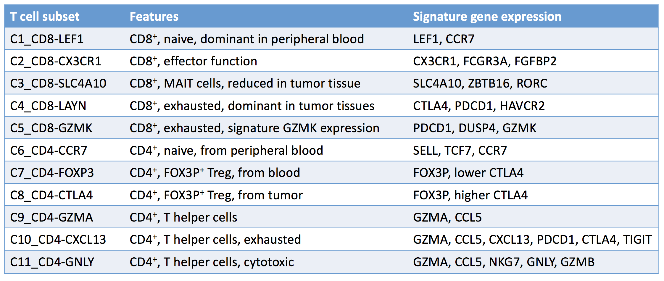
The more data you collect, the more details, and hopefully insights, you’ll uncover; that’s why Zheng et al. performed single-cell RNA sequencing and T cell receptor (TCR) profiling on over 5,000 individual T cells isolated from the tumors, adjacent normal tissue, and peripheral blood of six patients with hepatocellular carcinoma (HCC). From the mass of data collected, the researchers identified 11 unique T cell subsets with distinct patterns of T cell clustering and signature variable genes. Subsets were categorized based on their molecular and functional properties, as well as their developmental trajectories.

Tumor infiltration and TCR clonality

Overall, HCC had poor CD8+ T cell infiltration, making it a good model to study TIL dysregulation. Ten percent of CD8+ T cells found in normal tissue and blood had recurrently identified TCRs, while 30% of T cells from tumor tissues showed such TCRs, indicating clonal expansion, possibly within the tumor. Similar results were observed for T regulatory cells. Within the tumor, clonal CD8+ T cells were also more likely to be exhausted than T cells without TCR clones.

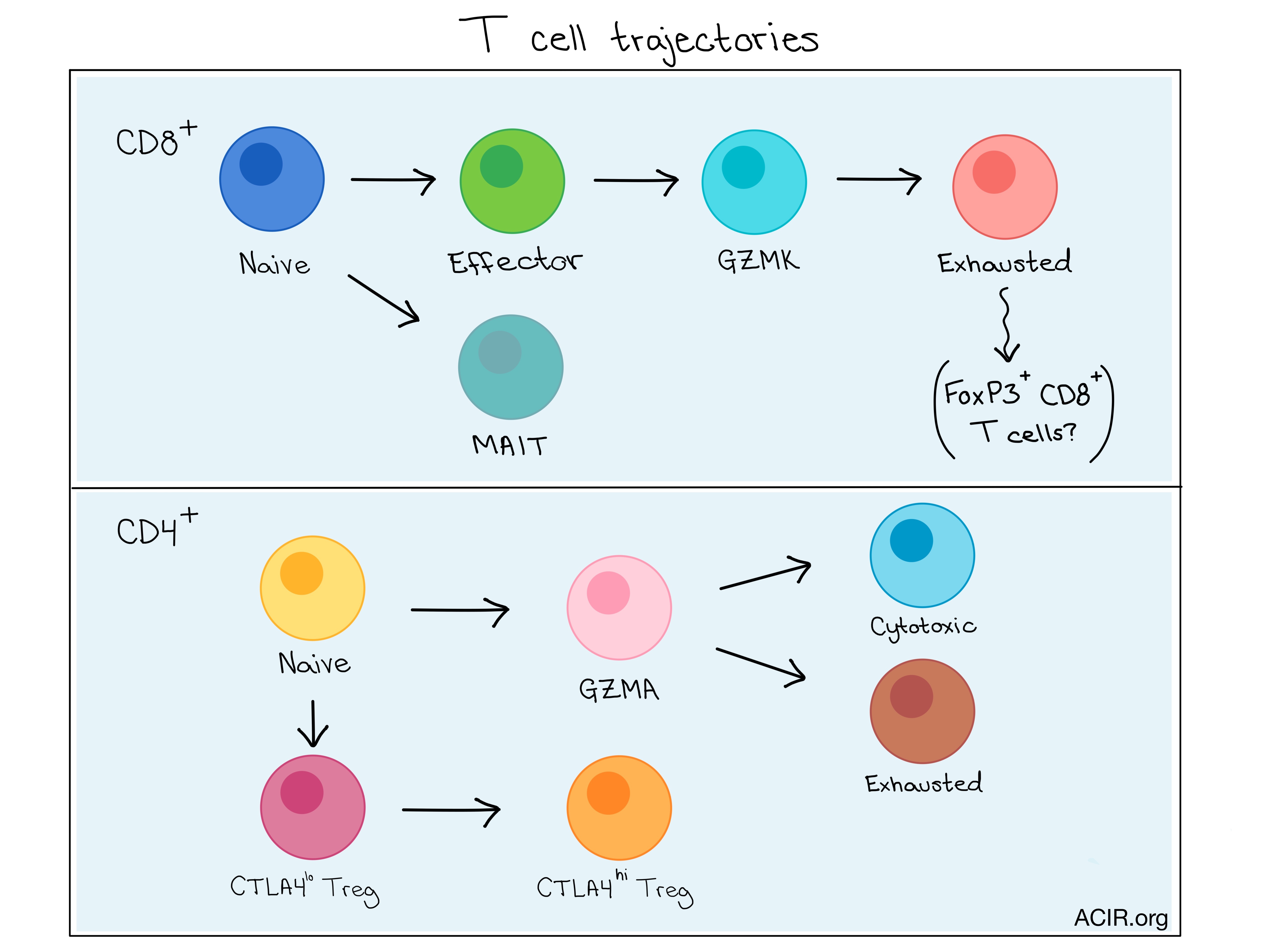
Exhausted CD8+ T cells (C4_CD8-LAYN) and active CTLA4hi T regulatory cells (C8_CD4-CTLA4) were significantly enriched in tumor samples and were potentially clonally expanded, and late stage cancers had higher exhausted CD8+ T cell populations. Meanwhile, percentages of MAIT cells were reduced in tumor samples compared to normal tissue. A small CD8+ cell subpopulation was also found to express the FOXP3 T regulatory marker, suggesting a possible developmental relationship between Tregs and exhausted CD8+ T cells.

Exhaustion

By comparing exhausted and non-exhausted cytotoxic CD8+ T cells, Zheng et al. identified 82 exhaustion-specific genes, including well-described markers like CTLA4, and PDCD1, along with less-described markers including MYO7A, WARS, and CXCL13, and novel exhaustion markers including LAYN, PHLDA4, and SNAP47. Interestingly, twenty-two of the exhaustion genes identified in cytotoxic T cells were also highly expressed on tumor-infiltrating Tregs.

Zheng et al. took a closer look at the LAYN gene and found that LAYN was upregulated both in activated Tregs, especially those with a more repressive phenotype, and exhausted CD8+ T cells isolated from the HCC tumor, and that high LAYN expression was associated with reduced disease-free survival. Through in vitro studies, they also showed that LAYN was only expressed following CD3 and CD28 activation of CD8+ T cells, and once expressed, it acted to repress CD8+ T cell functions. When CD8+ T cells were induced to overexpress LAYN, they produced significantly less IFN-γ. Interestingly, LAYN was expressed on LAG-3-, but not LAG3+, CD8+ T cells – a mutual exclusivity that could indicate distinct T cell subsets.

Differentiation trajectories

Combining the information from complete transcriptome and TCR analyses allowed the researchers to gain insights into the functional states of, and relationship between T cell subsets. Using algorithms to map the differentiation trajectories based on changes in expression over “pseudotime”, CD8+ T cells were shown to move sequentially from a naive to an effector state, then to an exhausted state expressing GZMK, and finally to a terminally exhausted state. This suggests that GZMK expression represents an intermediate state en route to exhaustion.

Analysis of the CD4+ T cell trajectory revealed that naive CD4+ and CD4+ T helper cells did not separate over pseudotime until T helper cells branched into either an exhausted or cytotoxic state, indicating functional divergence of these derivative subsets. The two end states were more closely related to the intermediate T helper state than to one another, as revealed by the number of shared TCRs between subsets.

In order to contribute further to the base of scientific knowledge and help other scientists out, Zheng et al. developed an interactive web-based tool for analyzing, visualizing, and downloading their single-cell data for single or multiple user-input genes. The tool can be found at: http://hcc.cancer.pku.cn.

Overall, exploring the function and dysfunction of T cells in the tumor microenvironment is critical to understanding mechanisms of action and identifying useful biomarkers and targets for cancer immunotherapies.

by Lauren Hitchings

References:

Zheng C., Zheng L., Yoo J.K., Guo H., Zhang Y., Guo X., Kang B., Hu R., Huang J.Y., Zhang Q., Liu Z., Dong M., Hu X., Ouyang W., Peng J., Zhang Z. Landscape of Infiltrating T Cells in Liver Cancer Revealed by Single-Cell Sequencing. Cell. 2017 Jun 15.

In the Spotlight...

In vivo CRISPR screening identifies Ptpn2 as a cancer immunotherapy target

Manguso et al. used an in vivo loss-of-function CRISPR-Cas9 genetic screen to confirm known immune evasion targets (PD-L1, CD47, IFN-γ signaling) and identify new genes in multiple biological pathways that were critical to immunotherapy, including the kinase PTPN2 which impacts IFN-γ sensing and antigen presentation.

CD20-Specific Immunoligands Engaging NKG2D Enhance gammadelta T Cell-Mediated Lysis of Lymphoma Cells

Peipp et al. used immunoligands consisting of a CD20 single-chain variable fragment linked to an NKG2D ligand (either MICA or ULBP2) to increase the surface density of NKG2D ligand on CD20+ lymphoma cell lines and CLL cells from patients. This engaged γδ T cells in target killing in a dose-dependent manner, and a combination with the γδ TCR agonist BrHPP further enhanced the efficacy in vitro.

Local Delivery of OncoVEXmGM-CSF Generates Systemic Anti-Tumor Immune Responses Enhanced by Cytotoxic T-Lymphocyte-Associated Protein Blockade

When Moesta et al. employed the oncoviral therapy OncoVEXmGM-CSF against A20 syngeneic tumors in mice, 100% of directly injected tumors were eliminated, while 50% of contralateral tumors were cleared. The anti-tumor effects depended upon CD8+ cells, could be adoptively transferred, and synergized with CTLA-4 blockade (90% of contralateral A20 tumors completely regressed and 60% regressed in the more challenging CT-26 model).

Imatinib and Nilotinib Off-Target Effects on Human NK Cells, Monocytes, and M2 Macrophages

To understand the efficacy of the tyrosine kinase inhibitors (TKIs) imatinib and nilotinib in cancer therapy, Bellora et al. explored their effects on immune cell subsets. Reduced monocyte viability, alteration in the chemokine receptor repertoires on NKs, and limited functional effects on NKs and macrophages suggest a tumor microenvironment tipped toward anti-tumor immunity.

PRKCI promotes immune suppression in ovarian cancer

Overexpression of PRKCI, which occurs in the early stages of human ovarian cancer, was confirmed as an oncogenic driver in an inducible mouse model. PRKCI, via YAP1, activates TNFα expression and leads to infiltration by myeloid-derived suppressor cells, promoting an immunosuppressive tumor microenvironment and suppressing cytotoxic CD8+ T cell infiltration – observations consistent with analysis of human ovarian tumors.

Increased expression of programmed cell death protein 1 on NK cells inhibits NK-cell-mediated anti-tumor function and indicates poor prognosis in digestive cancers

Upregulated PD-1 expression on peripheral and tumor-infiltrating NK cells from patients with digestive cancers led to functional inhibition and apoptosis of NK cells by interfering with the PI3K/AKT signaling, and correlated with poor prognosis. PD-1 blockade in nude mice xenografted with human esophageal cancer resulted in NK cell activation and suppressed tumor growth.

Insertion-and-deletion-derived tumour-specific neoantigens and the immunogenic phenotype: a pan-cancer analysis

Using the apparent immune sensitivity, but low single nucleotide variant (SNV) mutational burden of renal cell carcinomas (RCC) as a conundrum to investigate, Turajlic and Litchfield et al. provide evidence from analysis of multiple separate clinical cohorts suggesting that out-of-frame insertion and deletion variants, which are enriched in RCC, may have enhanced immunogenicity compared to SNVs and be better predictive biomarkers.

Intratumoral STING activation with T cell checkpoint modulation generates systemic antitumor immunity

Intratumoral injection of a combination of checkpoint modulators (antibodies to CTLA-4, PD-1, 4-1BB) and the STING agonist CDG in one flank of mice implanted with bilateral pancreatic tumors demonstrated tumor rejection and curative abscopal immunity. Mice had ulceration at the injection site; lower doses of CDG reduced toxicity without reducing survival benefit.

Ex vivo expanded natural killer cells from breast cancer patients and healthy donors are highly cytotoxic against breast cancer cell lines and patient-derived tumours

NK cells from peripheral blood of breast cancer patients and healthy donors that were expanded ex vivo exhibited similar expansion, phenotype, and cytotoxicity against breast cancer cell lines, and prevented tumor growth in a xenograft metastatic triple-negative breast cancer mouse model. Patient-expanded NK cells were cytotoxic against both autologous and allogeneic tumor cells in vitro.

HMGB1 promotes myeloid-derived suppressor cells and renal cell carcinoma immune escape

Li et al. explored in vitro and in vivo the mechanism by which HMGB1 promotes immune escape in renal cell carcinoma (RCC) and found that HMGB1 promotes differentiation and proliferation of myeloid-derived suppressor cells – critical contributors to immunosuppression in RCC – supporting evaluation of anti-HMGB1 or other anti-MDSC therapies.

Everything New this Week In...

Close Modal

Small change for you. Big change for us!

This Thanksgiving season, show your support for cancer research by donating your change.

In less than a minute, link your credit card with our partner RoundUp App.

Every purchase you make with that card will be rounded up and the change will be donated to ACIR.

All transactions are securely made through Stripe.