Weekly Digests
‹ Back to September

The Effect of Timing is Crucial in PD-1 Blockade/Agonist-OX40 Combination Immunotherapy

September 13, 2017

Timing is everything, especially when it comes to combination immunotherapy. This was the conclusion reached by two separate research groups that evaluated the combination of agonist-OX40 and anti-PD-1 antibody therapies in different mouse models.

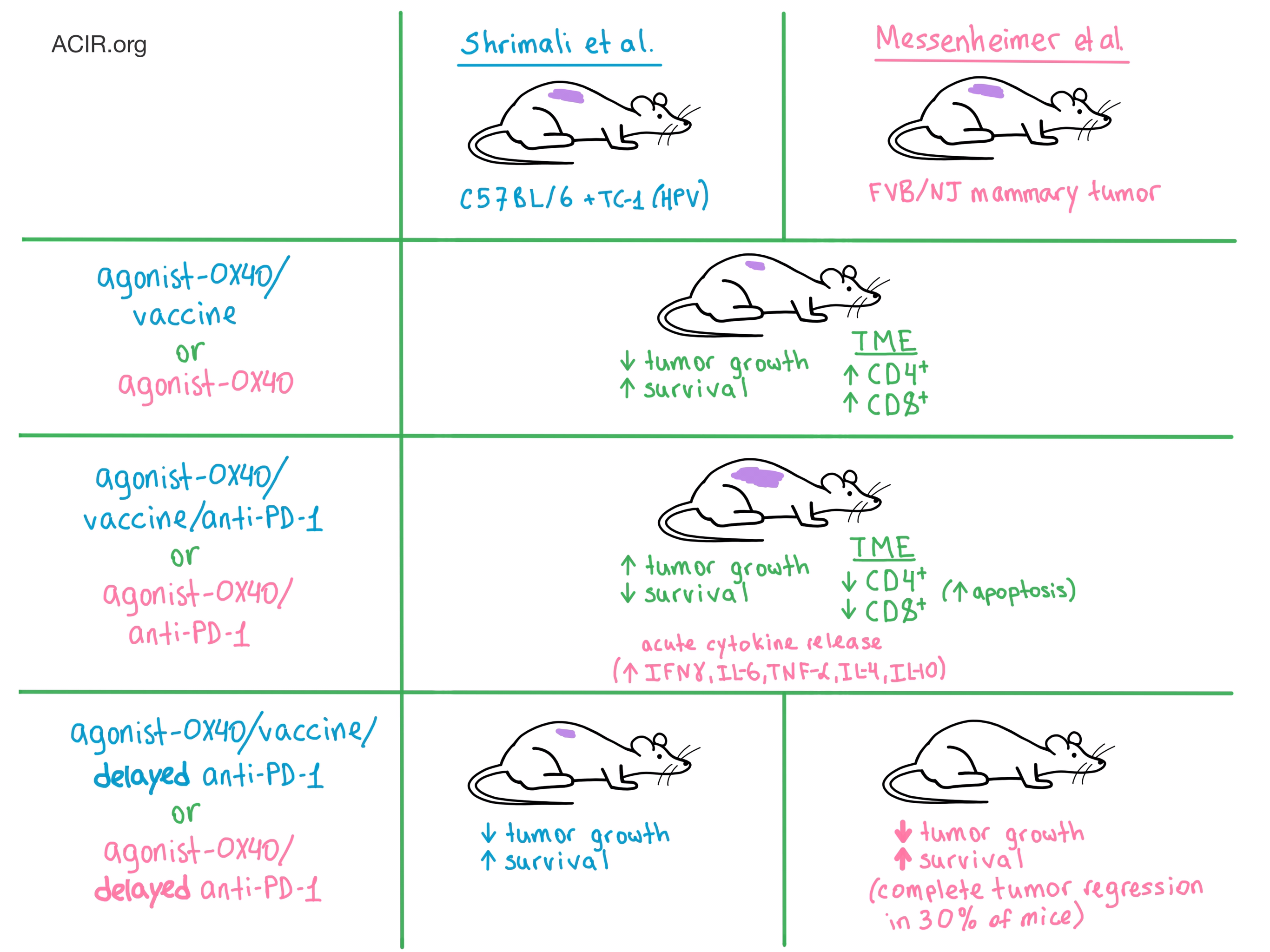
The researchers were inspired by the challenges of optimally combining immunotherapies in the clinical setting. To address this problem, Shrimali et al. utilized a syngeneic mouse model in which C57BL/6 mice were implanted with TC-1 cells (an HPV E6- and E7-driven tumor). This model requires a vaccine (an epitope from HPV protein E7) to create an effector immune response. Meanwhile, Messenheimer et al. orthotopically transplanted spontaneous tumors from FVB/NJ mice with mammary cancer into naive FVB/NJ mice. This mouse model does not utilize a vaccine, is refractory to PD-1 blockade, and was selected to represent the patients who do not respond to PD-1 blockade in the clinic.

OX40 is a costimulatory receptor that promotes T cell activation and expansion, enhanced effector function, immune memory generation, and inflammatory antitumor response. When Shrimali et al. administered agonist-OX40 in combination with vaccine, the rate of tumor growth was significantly reduced and mice experienced prolonged survival compared with untreated mice. Messenheimer et al. observed similar results with the administration of agonist-OX40 alone.

Intending to further improve the antitumor response, the researchers on both teams concomitantly added anti-PD-1 to the treatment. Surprisingly, both teams witnessed a significant reduction in tumor control and survival compared with agonist-OX40 or agonist-OX40/vaccine alone.

Next, the teams decided to delay the anti-PD-1 administration in order to allow the agonist-OX40 treatment to first boost T cell production. Messenheimer et al. observed that sequential dosing led to significant delay in tumor growth, as well as complete tumor regression in 30% of mice, compared with agonist-OX40 alone or concurrent combination therapy. They also witnessed a significant increase in survival compared with concurrent therapy. Reversing the order of therapy (anti-PD1 first followed by agonist-OX40) proved to be much less effective. Messenheimer et al. also confirmed these results in the poorly immunogenic 4T1 mammary tumor model. Meanwhile, Shrimali et al. observed that although delaying the addition of anti-PD-1 was not detrimental to the efficacy of the agonist-OX40/vaccine treatment, it did not provide any additional therapeutic benefit, either in tumor growth or survival.

To understand why the timing and order of administration of the two immunotherapy agents significantly influenced the therapeutic outcome, the researchers explored the effects of treatment on various T cell populations and secreted cytokines. Shrimali et al. noticed that the agonist-OX40/vaccine treatment increased the number of CD4+ and antigen-specific CD8+ T cells in the tumor, but the concomitant addition of anti-PD-1 significantly reduced the number of all tumor-infiltrating T cells, with the exception of CD4+ Tregs. Adding anti-PD-1 also led to significantly increased production of IFNγ, which resulted in enhanced CD8+ T cell proliferation and activation followed by increased apoptosis, both in the tumor and the spleen. High throughput immunosequencing demonstrated a decrease in T cell clonality after the addition of anti-PD-1 to agonist-OX40/vaccine, further suggesting apoptosis of antigen-specific T cells. In contrast, delaying the addition of anti-PD-1 did not alter the proliferation and apoptosis of CD8+ T cells compared to agonist-OX40/vaccine alone, which may help explain the distinct therapeutic results observed between concomitant and delayed administration of anti-PD-1.

Messenheimer et al. also observed a large increase in IFNγ production after concomitant combination therapy compared to either monotherapy and an increase in other cytokines, including IL-6, TNF-α, IL-4, and IL-10. Combined with certain physical symptoms (unkempt fur, lethargy), the researchers concluded that the animals experienced acute cytokine release. The researchers also saw that concurrent combination therapy increased T cell proliferation, but this effect was short-lived and did not correlate with improved therapeutic response due to rapid and increased apoptosis. In addition, concurrent combination treatment increased the frequency of exhausted TIM-3+CD8+ T cells in the tumor compared with agonist-OX40 alone. In contrast, sequential addition of anti-PD-1 did not lead to either abrogated T cell proliferation, increased T cell exhaustion, or spikes in cytokine production (indicating a safer, more effective profile). Additional depletion experiments demonstrated that both CD4+ and CD8+ T cells were required for optimal therapeutic effect: the former for immediate tumor control, and the latter for long-term memory response.

The results of these studies by Shrimali et al. and Messenheimer et al. exquisitely demonstrate the delicate “compensatory, regulatory, and homeostatic” balance that is maintained by the immune system, and underscore the importance of timing in the design of combination immunotherapy for cancer.

by Anna Scherer

References:

Shrimali R.K., Ahmad S., Verma V., Zeng P., Ananth S., Gaur P., Gittelman R.M., Yusko E., Sanders C., Robins H., Hammond S.A., Janik J.E., Mkrtichyan M., Gupta S., Khleif S.N. Concurrent PD-1 Blockade Negates the Effects of OX40 Agonist Antibody in Combination Immunotherapy through Inducing T-cell Apoptosis. Cancer Immunol Res. 2017 Sep.

Messenheimer D.J., Jensen S.M., Afentoulis M.E., Wegmann K.W., Feng Z., Friedman D.J., Gough M.J., Urba W.J., Fox B.A. Timing of PD-1 Blockade Is Critical to Effective Combination Immunotherapy with Anti-OX40. Clin Cancer Res. 2017 Aug 28.

In the Spotlight...

Transfer of in vitro-expanded naive T cells after lymphodepletion enhances antitumor immunity through the induction of polyclonal antitumor effector T cells

Tanaka et al. demonstrate in the mouse that transfer of ex vivo expanded naive CD4+ T cells into lymphodepleted hosts, coupled with in vivo depletion of Tregs, resulted in significant tumor control, induction of tumor cell-specific, recipient-derived CD8+ effector cells, and long-term persistence and activity of the transferred CD4+ T cells. Transfer of ex vivo-expanded naive CD8+ T cells had no effect. Addition of a dendritic cell vaccine enhanced CD4+ T cell efficacy.

Hit-and-run programming of therapeutic cytoreagents using mRNA nanocarriers

Moffett et al. developed targeted mRNA nanocarriers that can easily and robustly reprogram specific cell types to replace or complement the existing gene therapy toolbox (viral vectors and electroporation). To demonstrate the generalizability of this method, the team disrupted endogenous TCR expression in CAR T cells, reprogrammed effector T cells into a central memory-like state, and accelerated the in vitro expansion of hematopoietic stem cells.

Photodynamic-immune checkpoint therapy eradicates local and distant tumors by CD8+ T cells

In two murine tumor models, Kleinovink et al. evaluated the abscopal effect of local photodynamic therapy (PDT) with Bremachlorin. PDT effectively cleared primary tumors and slowed the growth of secondary tumors – an effect dependent on CD8+ T cells. Co-treatment with anti-CTLA-4 therapy complemented PDT, leading to a significant extension of survival, also dependent on CD8+ T cells.

Neoadjuvant Interferons: Critical for effective PD-1 based immunotherapy in TNBC

Brockwell et al. tested the Type I IFN-inducing TLR3 agonist poly(I:C) and anti-PD-1 against triple-negative breast cancer in preclinical models and found that while anti-PD-1 was ineffective as a single agent, it enhanced the antitumor effects of poly(I:C), activating NK cells, CD4+ T cells, and tumor-specific CD8+ T cells. Importantly, combination therapy prolonged survival only when administered as neoadjuvant therapy prior to primary tumor resection.

Antigen-presenting intratumoral B cells affect CD4+ TIL phenotypes in non-small cell lung cancer patients

Tumor infiltrating B cells (TIL-Bs) correlate with survival in patients with NSCLC, and Bruno et al. found that TIL-Bs efficiently present antigen to CD4+ T cells. “Activated” TIL-Bs led to activated (nonspecific) or antigen-associated effector CD4+ T cell responses, while “exhausted” B cells led to a non-responsive CD4+ Treg phenotype. Although the patient numbers were small, activated and exhausted TIL-B populations correlated with patient outcomes.

A Jak2-selective inhibitor potently reverses the immune suppression by modulating the tumor microenvironment for cancer immunotherapy

He et al. used a novel small molecule Jak2 kinase inhibitor (compound 9#) to suppress the Jak2-STAT3 signaling pathway. Compound 9# re-polarized macrophages from a pro-tumor (M2) to a proinflammatory (M1) phenotype, inhibited the induction of Treg cells, and increased the frequency and antitumor activity of CD4+ and CD8+ T cells, leading to effective suppression of tumor growth in two mouse models.

Role of Lymphocyte Subsets in the Immune Response to Primary B Cell-Derived Exosomes

Saunderson et al. explored the in vivo cellular and molecular requirements under which exosomes derived from antigen-exposed B cells present antigens and induce a cytotoxic CD8+ T lymphocyte response. They identified CD4+ T cells and, unexpectedly, NK cells, but not B cells, as crucial helper cells, and host MHC I molecules and splenic langerin+CD8α+ dendritic cells (DCs) as critical for antigen cross-presentation.

Lack of interleukin-6 in the tumor microenvironment augments type-1 immunity and increases the efficacy of cancer immunotherapy

Ohno et al. showed that tumor growth in IL-6-deficient mice was suppressed compared to wild-type mice due to augmented infiltration of cytotoxic T cells and mature dendritic cells into the tumor microenvironment. Lack of IL-6 further enhanced the antitumor effect of anti-PD-L1 therapy, suggesting that blocking IL-6 signaling may be a promising approach in cancer immunotherapy.

Everything New this Week In...

Close Modal

Small change for you. Big change for us!

This Thanksgiving season, show your support for cancer research by donating your change.

In less than a minute, link your credit card with our partner RoundUp App.

Every purchase you make with that card will be rounded up and the change will be donated to ACIR.

All transactions are securely made through Stripe.