Weekly Digests
‹ Back to December

The battle of receptors: CAR vs TCR

December 13, 2017

Chimeric antigen receptor (CAR) T cells have successfully induced remission in patients with relapsed or refractory B cell malignancies, but a substantial portion of patients eventually relapse. Although CAR expansion and persistence are considered important attributes for long-term benefit, systematic evaluation of contributing factors has been complicated by reliance on xenograft models. In a paper published in Science Translational Medicine, Yang et al. overcame this limitation by using transgenic TCR mice and uncovered some unexpected findings surrounding the biology of the dual antigen specificity (CAR and TCR) of CAR T cells and how it affects their antitumor response.

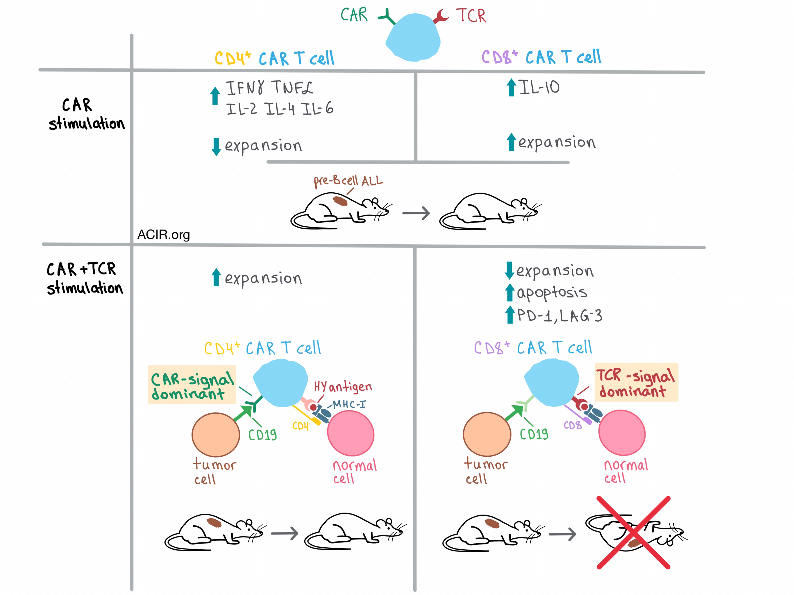
In the initial experiments, the team found that CD4+ and CD8+ T cells transduced with a murine CD19 CAR (CAR4 and CAR8 cells, respectively) were equivalently cytotoxic in vitro against syngeneic CD19+ pre-B cell acute lymphoblastic leukemia (ALL). A closer look, however, revealed important differences between the two CAR T cell types stimulated through the CAR. For example, CAR4 cells produced significantly more immunostimulatory cytokines, such as IFNγ, TNFα, IL-2, IL-4, and IL-6, than CAR8 cells, while CAR8 cells produced more immunosuppressive IL-10. On the other hand, the expansion of CAR4 cells was reduced in the presence of CAR8 cells, which were preferentially expanded.

To explore the effect of dual activation of CAR T cells on antitumor efficacy in vivo, Yang et al. utilized their CD19+ pre-B cell ALL model together with syngeneic CAR4 and CAR8 cells from transgenic mice with endogenous TCRs specific for a male minor histocompatibility antigen (HY). In a clever experiment, the team adoptively transferred HY-specific CAR4 or CAR8 T cells from female donors into ALL male (HY+) and female (HY-) mice, thus allowing the researchers to isolate the effects of CAR stimulation from the influence of TCR signaling. They found that CAR4 cells eliminated leukemia in both female and male mice and improved survival to >100 days. While CAR8 cells had a similar effect on female mice, they failed to clear leukemia and prolong survival in males relative to control mice. These results indicate that TCR signaling abrogates the ability of CAR8, but not CAR4 cells to eradicate leukemia, hinting at innate biological differences between the two types of CAR T cells.

In an attempt to figure out why CAR8 cells did not eliminate leukemia in HY+ mice, Yang et al. analyzed the adoptively transferred CAR T cells and found that the presence of the TCR antigen preferentially reduced the expansion of the CAR8 and not CAR4 cells, and that this was due to increased CAR8 apoptosis. Further analysis revealed that in female mice, both CAR4 and CAR8 cells had elevated PD-1 and LAG-3 exhaustion markers upon CAR stimulation. While the presence of TCR antigen in male mice did not further increase these markers on CAR4 cells, CAR8 cells became even more exhausted.

The team then created female/male hematopoietic chimeras via CD3-depleted bone marrow transplantation and found that even when the HY antigen was restricted to the hematopoietic tissues, CAR8 cells failed to eliminate leukemia, whereas CAR4 cells cleared the tumor and prolonged survival regardless of where HY was expressed. Furthermore, the hematopoietic restriction did not prevent CAR8 exhaustion. Similar results were observed in the OVA-specific OT1 transgenic mouse model, suggesting that these results apply to TCR-stimulated CD8+ CAR T cells in general. These experiments provide further evidence of an inherent, selective resistance of CAR4 cells to the negative effects of TCR stimulation. However, it is important to note that although CAR4 cells were successful in initial expansion and tumor clearance, they did eventually become exhausted and apoptotic upon chronic TCR antigen exposure, suggesting that long-term persistence of CAR4 cells and surveillance of leukemia may still be impaired.

In order to get to the root of the biological differences between CAR4 and CAR8 cells, Yang et al. examined the differential gene expression profiles of these cells after stimulating them with either CAR antigen, TCR antigen, or a combination of the two. Stimulation through either receptor alone generated distinct patterns in both CAR4 and CAR8 cells, but dual stimulation demonstrated that the TCR signal dominated in CAR8 cells, while the CAR signal dominated in CAR4 cells. In addition, CAR8 cells exhibited an increased expression of proapoptotic and inhibitory receptor genes after TCR stimulation, consistent with the phenotypic results.

Overall, these results raise awareness of the importance of TCR specificity and antigen presentation to CAR cells with endogenous TCRs, and indicate multiple pathways that could be targeted to improve the performance and persistence of CAR T cells and enhance the therapeutic potential of CAR T cell therapy.

by Anna Scherer

References:

Yang Y., Kohler M.E., Chien C.D., Sauter C.T., Jacoby E., Yan C., Hu Y., Wanhainen K., Qin H., Fry T.J. TCR engagement negatively affects CD8 but not CD4 CAR T cell expansion and leukemic clearance. Sci Transl Med. 2017 Nov 22.

In the Spotlight...

STAT5B: A Differential Regulator of the Life and Death of CD4(+) Effector Memory T Cells

To understand the mechanism underlying antigen restimulation-induced T cell death (RICD), a critical control to dampen T cell effector memory (TEM) levels, Majri et al. observed that STAT5B signaling drives RICD in TEM cells. A genetic screen of a patient with an autoimmune disorder revealed a missense mutation in the dimerization domain of STAT5B, causing lack of transcriptional activity. The patient’s CD4+ TEM cells were significantly increased and were highly resistant to cell death by in vitro TCR restimulation.

Delivering type I interferon to dendritic cells empowers tumor eradication and immune combination treatments

To reduce systemic side effects of alpha interferon but retain immune cell stimulating activity, Cauwels et al. constructed a mutated cytokine with >100x lower potency targeted to Clec9A+ cross-presenting dendritic cells (Clec9A-mAFN). In vivo, peri-tumoral Clec9A-mAFN stimulated Clec9A+ DCs in tumor-draining lymph nodes, controlled tumor growth in multiple models as well as full strength interferon but without toxicity, and synergized with other therapies, including checkpoint blockade. Activity depended on Batf3+ DCs and CD8+ T cells.

Elimination of tumor by CD47/PD-L1 dual-targeting fusion protein that engages innate and adaptive immune responses

Building on prior evidence that anti-PD-1 and anti-CD47 antibodies can synergize by stimulating both the innate and adaptive immune systems and that tumor cells co-express both targets, Liu et al. developed a bispecific antibody (IAB) targeting both molecules. In vitro, IAB promoted T cell activation and macrophage phagocytosis. In an immunocompetent MC38 mouse model, intratumoral injection of IAB led to partial or complete antitumor responses in most mice. Both CD8+ T cells and macrophages contributed to the anti-tumor effect of IAB.

TEM8/ANTXR1-specific CAR T cells as a targeted therapy for triple-negative breast cancer

Byrd et al. engineered CAR T cells targeting TEM8, a marker on triple-negative breast cancer (TNBC), breast cancer stem-like cells (BCSCs), and tumor endothelial cells (TEC). The CAR T cells effectively killed TEC and TNBC cells and reduced the formation of mammospheres by BCSCs in vitro. In vivo, the CAR T cells induced regression of established cell line- and patient-derived xenograft tumors, disrupted tumor vascularization and infrastructure, blocked metastasis in xenograft tumors derived from a lung metastatic TNBC cell line, and improved survival.

Engagement of MHC class I by the inhibitory receptor LILRB1 suppresses macrophages and is a target of cancer immunotherapy

Barkal et al. discovered that a high level of expression of MHC class I molecules on the surface of tumor cells provides protection against phagocytosis by macrophages, mediated by interaction between the LILRB1 inhibitory receptor on the surface of macrophages and the β2-microglobulin component of the MHC class I molecule, independent of the MHC-I allele. Disrupting the MHC class I/LILRB1 axis, similar to disruption of the CD47/SIRPα axis, enhances phagocytosis of tumor cells in vitro and in vivo.

Single-cell multiplexed cytokine profiling of CD19 CAR-T cells reveals a diverse landscape of polyfunctional antigen-specific response

Xue et al. used a single-cell microfluidics device to simultaneously measure 16 cytokines released by CD19 CAR T cells in response to target-specific stimulation, and developed two novel bioinformatics tools to visualize the data. CD19 CAR T cells exhibited a high degree of polyfunctionality (production of multiple cytokines, a predictor of therapeutic efficacy) dominated by antitumor effector and stimulatory functions, and significant heterogeneity, both within cells from the same donor and between donors.

Hypoxia and hypoxia-inducible factor (HIF) downregulate antigen-presenting MHC class I molecules limiting tumor cell recognition by T cells

Sethumadhavan et al. discovered that hypoxia in the tumor microenvironment, and in 3D cultures, leads to decreased surface expression of MHC class I molecules by downregulating transcription of MHC class I heavy chains via the hypoxia-inducible factor (HIF) and by decreasing the levels of TAP1, TAP2, and LMP7, critical components of the antigen processing pathway. These results suggest that cancer immunotherapy may be improved by the addition of oxygenation or HIF inhibition.

Anti-SIRPalpha antibody immunotherapy enhances neutrophil and macrophage antitumor activity

Ring et al. developed KWAR23, an antibody that targets the SIRPα receptor on myeloid cells, to disrupt binding to the CD47 “don’t eat me” signal expressed on tumor (and normal) tissue to prevent macrophage phagocytosis. In combination with tumor-opsonizing antibodies, KWAR23 induced macrophages and neutrophils to phagocytose target tumor cells (from various tumor cell lines) in vitro. In human sirpa knock-in mice on an SRG background (lacking T, B, and NK cells), this combination elicited partial and complete responses.

PD-1 is a haploinsufficient suppressor of T cell lymphomagenesis

In T cell non-Hodgkin lymphoma (NHL), oncogenic mutations to the TCR pathway induce chronic proliferation, however, Wartewig et al. found that PD-1 receptor-driven T cell inhibition suppresses oncogenic signaling in premalignant cells. In this context, total or hemizygous knockout of the PD-1 gene led to T cell hyperproliferation and rapid lymphomagenesis; treatment with anti-PD-1 antibodies led to lethal hyperproliferation, but the expanded cells were not lymphomagenic. Caution is warranted in employing checkpoint inhibition in T cell NHL.

An Efficient Single-Cell RNA-Seq Approach to Identify Neoantigen-Specific T Cell Receptors

Lu et al. developed a new approach to identify neoantigen-specific TCRs for use in adoptive T cell transfer. The process involves briefly co-culturing expanded tumor-infiltrating lymphocytes with autologous APCs that have been transfected with tandem minigenes containing patient-specific mutations or pulsed with mutant peptide, and using single-cell RNA sequencing to identify TCR sequences on activated T cells that express high levels of IFNγ and/or IL-2. A clinical trial to test this approach is being planned.

Everything New this Week In...

Close Modal

Small change for you. Big change for us!

This Thanksgiving season, show your support for cancer research by donating your change.

In less than a minute, link your credit card with our partner RoundUp App.

Every purchase you make with that card will be rounded up and the change will be donated to ACIR.

All transactions are securely made through Stripe.