Dendritic cell (DC)-targeting vaccines directed at inducing an immune response to tumor antigens can induce an antitumor immunity, but are limited in their activity and efficacy. Inducing more robust DC responses might enhance the immune response to these vaccines. Bhardwaj et al. evaluated the ability of the dendritic cell stimulator Flt3 ligand to enhance responses to a DC-targeted antigen in a phase II trial (NCT02129075) and reported the results in Nature Cancer.
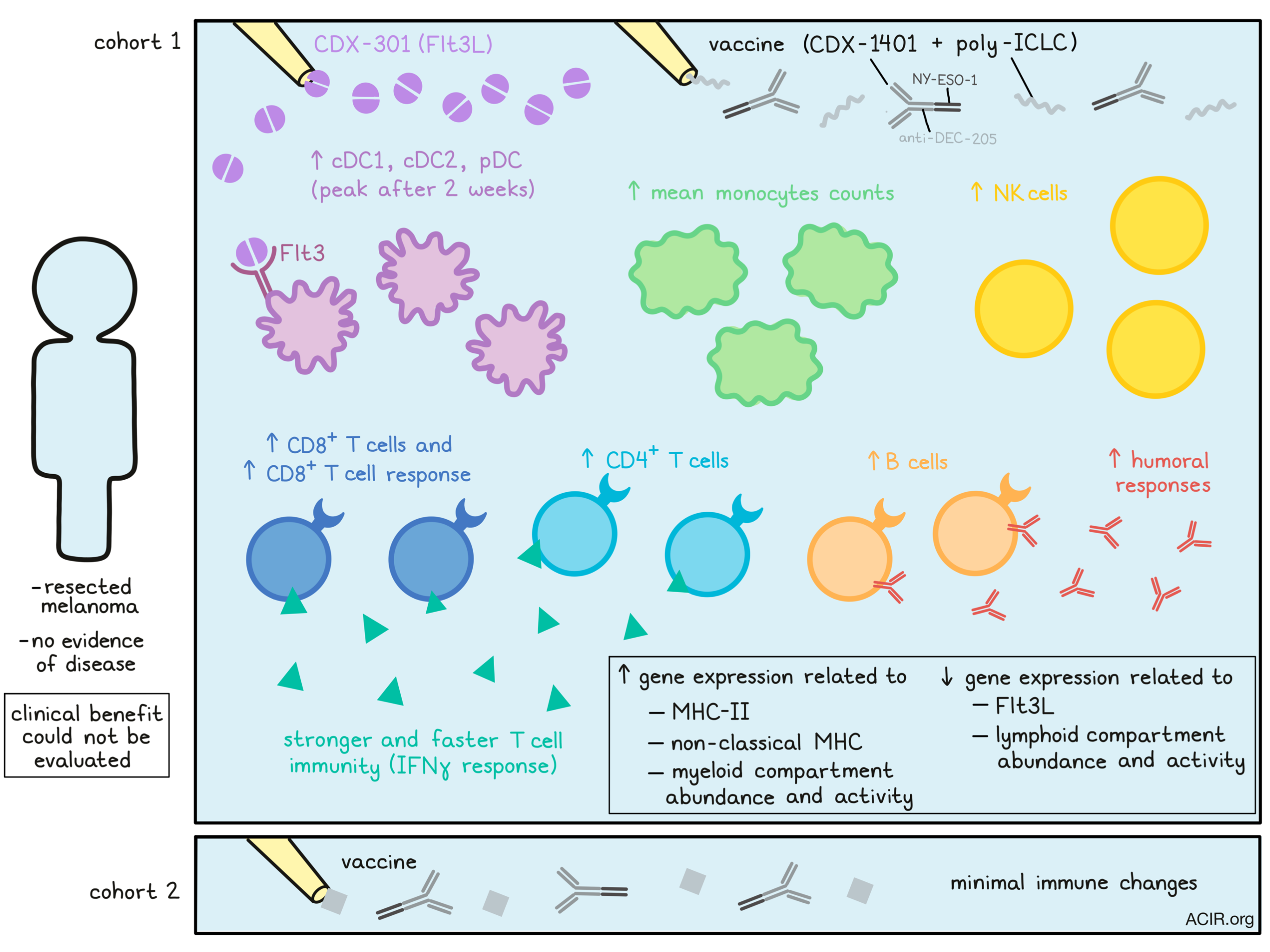
CDX-301 is a soluble form of the homodimeric surface-bound ligand for Flt3 and has been shown to induce proliferation, differentiation, and mobilization of DC precursors in the bone marrow, resulting in enhanced DC numbers in the blood and lymphoid organs. Study treatment also included a vaccine consisting of CDX-1401, a fusion protein of the human anti-DEC-205 monoclonal antibody linked to the NY-ESO-1 antigen, and poly-ICLC, a TLR3 and MDA-5 agonist. The trial included 60 patients with resected melanoma without evidence of disease at enrollment. Patients were randomized into two cohorts; cohort 1 received the vaccine supplemented with CDX-301, and cohort 2 received the vaccine alone.
Bhardwaj et al. started by analyzing specific T cell responses to NY-ESO-1. Using IFNγ ELISpot, the researchers showed that T cell immunity was induced faster and with a higher magnitude in cohort 1 (which included CDX-301) than in cohort 2. All patients in cohort 1 had responses at at least one of the measured time points, which was only found for 27% of cohort 2. Responses were detected until the latest time point, 12 weeks after vaccination; in a sub-analysis a few months later, patients still had detectable responses. The researchers also measured intracellular IFNγ, TNFα, and IL-2 production in CD4+ and CD8+ T cell subsets. All patients assessed had detectable CD4+ T cell responses in both cohorts, while CD8+ T cell responses were found in 86% of patients in cohort 1 and 29% of patients in cohort 2. Patients were enrolled independently of the NY-ESO-1 status of their tumors, and archival sample analysis revealed that only 19% of patients had NY-ESO-1-expressing tumors. This expression correlated with higher baseline T cell responses, but not with T cell responses to vaccination.
Moving to assess the humoral response, the researchers measured NY-ESO-1 antibodies in the patient’s serum. Patients in cohort 1 all achieved humoral responses, while those in cohort 2 required more vaccinations for detectable titers. Titers in cohort 1 were also higher than those detected in cohort 2, while baseline levels were low in all patients; titers were independent of tumoral NY-ESO-1 expression.
The researchers then assessed the quantity and phenotypes of monocytes, DCs, and lymphocytes in patients' blood by performing whole-blood phenotyping using flow cytometry. The DC subsets cDC1, cDC2, and pDC increased to peak levels two weeks after the start of CDX-301 administration, and these levels decreased back to baseline levels over 10-20 days, similar to results obtained in healthy individuals treated with CDX-301 alone, suggesting a specific vaccine-independent effect. While no differences in monocyte counts were observed in cohort 2, patients who received CDX-301 had a six-fold rise in mean monocyte counts.
Lymphocyte counts also significantly changed after CDX-301 treatment, while no changes occurred in cohort 2. NK cells increased in the first cycle, and immature (CD56bright) NK cells increased even more during the second cycle. Only modest increases were found for B cells, CD4+ T cells and CD8+ T cells over the two cycles. HLA-DR was used to quantify activation levels of these subsets. In cohort 1, there was an increase in the number of cDCs, NK cells, and CD4+ and CD8+ T cells with higher HLA-DR levels, which was not seen in cohort 2. After treatment, all levels returned to baseline.
The researchers then assessed transcriptional changes in PBMC in response to CDX-301 treatment. For this, the NanoString PanCancer Immunology Panel was applied to RNA extracted from PBMCs at 12 timepoints. CDX-302 induced extensive and durable changes in gene expression, which were greatest on the eighth day after cycles 1 and 2, and most changes returned to baseline after cycle 2. Some of the highly upregulated genes included those involved in antigen presentation by MHC class II (HLA-DM, HLA-DP, HLA-DR), TLR, IL13RA1, non-classical MHC-related genes (CD1C and CD1D), and multiple genes related to myeloid compartment abundance and activity. On the other hand, Flt3LG and genes associated with lymphoid compartment abundance and activity were downregulated.
Using the Nanostring panel, the researchers inferred cell composition by computing cell type abundance scores using the mean expression of cell type-specific marker genes. Monocyte and DC scores increased after CDX-301 treatment, while lymphoid scores dropped, except for the NK cell score, which was elevated 15 days after cycle 2. These changes were consistent with the flow cytometry data, except for the drop in lymphoid scores, which might be explained by the strong increase in the myeloid compartment.
Finally, the NanoString expression data were used to create a modular repertoire framework. Modules were formed based on sets of genes with similarities in abundance patterns among the datasets. Again, changes in immune populations were abundant in cohort 1 and absent in cohort 2. All changes were temporary, peaking at day 8 after treatment and decreasing back to baseline levels at day 22. Modules associated with inflammation, cell death, apoptosis/survival, proliferation, immune suppression, monocytes, and neutrophils were upregulated. Ten modules were downregulated and included T cells, B cells, platelets, leukocyte activation, cell cycle, proliferation, cytotoxic, and NK cells modules. Within the IFN modules, modules associated with IFNα and IFNβ were upregulated at day 8 of both cycles, while the module related to IFNγ was not affected. Together, these changes suggest potentially useful perturbations in innate and adaptive aspects of the immune system.
The regimen was well-tolerated, and there was no dose-limiting toxicity. The study design prevented clinical benefit measurement, but the monocyte compartment changes and the elevated specific T cell responses suggest increases in immunogenicity and warrant clinical effects assessment. Since many solid tumors express NY-ESO-1, this vaccine strategy is of interest to multiple cancer types, and could be extended to other antigens. Finally, combining this T cell-stimulating vaccine strategy with checkpoint blockade has the potential to enhance antitumor responses further.
Write-up by Maartje Wouters, image by Lauren Hitchings
Meet the Researcher
This week, corresponding authors Steven Fling and Nina Bhardwaj answered our questions.
What prompted you to tackle this research question?
This study was the brainchild of my colleague and co-corresponding author Dr. Nina Bhardwaj. Most importantly, the trial was prompted by the critical need to increase effective cancer antigen-specific immune responses in patients. While T cells are vital in so many respects to cancer immunotherapy, actual control of such effective adaptive immunity resides with professional antigen presenting cells. In this, dendritic cells are essential at several levels in controlling this aspect of immunity – productively regulating immune reactivity to “self” versus “non-self”. Flt3 ligand was discovered ~27 years ago at Immunex Corp. as a hematopoietic factor essential to regulating dendritic cell development, and researchers there had prescient hopes for this molecule as a cancer treatment. The hypothesis that expanding dendritic cells with Flt3L, targeting antigen to their antigen-presenting machinery via a cell surface marker (CD205), and activating those cells for antigen presentation would lead to an improved immune response was a logical and reasonable therapeutic hypothesis.
What was the most surprising finding of this study for you?
There are, for me, three things perhaps most enlightening (surprising) in this study. Scientifically, while we were confident in the hypothesis described above, I was surprised in the strength of the immune response within Flt3L-treated subjects. Significant T cell responses to a quasi-self-antigen were invoked and observed directly ex vivo after only ONE vaccination in a high percentage of those receiving Flt3L, and ultimately all subjects responded strongly. This is truly a very significant observation, not only for cancer patients, but for the potential of vaccine strategies in general. I was also surprised, and grateful, for the incredible adherence to protocol from all the patient participants in this trial. The protocol was demanding, and none of the patients deviated from the rigors of the study. We are grateful. Finally, I have been surprised by the complexity of dendritic cell biology, which has been revealed during the period of this study, and which we are now only beginning to investigate within this study to determine correlates of the productive immune response.
What was the coolest thing you’ve learned (about) recently outside of work?
These have been unprecedented times during COVID-19, so outside of work, I would say that the ‘coolest’ thing I’ve learned/observed recently is the incredibly resilient, healing potential of nature when civilization pauses expansion. The speed at which nature recovers to re-establish balance is remarkable, encouraging… and enlightening. Besides this critical need to pause in general and “smell the roses you have” even in the worst of times, I realized how important it was to be with family. So I am doing my best now to reengage with those I have not in a while, even though it is by zoom.


