
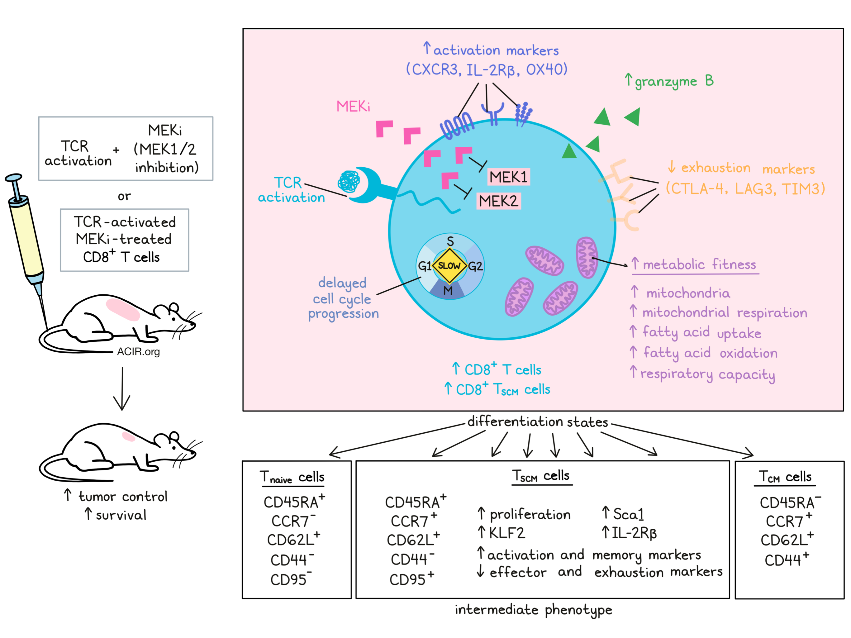
In the past, studies have shown that inhibition of MEK (MEKi) increases antitumor immunity and leads to the accumulation of activated CD8+ T cells in the TME. While this strategy is known to synergize with checkpoint inhibitors and adoptive cell transfer, little is known regarding exactly how MEKi affects T cell function, differentiation, or memory generation. In a study recently published in Nature Immunology, Verma et al. investigated the effects of MEKi on T cells and how it contributes to antitumor immunity.
To begin, Verma et al. showed that the addition of MEKi (using the MEK1/2 inhibitor selumetinib) to vaccination against known antigens (gp100 in B16F10 melanoma models and HPV16E7 in TC-1 models) in tumor-bearing mice increased total CD8+ T cells (many of which were antigen-specific and expressed granzyme B), reduced tumor burden, and improved survival. The CD8+ T cell population showed increased markers of activation (CXCR3, IL-2Rβ, OX40) and reduced markers of inhibition (CTLA4, TIM3, LAG3), though differences in PD-1 expression were not observed with the addition of MEKi to vaccine.
Investigating how MEKi enhances CD8+ T cells, Verma et al. turned to metabolic fitness as a possible explanation. MEKi treatment in mice increased mitochondrial mass in CD8+ T cells from the TME, and ex vivo, MEKi treatment of pMEL-1 cells increased the number of mitochondria per cell. MEKi-treated cells had intact redox machinery, tightly packed cristae in mitochondria, higher oxygen consumption rates, and lower extracellular acidification rates, indicative of enhanced mitochondrial fitness and respiratory capacity. Further analysis of metabolic activity showed that glucose uptake did not change with MEKi treatment; however, fatty acid uptake and fatty acid-mediated metabolism were increased.
Given that metabolic fitness, respiratory capacity, and fatty acid oxidation have previously been associated with immune memory, Verma et al. investigated whether MEKi might have affected the development of memory phenotypes in CD8+ T cells. Within MEKi-treated T cells, they observed markers of memory generation and identified populations of central memory-like (CD62L+CD44+) and naive-like (CD62L+CD44-) CD8+ T cells; however, within the naive-like population, many cells expressed high levels of memory markers, including CD95 and CCR7, in line with recent observations of a population of minimally differentiated stem cell-like memory T (TSCM) cells. Expression of IL-2Rβ provided additional evidence of stem-like characteristics in these cells, suggesting that MEKi enriches CD8+ TSCM cells in tumors.
Next, the researchers characterized these cells in vitro by activating CD8+ T cells with gp100. Without MEKi, most of the CD8+ T cells attained a TCM phenotype, while a minor portion remained naive. When MEKi was added, a large portion of CD8+ T cells attained a TSCM phenotype; these cells were naive-like and showed lower mitochondrial potential, higher proliferative capacity, higher expression of the stem cell/T cell memory marker Sca1, higher expression of activation and memory markers, lower expression of effector and exhaustion markers, and higher expression of KLF2, which is associated with self-renewability, prolonged survival, and reduced apoptosis. Further studies showed that simultaneous inhibition of both MEK1 and MEK2 was required for TSCM generation.
To further establish that TSCM cells were distinct from Tnaive and TCM cells, Verma et al. analyzed human MEKi-treated CD3/CD28-activated CD8+ T cells. Based on methylation profiles and CpG DNA methylation of key genes dynamically regulated during T cell memory development, the researchers showed that MEKi-induced TSCM cells (CD45RA+CCR7+CD95+) were distinct from both TCM cells (CD45RA-CCR7+) and Tnaive cells (CD45RA+CCR7-CD95-), and were most similar to freshly isolated bona fide TSCM cells. The phenotypes were also functionally differentiated by their multipotency/plasticity proliferation indices and self-renewal capacities. Based on their observations, Verma et al. determined that the TSCM phenotype is an intermediate between Tnaive and TCM phenotypes.
To determine when and how MEKi induces the generation TSCM cells, Verma et al. pre-treated naive T cells with gp100, then later treated them with MEKi. This failed to induce the TSCM cell generation seen when gp100 and MEKi were administered simultaneously, suggesting that MEKi acts during TCR-mediated cell priming. Looking at cell cycle progression and differentiation, the researchers found that MEKi delayed cell cycle progression, leading to an accumulation of activated cells in earlier stages of cell division. MEKi did not interfere with PI3K- or Akt-mediated T cell activation, suggesting that it inhibits the cell cycle at an early phase while allowing for antigen-mediated activation. By knocking out or inhibiting various molecules along different pathways, Verma et al. showed that both the delay in cycle progression and the enhanced metabolic fitness induced by MEKi contributed to the generation of TSCM cells, and that both effects were required for optimal TSCM cell generation.
Testing whether MEKi treatment might be able to enhance CD8+ T cells for adoptive cell transfer (ACT), Verma et al. found that transfer of MEKi-treated, gp100-stimulated pMEL-1 CD8+ T cells prepared in vitro showed stronger effector recall responses than controls stimulated with gp100 alone, producing higher levels of IFNγ and granzyme B after antigenic rechallenge. ACT of gp100- and MEKi-treated cells into mice bearing B16F10 melanoma tumors resulted in a stronger antitumor response and prolonged survival compared to ACT of CD8+ T cells activated with gp100 alone. To determine the contribution of TSCM cells, the researchers depleted the cell product of CD62L+CD44-Sca1+ cells and repeated the experiment. While the gp100-treated cells showed similar efficacy upon adoptive cell transfer, the gp100- and MEKi-treated, TSCM-depleted cell product was less effective. Interestingly, these cells were still more effective than those treated with gp100 alone, suggesting that MEKi also enhances the antitumor efficacy of TCM cells. Long term, adoptively transferred MEKi-treated cells maintained a memory phenotype more than non-treated cells.
Overall, Verma et al. showed that MEKi enhances the generation of TSCM cells by enhancing metabolic fitness and delaying cell cycle progression. MEKi could be particularly useful in enhancing CD8+ T cell products for ACT, as MEKi-treated T cells were more effective against tumors and prolonged survival in mice.
by Lauren Hitchings
Meet the researcher
This week, lead author Samir Khleif answered our questions.
What prompted you to tackle this research question?
T cell differentiation is highly important for proper immunologic responses. Accordingly, understanding the mechanism that regulates such differentiation is crucial not only to properly appreciate the fundamental biology of the T cells, but also to be able to develop effective strategies for immunotherapy of cancer. Furthermore, stem cell memory CD8+ T cells (TSCM) are highly potent and regenerative T cells. Understanding the signaling pathways that control the generation of TSCM cells would provide effective tools to pharmacologically reprogram CD8+ T cells at the appropriate time and conditions into this highly effective “super” T cell, and hence generate more effective immune therapeutic approaches.
What was the most surprising finding of this study for you?
The ability of the MEK pathway to highly and effectively control the differentiation of CD8+ T cells towards terminal differentiation, especially by enhancing their metabolic fitness. Also surprising was the high potential for such reprogramming to enhance the immunotherapy of cancer in a direct pharmacologic, therapeutic approach, and/or to enhance the potency of CD8+ ex vivo for use in an adoptive T cell approach.
What was the coolest thing you’ve learned (about) recently outside of work?
We may be able to do many things from a distance as effectively, or in some cases more effectively than in-person; however, human contact – discussion over a dinner or a drink – especially in science is highly missed as part of the creative cycle of our lives. This last year has been very special by all measures; however, one of the coolest things that we have lived through as scientists is the amazing dedication of our community, and the remarkable international collaboration leading to historical unprecedented discoveries. That was soooo COOL!