Weekly Digests
‹ Back to December

Antibodies, targets, and Fcγ receptors: a three-way tango

December 19, 2018

Given that antibodies to 4-1BB (a member of the tumor necrosis factor receptor superfamily that is expressed on activated T cells and other activated immune cells) have demonstrated antitumor effects in preclinical cancer models, Buchan, Dou, and Remer et al. set out to delineate the ways in which these antibodies promote antitumor immunity, and how the specific mechanisms depend on the antibodies’ Fab and Fc regions. The results were recently published in Immunity.

The researchers began by generating murine mIgG1 and mIgG2a versions of a anti-4-1BB monoclonal antibody (mAb). When tested in three solid tumor models (CT26 colon carcinoma, EG7 thymoma, NXS2 neuroblastoma), the mIgG2a antibody resulted in 80% long-term survival while the mIgG1 antibody did not produce much long-term benefit.

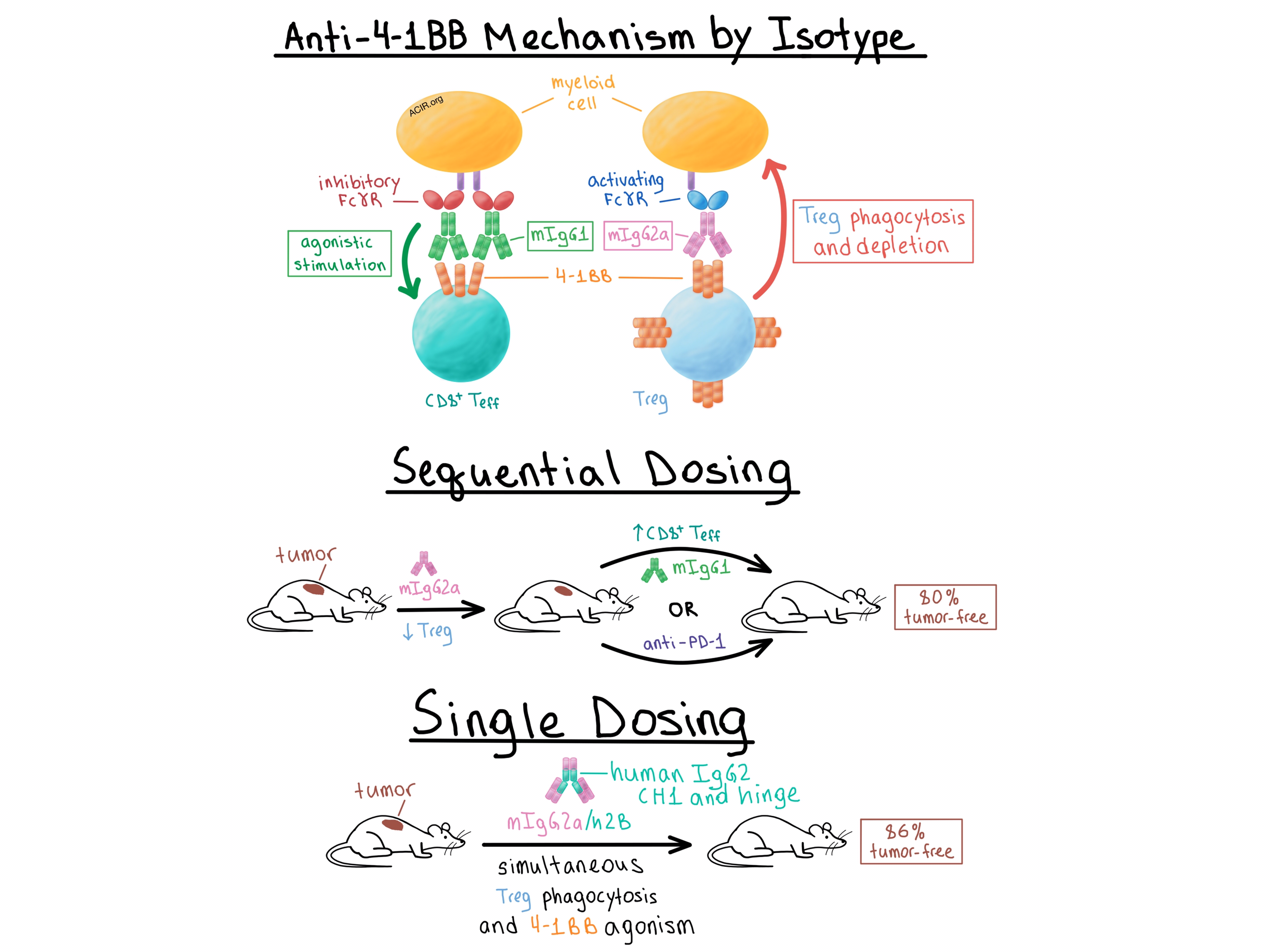
Buchan, Dou, and Remer et al. sought to understand the mechanistic differences between the two antibody isotypes that might explain their differential efficacy. Analysis of the antibodies’ binding to activating and inhibitory Fcγ receptors (FcγRs) showed that, as expected, mIgG1 was more likely to bind inhibitory FcγRs and mIgG2a was more likely to bind activating FcγRs, while both isotypes bound 4-1BB equally. Aware that 4-1BB is upregulated at much higher levels on intratumoral Tregs than on effector T cells in murine and human solid cancers, the researchers looked into Treg depletion.

The team found that mIgG2a, but not mIgG1, efficiently reduced the percentage of intratumoral Tregs in CT26 tumor-bearing wild-type mice. However, in mice that lacked the inhibitory FcγRIIB, mIgG1 also became quite effective at depleting intratumoral Tregs. In order to confirm these results, the researchers utilized bone marrow-derived macrophages from wild-type mice and showed that mIgG2a induced phagocytosis of 4-1BB-expressing target T cells, while mIgG1 did not. A separate experiment found that the efficiency of target cell depletion was directly correlated with the level of 4-1BB expression. Furthermore, in EG7 tumor-bearing mice with a selective knockout of 4-1BB only on Tregs, mIgG2a lost its Treg-depleting activity and long-term survival benefit. Together, these experiments demonstrated that Treg depletion depended on the binding of the antibody to both the 4-1BB on the target cell and an FcγR, presumably on a myeloid cell.

Diving deeper into the role of FcγRs in the therapeutic effect of anti-4-1BB mAbs, the researchers noted that while mIgG2a appeared to rely on activating FcγRs for its depleting functionality, it retained its antitumor effect in mice lacking activating FcγRs. In addition, mIgG1 improved its antitumor effect in the absence of activating FcγRs. When all FcγRs were absent, neither isotype provided any therapeutic effect. Together, these results suggested that the FcγRs compete for binding of both antibody isotypes, and in the absence of activating FcγRs, both isotypes successfully bind the inhibitory FcγRIIB and deliver the costimulatory signal.

Next, Buchan, Dou, and Remer et al. explored how isotype affects agonist activity. In vitro and in vivo, mIgG1 was more effective than mIgG2a in stimulating T cells. This effect was not dependent on the presence of Tregs, indicating that mIgG1 directly targets 4-1BB on effector T cells. When the two antibody isotypes were mixed, the costimulatory effect of mIgG1 disappeared, demonstrating that the two isotypes equivalently compete for binding of 4-1BB on T cells. Mechanistically, knockout experiments revealed that the agonistic activity of mIgG1 was dependent on the inhibitory FcγRIIB. Furthermore, mIgG2a demonstrated agonistic activity in the absence of activating FcγRs, suggesting that it bound the inhibitory FcγRIIB in these circumstances. In tumor-bearing mice, both isotypes led to an increase in proliferation and activation of tumor-infiltrating CD8+ T cells, although the magnitude of this effect was greater with mIgG1. Overall, these experiments showed that the therapeutic efficacy of mAbs was dependent on the mAb Fc binding profile and the availability of particular FcγRs. Specifically, mIgG1 exerted its effect primarily by directly activating CD8+ T cells following cross-linking via the inhibitory FcγRIIB, while mIgG2a preferentially bound activating FcγRs and promoted antitumor immunity primarily by depleting Tregs.

The team then considered that the differential mechanisms of action of the two isotypes could be combined to maximize the antitumor effect. While concurrent administration of mIgG1 and mIgG2a led to reduced antitumor efficacy compared with mIgG2a alone, sequential administration of mIgG2a first to deplete Tregs, followed by mIgG1 to stimulate effector T cells, reduced tumor size and increased the percentage of tumor-free mice to 80%. Sequentially dosing mice with mIgG2a followed by anti-PD-1 similarly increased the long-term response rate to 80%, compared with 20-25% response with either monotherapy. Meanwhile, combining mIgG1 with anti-PD-1 did not improve the response rate, once again demonstrating the importance of understanding the effect of isotype.

Finally, the researchers created a single anti-4-1BB antibody capable of simultaneously depleting Tregs and stimulating effector T cells by substituting a modified human IgG2 CH1 and hinge regions for the equivalent domains in the murine mIgG2a constant region – a change known to enhance FcγR-independent agonistic properties for other TNFR family members. The resulting anti-4-1BB mIgG2a/h2B antibody was more agonistic in vitro than the mIgG2a isotype while retaining its phagocytosis-inducing properties. In vivo, monotherapy with the antibody depleted intratumoral Tregs, stimulated tumor-infiltrating CD8+ T cells, and led to cure of 86% of EG7 tumor-bearing mice compared with 53% cure rate with mIgG2a. Thus, the team demonstrated that a single mAb could be rationally engineered, based on an understanding of the contributions of Fc binding to FcγR, to effect both depletion and agonism in order to improve the antitumor response of mAbs targeting 4-1BB and potentially other targets.

by Anna Scherer

References:

Buchan S.L., Dou L., Remer M., Booth S.G., Dunn S.N., Lai C., Semmrich M., Teige I., Mårtensson L., Penfold C.A., Chan H.T.C., Willoughby J.E., Mockridge C.I., Dahal L.N., Cleary K.L.S., James S., Rogel A., Kannisto P., Jernetz M., Williams E.L., Healy E., Verbeek J.S., Johnson P.W.M., Frendéus B., Cragg M.S., Glennie M.J., Gray J.C., Al-Shamkhani A., Beers S.A. Antibodies to Costimulatory Receptor 4-1BB Enhance Anti-tumor Immunity via T Regulatory Cell Depletion and Promotion of CD8 T Cell Effector Function. Immunity. 2018 Nov 20.

In the Spotlight...

Batf3+ DCs and type I IFN are critical for the efficacy of neoadjuvant cancer immunotherapy

To understand the role of innate immunity, particularly dendritic cells, in neoadjuvant checkpoint immunotherapy, Liu et al. evaluated the impact of knockout of batf3, a transcription factor that identifies a key subset of cross-presenting DCs, and blockade of type I IFN signaling. Batf3+ DCs and IFN signaling were required for efficacy of neoadjuvant immunotherapy in a mouse breast cancer model, impacting increases in antigen-specific TILs. Neoadjuvant immunotherapy increased the number and activation of CD103+ DCs in the draining, but not non-draining, lymph node. Human correlates with efficacy and batf3-associated genes were observed in a small trial.

Intracellular activation of complement C3 leads to PD-L1 antibody treatment resistance by modulating tumor-associated macrophages

Zha et al. showed that activation of complement factor C3 within tumor cells promoted an immunosuppressive TME by mediating the accumulation of TAMs and promoting their polarization towards an M2 (pro-tumor) phenotype. M2 polarization was induced by C3a binding to C3aR on TAMs, thereby activating PI3Kγ signaling and subsequent Akt phosphorylation. Deletion of C3 in various murine tumor models reduced tumor infiltration by TAMs, promoted M1 polarization, increased CD8+ T cell infiltration and proliferation, and slightly delayed tumor growth. Survival was significantly improved in conjunction with anti-PD-L1.

Semaphorin4D inhibition improves response to immune checkpoint blockade via attenuation of MDSC recruitment and function

Clavijo et al. found that use of a semaphorin4D (Sema4D)-blocking antibody enhanced the antitumor efficacy of anti-CTLA-4 or anti-PD-1 checkpoint blockade and improved survival in mice bearing MOC1 or LCC tumors. Mechanistically, anti-Sema4D prevented the recruitment of Ly6Ghi MDSCs by inhibiting MAPK-dependent production of CXCL1, 2, and 5 by tumor cells. Anti-Sema4D also decreased the suppressive capacity of MDSCs by blocking Sema4D-driven arginase expression. Together these effects increased tumor infiltration and expansion of CD8+ (and to a lesser extent, CD4+) T cells, which demonstrated an enhanced response to tumor antigen.

An antibody designed to improve adoptive NK-cell therapy inhibits pancreatic cancer progression in a murine model

To enhance the infiltration of adoptively transferred NK cells into pancreatic cancer (PC), Lee and Kang et al. developed the “NRP-body” – a fusion protein consisting of a mesothelin-targeting domain and the chemokine CXCL16 flanked by a furin cleavage site. In vitro and in vivo studies confirmed the release of CXCL16 from the NRP-body within the tumor tissue and the recruitment of transferred NK cells via the ERK-RhoA signaling cascade. In murine primary orthotopic and metastatic PC models treatment with NRP-body followed by NK cell therapy increased NK cell infiltration, reduced tumor burden, and increased overall survival.

T cell receptor fingerprinting enables in-depth characterization of the interactions governing recognition of peptide-MHC complexes

To derive a “TCR fingerprint” (amino acid residues that determine interactions between the TCR and the peptide-MHC [pMHC] complex), Bentzen and Such et al. measured the relative affinities of a TCR to DNA barcode-labeled pMHC multimers. Fingerprints differed substantially among TCRs recognizing the same epitope (even when isolated from the same patient) and the number of potential pMHC targets for a given TCR inversely correlated with the functional avidity of the corresponding T cell clone. TCR fingerprinting could be used to assess potential cross-recognition of TCRs selected for clinical development to reduce the risk of autoimmune reactions.

Genome-wide CRISPR Screens in Primary Human T Cells Reveal Key Regulators of Immune Function

Shifrut and Carnevale et al. developed a high-throughput CRISPR screening platform that works directly on ex vivo human T cells by combining sgRNA lentiviral infection with Cas9 protein electroporation (SLICE). Genome-wide specific disruption of target genes led to the identification of positive and negative regulators of proliferation following TCR stimulation and regulators of suppression following adenosine signaling. Knockdown of some negative regulator genes identified by SLICE enhanced target cell killing by T cells in vitro. Combining SLICE with RNAseq revealed how each gene manipulation can distinctly shape the cell state.

Lineage tracking reveals dynamic relationships of T cells in colorectal cancer

Analyzing single-cell transcriptome and TCR sequences from 12 colorectal cancer patients, Zhang, Yu, Zheng, and Zhang et al. categorized T cell subsets by unbiased clustering and phenotyping by localization, migration, and proliferation. A TCR clonotype-dependent developmental lineage linked CD8+ effector memory cells to recently activated effector memory and exhausted T cells (TEX). Microsatellite-unstable tumors were enriched in a clonally expanded and proliferative CXCL13+ BHLHE40+ Th1-like CD4+ T cell subset with upregulated IGFLR1, which was also upregulated in TEX . In vitro experiments suggested a role of IGFLR1 in TCR costimulation.

CD8+ T Cell Priming in Established Chronic Viral Infection Preferentially Directs Differentiation of Memory-like Cells for Sustained Immunity

To find out whether chronic viral infection, conceptually similar to cancer, quantitatively or qualitatively impacts T cell priming, Snell et al. transferred naive LCMV GP33-41-specific T cells to acutely or chronically LCMV-infected mice. Interestingly, transfer to chronically infected mice resulted in a greater self-renewing, memory-like expression pattern among CD8+ T cells, with more TCF1+ and fewer “exhausted” effector cells compared to acutely infected mice, apparently due to decreased TCR and co-stimulatory signaling by APCs. T cells primed during chronic infection were more responsive to anti-PD-1 and dependent on IL-21 for stimulation of T cell help.

Everything New this Week In...

Close Modal

Small change for you. Big change for us!

This Thanksgiving season, show your support for cancer research by donating your change.

In less than a minute, link your credit card with our partner RoundUp App.

Every purchase you make with that card will be rounded up and the change will be donated to ACIR.

All transactions are securely made through Stripe.