Weekly Digests
‹ Back to January

The shape of intratumor heterogeneity

January 9, 2019

Most tumors exhibit a high degree of genetic and functional heterogeneity, but how that heterogeneity is established and evolves in situ under immune pressure has been difficult to study. To tackle this obstacle, Milo et al. established a mouse model of MYC-driven B cell lymphoma with a large degree of genetic diversity, both between and within individual mice. In this model, chromosomes are frequently lost, which contributes to genetic diversity and subsequent mosaic expression of tumor antigens. In order to study tumor heterogeneity in this mouse model, the researchers developed a multicolor barcoding strategy that allows for analysis and visualization of marked tumor cells from clonally diverse tumors.

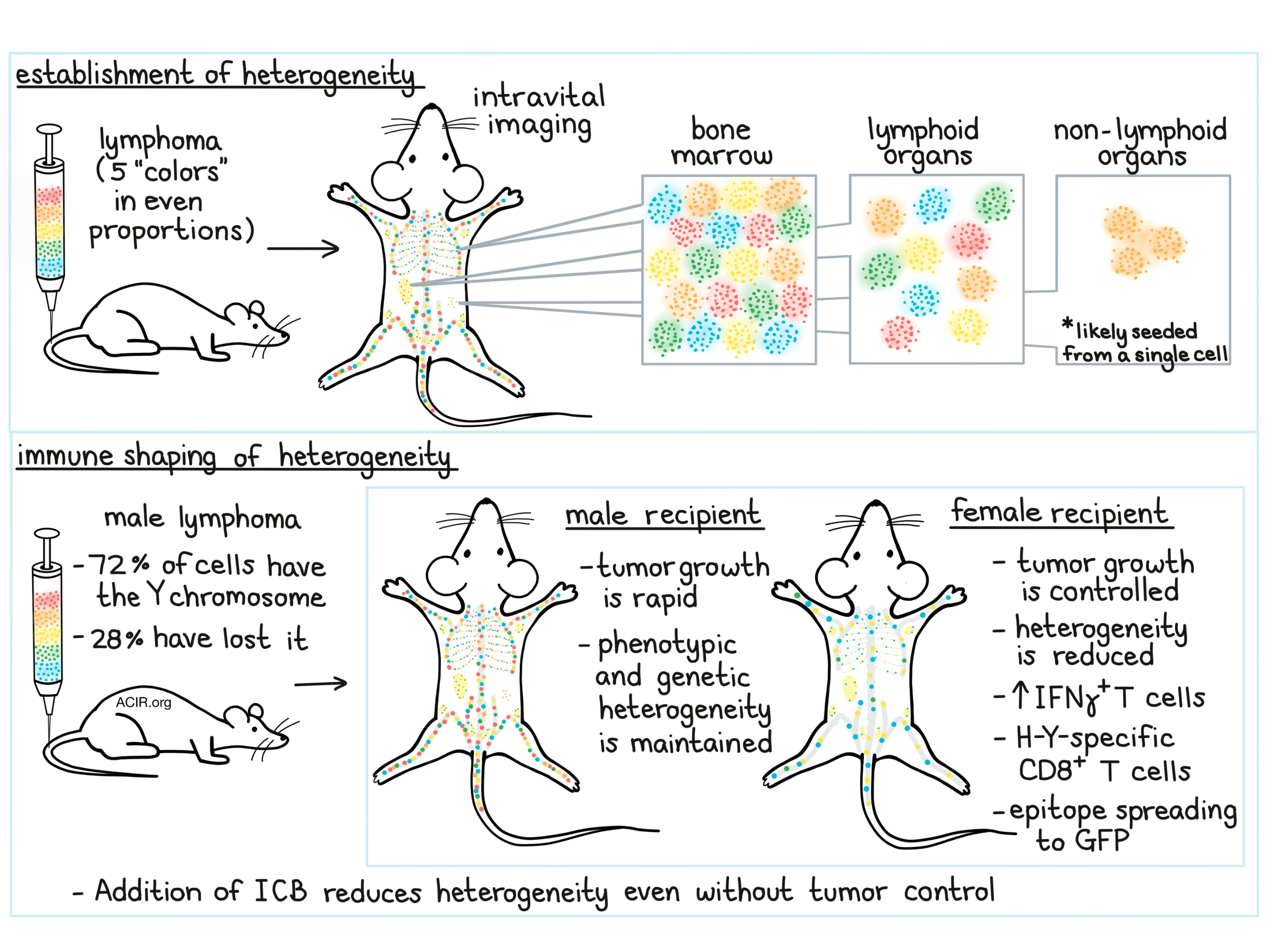
To create their multicolor barcode for lymphoma, Milo et al. used fluorescent proteins (alone or in combination) to tag tumor cell lines from individual tumor-bearing mice. They then created a mixture with equal proportions of five “colors” to be transferred into mice. Using flow cytometry, the researchers tracked the proportions of each color on an “evenness” index to determine whether the proportions remained even, showing maintenance of heterogeneity, or skewed, showing loss of heterogeneity. This multiciolor system also lends itself to intravital imaging, which allowed researchers to visualize spatial organization of individual progeny at different stages of tumor development.

In mice, B cell lymphoma growth was observed primarily in the bone marrow, to a lesser extent in the spleen and some lymph nodes, and occasionally in non-lymphoid tissue. Using the multicolor barcoding system to study the extent of tumor heterogeneity, the researchers found that in most lymphoid locations, all 5 colors could be recovered with comparable “evenness”. Tumor nodules that developed in non-lymphoid tissues, however, were usually only one color. This effect was found to be independent of immune editing (as it also occurred in Rag-/- mice), suggesting that masses in non-lymphoid tissues were seeded from a single cell. Looking closer at heterogeneity just within bone marrow, the researchers found that early on, small patches of cells of each color were established in distinct locations, forming individual niches containing progeny from a single cell. These patches grew over time but showed little migration or overlap.

To better understand how the immune system responds to and shapes intratumor heterogeneity, the researchers utilized a male mouse tumor model in which 28% of tumor cells had lost the Y chromosome. This male lymphoma line was then transferred into either male or female mice, with the expectation that in females, cells expressing the Y chromosome would be highly antigenic. As expected, tumors grew rapidly in males, but were well-controlled for a period of time in females. Using the multicolor barcoding strategy, the researchers found that male mice maintained tumor heterogeneity over time, while females showed a dramatic loss of heterogeneity and the emergence of a few dominant clones. This effect was not observed in Rag2-/- mice, suggesting that it was likely caused by immunoediting. As further evidence of this, the researchers observed an increase in IFNγ-producing T cells in females and identified CD8+ T cells specific to H-Y – a Y chromosome antigen. Further analysis of cell karyotypes showed that tumor cell clones that had maintained the Y chromosome were efficiently eliminated by CD8+ T cells in female mice.

When the researchers restricted karyotype analysis to only the subset of tumor cells that had lost the Y chromosome, they still saw a reduction in diversity in females compared to males. Further, they found that female mice mounted a T cell response to green fluorescent protein (GFP, typically poorly immunogenic), suggesting that epitope spreading – which occurs when the response to strongly immunogenic tumor antigens (such as H-Y on the Y chromosome) enhances responses to poorly immunogenic tumor antigens – may have contributed to the loss of tumor heterogeneity, even among tumor cells lacking a strong immunogenic target.

In order to better quantify genetic changes in intratumor heterogeneity, Milo et al. utilized whole-exome sequencing to identify tumor mutations and predict neoantigens and to track possible changes in these targets under immune pressure. Using the distribution of allele frequencies among targets to measure and calculate heterogeneity, the researchers found that genetic diversity was high in tumors isolated from bone marrow and low in tumors isolated from non-lymphoid tissues, confirming the multicolor barcoding results. They also found that tumors that had been growing under high immune pressure in female mice were less genetically diverse than tumors that had been growing under low immune pressure in males.

Having shown that immune pressure restricts intratumor heterogeneity, Milo et al. investigated the effect of checkpoint blockade on heterogeneity. Treatment with anti-PD-1 in male mice inoculated with male lymphoma had little effect on tumor growth, but significantly reduced tumor diversity and favored clonal dominance. Anti-CTLA-4 alone had little effect, but combination anti-PD-1/anti-CTLA-4 increased mouse survival and significantly decreased tumor heterogeneity, likely through broadening of the immune response. These results were confirmed in a second B cell tumor model. Together, this suggests that checkpoint blockade can impact diversity and tumor composition even in the absence of strong tumor control.

Overall, Milo et al. show that multicolor tumor barcoding can be used to study genetic intratumor heterogeneity and that heterogeneity can be shaped and restricted by the immune response. This strategy also represents a powerful new tool to further study tumor heterogeneity in a broad range of cancers and contexts.

by Lauren Hitchings

References:

Milo I., Bedora-Faure M., Garcia Z., Thibaut R., Périé L., Shakhar G., Deriano L., Bousso P. The immune system profoundly restricts intratumor genetic heterogeneity. Sci Immunol. 2018 Nov 23.

In the Spotlight...

Tumor-conditional anti-CTLA4 uncouples antitumor efficacy from immunotherapy-related toxicity

To reduce the toxicity of anti-CTLA-4, Pai and Simons et al. created an anti-CTLA-4 with dual variable domains (DVD), in which the inner variable domain (ID) targeting CTLA-4 was connected via a cleavable linker to the outer domain (OD) specific for the prostate stem cell antigen; the OD blocked binding by the ID. DVD ameliorated toxicities observed with anti-CTLA-4 by preserving tissue-resident Tregs and preventing systemic T cell activation. DVD accumulated at higher levels in TRAMP-C2 tumors than anti-CTLA-4, and linker cleavage by protease present in the tumor led to depletion of intratumoral Tregs and effective antitumor immunity.

In situ sprayed bioresponsive immunotherapeutic gel for post-surgical cancer treatment

Chen et al. developed an in situ-formed biodegradable gel comprising biocompatible fibrinogen and thrombin, as well as calcium carbonate nanoparticles that scavenge H+ and slowly release encapsulated anti-CD47 to block the “don’t eat me” signal on tumor cells. When sprayed onto an incomplete tumor resection site, the gel reduced the acidity of the TME (thus promoting M1-polarization of TAMs), enhanced phagocytosis of tumor cells by macrophages and DCs, and mounted a systemic immune response, resulting in inhibition of local and distant tumor growth. Tumors showed an increase in TILs and a decrease in intratumoral MDSCs and Tregs.

Tumor-draining lymph nodes are pivotal in PD-1/PD-L1 checkpoint therapy

In mice with either MC38 or CT26 colon tumors, the presence of the tumor-draining lymph nodes (TDLNs) was crucial to the antitumor efficacy of anti-PD-1/anti-PD-L1 treatment. PD-1 blockade led to an increase in total as well as activated CD8+ T cells in the TDLNs. Resection of the TDLNs before anti-PD-1/anti-PD-L1 treatment strongly reduced survival, while removal of the TDLNs prior to tumor inoculation, or locking T cells within lymphoid organs completely abrogated the efficacy of PD-1/PD-L1 blockade, suggesting that the success of this treatment relies on T cells from outside of the TME and on reinvigoration of antigen-specific T cells in TDLNs.

CD4+ T cells induce rejection of urothelial tumors after immune checkpoint blockade

Sato et al. developed mouse tumorigenic organoid lines that resembled the basal-squamous subtype of the human urothelial carcinoma. In mice implanted with the organoids, anti-PD-1 and anti-CTLA-4 combination (ICB) led to tumor rejection and formation of immunological memory in a manner dependent on CD4+ T cells, but not CD8+ T cells. Tumor rejection did not rely on the expression of MHC class I, MHC class II, or Ifngr1 in the tumor. Mechanistically, ICB expanded IFNγ-producing Th1 CD4+ T cells in the tumor and draining lymph nodes, and IFNγ impaired tumor growth independent of other CD4+ T cell functions.

Generation and molecular recognition of melanoma-associated antigen-specific human γδ T cells

Benveniste and Roy et al. were able to generate classical MHC-restricted γδ T cells from hematopoietic stem/progenitor cells and to detect such cells in the naive peripheral human T cell repertoire. Transfer of a melanoma-associated antigen MART-1-specific γδ TCR to Jurkat cells confirmed that activation of these cells was HLA-A2/MART-1-restricted. The affinity ranges of γδ TCRs to HLA-A2/MART-1 were similar to those of αβ TCRs. Crystal structures revealed both similarities and differences between binding of γδ TCRs versus αβ TCRs to HLA-A2/MART-1.

Magnitude of Therapeutic STING Activation Determines CD8+ T Cell-Mediated Anti-tumor Immunity

Intratumoral administration of S100 – a STING agonist currently in clinical trials – at relatively low, but not high, doses led to rejection of injected tumors and durable immune protection. Mechanistically, hematopoietic cells contributed to STING signaling, and intratumoral monocytes produced STING pathway cytokines IFNβ and TNFα; type I IFN, but not TNFα, was required for CD8+ T cell activation and tumor control. Combination of low-dose S100 with anti-PD-1 and/or anti-CTLA-4 eliminated both injected and non-injected tumors and increased survival. High doses of S100 ablated the injected tumors, but resulted in poor immunological memory.

Noncoding regions are the main source of targetable tumor-specific antigens

Laumont and Vincent et al. developed a proteogenomic approach to identify tumor specific antigens (TSAs) as MHC-bound peptides derived not only from mutated coding regions but also from aberrantly expressed noncoding regions of the genome (aeTSAs). In 2 murine cancer cell lines (CT26 and EL4) and 7 primary human tumors (4 B-ALL, 3 lung), the majority of MHC-presented TSAs originated from noncoding regions. In mice, the antitumor efficacy of vaccination with aeTSA-pulsed DCs largely depended on the level of aeTSA expression and the frequency of aeTSA-specific T cells – markers which could be evaluated in humans.

Toll like receptor 7 expressed by malignant cells promotes tumor progression and metastasis through the recruitment of myeloid derived suppressor cells

Dajon and Iribarren et al. used a murine lung adenocarcinoma model to show that stimulation of TLR7 on malignant, but not stromal, cells leads to secretion of CCL2 and GM-CSF in the TME and to recruitment of MDSCs, which promotes accelerated tumor growth and metastasis. In adenocarcinoma patients, high TLR7 expression on tumor cells was an independent prognostic marker for poor clinical outcome, and was associated with a gene expression profile of metastasis (overexpression of ICAM-1, KRT-7, KRT-19, NME1, SDC4, and TP53) and a phenotype of epithelial-mesenchymal transition (increased vimentin and decreased E-cadherin).

Everything New this Week In...

Close Modal

Small change for you. Big change for us!

This Thanksgiving season, show your support for cancer research by donating your change.

In less than a minute, link your credit card with our partner RoundUp App.

Every purchase you make with that card will be rounded up and the change will be donated to ACIR.

All transactions are securely made through Stripe.