Weekly Digests
‹ Back to January

Classical monocytes may predict response to anti-PD-1 immunotherapy

January 24, 2018

Anti-PD-1 therapy has been clinically effective against a wide range of cancers, but still only a fraction of patients achieve a durable response. Researchers have been on the hunt for biomarkers that could predict which patients are likely to respond to this therapy, and Krieg et al. may have finally found their mark. The team utilized peripheral blood mononuclear cells (PBMCs) from samples from twenty patients with metastatic melanoma before and twelve weeks after the initiation anti-PD-1 immunotherapy to compare responders and non-responders. They used high-dimensional, single-cell mass cytometry with three optimized immune marker panels and several bioinformatics tools to identify differences between responders and non-responders to anti-PD-1 immunotherapy.

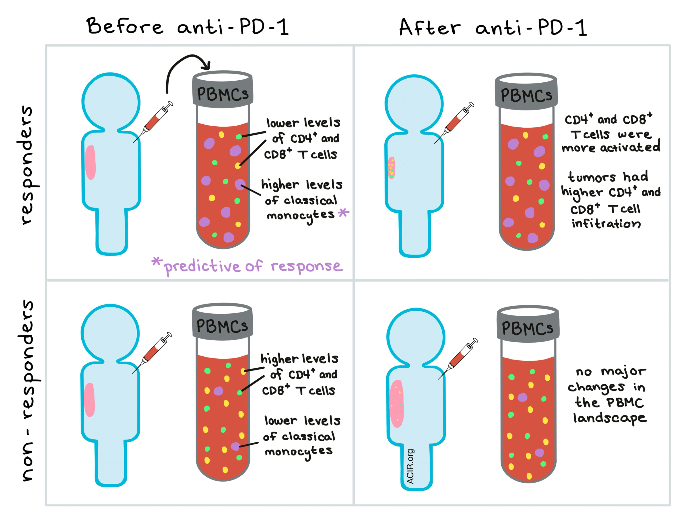
In an initial ‘cytometry by time of flight’ (CyTOF) analysis of patient PBMCs, hierarchical clustering by marker expression on all leukocytes in each patient before and after therapy stratified patients into responders and non-responders, indicating a possible pre-therapeutic biomarker. Analysis of the first cyTOF panel revealed that responders had lower frequencies of CD4+ and CD8+ T cells and higher frequencies of CD19-HLA-DR+ myeloid cells in the peripheral blood both before and after therapy.

Expression profiles of CD8+ T cells before therapy suggested that CD8+ T cells from responders had improved migratory capabilities compared to CD8+ T cells from non-responders and the second CyTOF panel revealed that CD4+ and CD8+ T cells expanded in responders and shifted towards a more activated and cytotoxic state; at the time the samples were taken, most CD4+ and CD8+ T cells fell into memory phenotype categories.

While the lymphocyte landscape provided some insight into patients’ responsiveness to anti-PD-1, it did not provide any robust predictive biomarkers. As earlier results from the CyTOF analysis indicated higher levels of CD14+ myeloid cells in responders before therapy, the researchers performed an in-depth characterization of the myeloid cell compartment with a third ‘myeloid-centric’ CyTOF panel. Unsupervised clustering of marker expression again stratified the patients and led to identification of 16 markers that were upregulated in responders compared to non-responders. FlowSOM analysis confirmed the elevated levels of classical CD14+CD16-HLA-DRhi monocytes in responders and implicated this signature as a robust predictive biomarker of responsiveness to anti-PD-1 immunotherapy. RNA sequencing of myeloid cells revealed no major differences in function, phenotypic state, or polarization of these cells between responders and non-responders, indicating that the frequency of CD14+CD16-HLA-DRhi cells in circulation before therapy was the most important factor to consider as a predictive biomarker.

To make their analysis process simpler for clinical translation, the researchers designed a flow-cytometry-based validation panel using fewer markers and curated combinations of markers that confirmed prior observations of lower T cell frequencies and higher CD14+ monocyte frequencies in peripheral blood of patients who would respond to therapy. Finally, they assessed the prognostic value of the frequency of classical monocytes in peripheral blood and found that the frequency of classical monocytes was robustly associated with progression-free and overall survival in response to anti-PD-1.

While no mechanistic studies were performed, Krieg et al. did speculate on what the mechanism might be based on their data. They propose that in patients who would ultimately respond to therapy, T cells are low in the peripheral blood because they have superior migratory capacity and have already begun to move out of the blood and into the tumor, producing IFNγ that could trigger the release of myeloid cells from bone marrow and support their expansion. Whether the expanded myeloid cells actively contribute to sustaining the immune response is uncertain. Determining whether this presumptive mechanism is accurate will require significantly more testing.

In addition to experimentally exploring the mechanism, the researchers intend to study whether the response-associated immune signature identified in this study will hold true in larger cohorts and across a range of other cancers. If so, a prediction signature based on the frequency of classical monocytes in peripheral blood might be used in clinical practice to tailor the application of anti-PD-1 immunotherapy to those who are most likely to benefit from it.

by Lauren Hitchings

References:

Krieg C., Nowicka M., Guglietta S., Schindler S., Hartmann F.J., Weber L.M., Dummer R., Robinson M.D., Levesque M.P., Becher B. High-dimensional single-cell analysis predicts response to anti-PD-1 immunotherapy. Nat Med. 2018 Jan 8.

In the Spotlight...

Blockade of Tumor-expressed PD-1 promotes lung cancer growth

Du et al. describe a patient with non-small cell lung cancer who rapidly progressed after treatment with radiotherapy and anti-PD-1, and was found to have PD-1 expressed on the tumor. In a murine PD-1-expressing M109 lung cancer cell line, knocking out or blocking PD-1 increased cell viability, while overexpressing PD-1 or treating with soluble PD-L1 reduced viability in vitro; PD-1 blockade accelerated M109 tumor growth in vivo. Tumor-intrinsic PD-1 expression was observed in about 3-10% of lung cancer tissue samples examined.

Tumor-Specific T-Cells Engineered to Overcome Tumor Immune Evasion Induce Clinical Responses in Patients With Relapsed Hodgkin Lymphoma

To circumvent the immunosuppressive effects of TGFβ in the tumor microenvironment of relapsed Hodgkin lymphoma, Bollard et al. engineered enriched EBV-specific T cells to express a dominant-negative TGFβ receptor type 2 (DNRII). When adoptively transferred without lymphodepletion into seven relapsed patients and one patient in remission, the cells expanded and persisted up to four years, yielding positive clinical responses, including two complete responses ongoing after four years.

Diverse genetic-driven immune landscapes dictate tumor progression through distinct mechanisms

Bezzi et al. discovered that the type and number of immune cells infiltrating human or murine prostate cancer, and their tumor-inhibitory mechanism, depend on the specific gene pathways that are mutated in the tumor, and on the resulting differential cytokine and chemokine population. The tumor microenvironments ranged from immune deserts to highly infiltrated landscapes, suggesting that stratification of patients based on integrated genotypic-immunophenotypic analysis may be required for the success of immunotherapy in prostate cancer.

Genomic correlates of response to immune checkpoint therapies in clear cell renal cell carcinoma

Using genomic analysis of metastatic clear cell renal cell carcinoma (ccRCC), Miao et al. found that patients with biallelic loss of PBRM1, a gene encoding part of the PBAF chromatin remodeling complex, experienced sustained tumor reduction and had significantly prolonged overall and progression-free survival following immune checkpoint blockade therapy. PBRM1 deficiency was associated with enhanced expression of genes involved in JAK/STAT3/IFNγ and hypoxia pathways, providing a potential mechanism of ccRCC response to immunotherapy.

A major chromatin regulator determines resistance of tumor cells to T cell-mediated killing

Pan et al. used a CRISPR/Cas9 knockout screen, specific gene knockouts, and ATAC-seq to show that inactivating Pbrm1, Arid2, or Brd7 – genes encoding components of the PBAF chromatin remodeling complex – enhanced melanoma cell sensitivity to IFNγ and increased the release of chemokines that attract cytotoxic CD8+ T cells, thus increasing effector T cell infiltration into the tumor and allowing the previously treatment-resistant tumors to respond to immune checkpoint blockade.

Intravenous delivery of oncolytic reovirus to brain tumor patients immunologically primes for subsequent checkpoint blockade

In a small, window-of-opportunity clinical trial in patients with primary or metastatic brain disease prior to scheduled surgery, Samson et al. demonstrated that intravenous delivery (to overcome the practical difficulties of direct intracranial delivery) of oncolytic reovirus resulted in infection of brain tumor cells, and initiated one or more immune-related events, including PD-L1 expression. In an orthotopic murine model, intravenous reovirus followed by anti-PD-1 therapy significantly increased CD4+ and CD8+ T cell infiltration into the tumor and extended survival.

Neoadjuvant oncolytic virotherapy before surgery sensitizes triple-negative breast cancer to immune checkpoint therapy

Based on the hypothesis that early disease treatment will be a more effective immunotherapy strategy, Bourgeois-Daigneault et al. demonstrated the long-term protective impact of the oncolytic Maraba virus delivered as neoadjuvant therapy to mice bearing syngeneic TNBC tumors. Gene expression and secreted cytokine analysis of infected tumor cells demonstrated upregulation of key inflammatory pathways, including PD-L1. Treatment with anti-PD-1 and anti-CTLA-4 post-surgery dramatically improved overall survival following rechallenge.

A novel immunization strategy using cytokine/chemokines induces new effective systemic immune responses, and frequent complete regressions of human metastatic melanoma

In a Phase I clinical trial of 87 patients with multiple metastatic subcutaneous melanoma lesions, Valentine et al. stimulated autologous PBMCs with a recall microbial antigen ex vivo and repeatedly injected the resulting cytokine mixture into a subset of lesions. Complete responses lasting 2 - 334 months (median 59) with regression of injected and never-injected metastases was observed in 38% of patients. Dense infiltrates of CD8+ T cells and isolation of cells cytotoxic to autologous melanoma cells in vitro indicated induction of a systemic immune response.

Everything New this Week In...

Close Modal

Small change for you. Big change for us!

This Thanksgiving season, show your support for cancer research by donating your change.

In less than a minute, link your credit card with our partner RoundUp App.

Every purchase you make with that card will be rounded up and the change will be donated to ACIR.

All transactions are securely made through Stripe.