Over the years, dendritic cell (DC) vaccines have consistently shown promise, but have come up short in the clinic, potentially due to poorly presented antigens, limited in vivo persistence, or impaired function when derived autologously from patients with cancer. As an alternative to DCs, Abusarah et al. engineered mesenchymal stromal cells (MSCs) to express the immunoproteasome (IPr) to promote their potential as antigen-presenting cells (APCs), and subsequently, as cancer vaccines. The results of their studies were recently published in Cell Reports Medicine.
MSCs were chosen for genetic engineering based on their known capacity for antigen presentation when primed with low levels of IFNγ. To force more effective antigen presentation in MSCs, the researchers introduced retroviral vectors encoding subunits for the IPr, which has peptide cleavage properties capable of generating stable and immunogenic peptide–MHC complexes; the researchers hypothesized that would convert MSCs into antigen-presenting cells. The resulting MSC-IPr expressed MHC-I, CD80, and IL-12, without expression of PD-L1, IL-4, or IL-10. Their chemokine expression profile was similar to that of conventional bone marrow-derived DCs. Processes related to antigen presentation, immune responses, and metabolism were upregulated, while pathways associated with protein folding, amino acid turnover, ER stress, and pH reduction were downregulated.
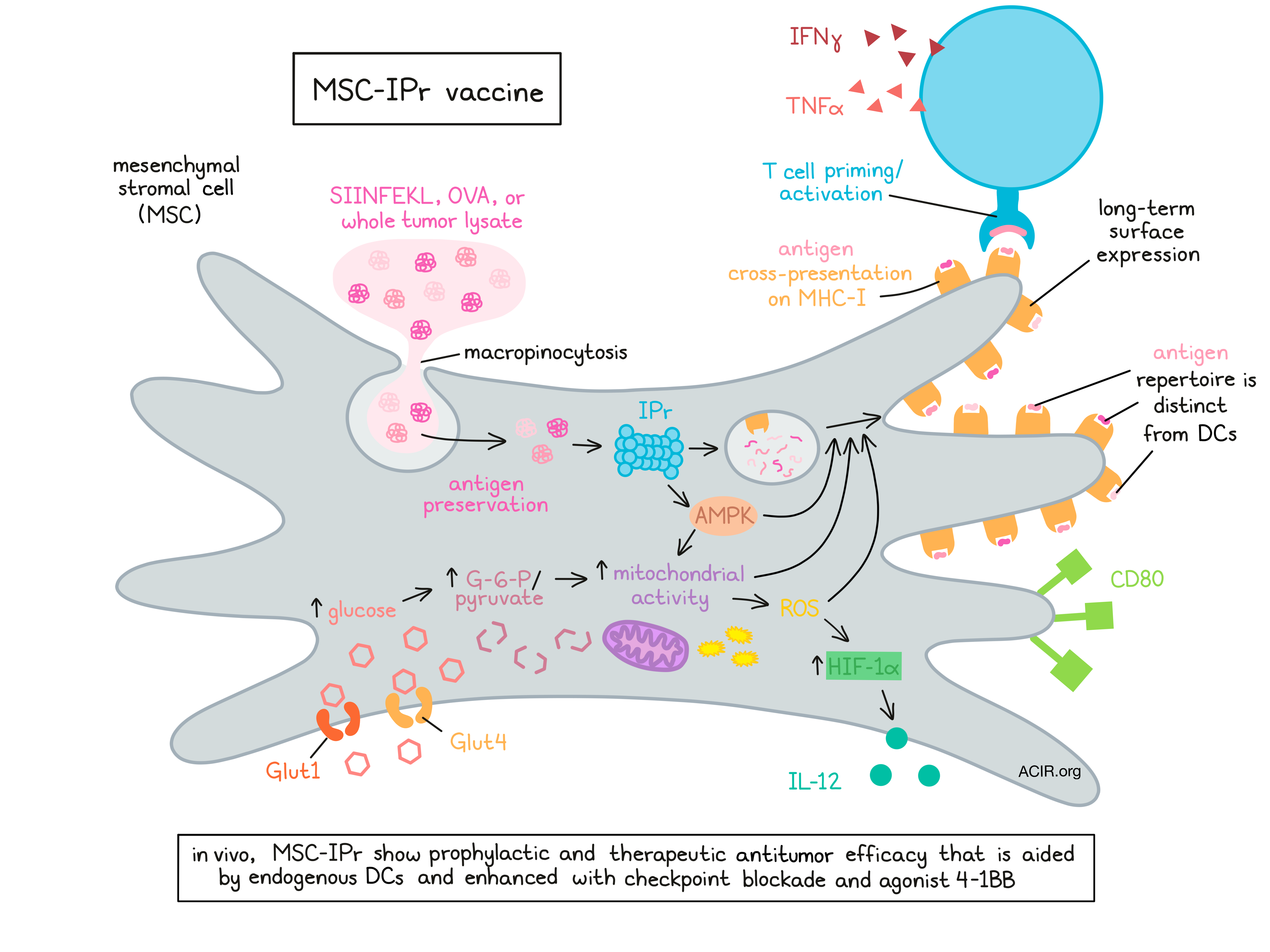
To monitor for effective antigen presentation, the researchers pulsed MSC-IPr with SIINFEKL or full-length OVA and evaluated the kinetics of SIINFEKL–MHC-I complex formation at the cell surface. While these complexes turned over more rapidly on DCs, they were sustained for much longer on the cell surfaces of MSC-IPr. An antigen presentation assay using soluble antigens showed that MSC-IPr primed T cell responses that were superior to those primed by DCs, and almost as strong as those primed by ex vivo-sorted cDC1s. Interestingly, inducing PD-L1 expression on MSC-IPr did not impair their ability to induce T cell activation.
Looking at how MSC-IPr capture extracellular antigens, the researchers observed that MSC-IPr showed a moderate increase in OVA internalization over BM-derived DCs, and that they captured antigens by macropinocytosis. Antigen cross-presentation was not dependent on late endosomal transport, vacuolar acidification, or ER-Golgi vesicular transport, suggesting that antigens were not sorted into mature endosomes or lysosomes. From this, the researchers concluded that MSC-IPr may not rely exclusively on cytosolic or vacuolar pathways, and may instead rely on a route involving early recycling endosomes that preserves internalized antigens and sustains antigen presentation.
Next diving into their observation that engineering MSCs with the IPr induces metabolic reprogramming, Abusarah et al. showed that although the expression of genes encoding Glut1 and Glut4 remained unchanged, these glucose transporters were upregulated on the cell surface, suggesting decreased degradation or increased recycling. Further, the oxygen consumption rate was also increased, percentages of activated/functional mitochondria were high, and AMPK and two of its target genes were highly activated in MSC-IPr. Mitochondrial functions and AMPK were both required for antigen cross-presentation functions and T cell activation, suggesting that IPr expression is linked to metabolic reprogramming that favors oxidative phosphorylation.
Evaluating the ATP metabolic pathway, the researchers found that MSC-IPr absorbed more glucose and contained higher levels of the glycolysis intermediates G-6-P and pyruvate. The excess of pyruvate was not entirely converted into lactate, and was instead used by TCA cycle, increasing mitochondrial activity. In addition to producing ATP, the increased mitochondrial activity increased superoxide anion production and HIF-1α levels. Efforts to reduce various reactive oxygen species (ROS) derived from mitochondrial activity negatively impacted T cell activation, indicating an association between the TCA cycle intermediates and antigen cross-presentation.
Armed with a more thorough understanding of MSC-IPr as APCs, Abusarah et al. investigated their use as cancer vaccines. MSC-IPr pulsed with OVA were well tolerated in mice, induced T cell responses (measured by production of IFNγ and TNFα), and completely protected mice from OVA-expressing E.G7 tumors delivered either subcutaneously (s.c.) or systemically; similar DC vaccines only protected against s.c. tumors. MSC-IPr vaccines also protected against OVA-expressing E.G7 in the presence of non-OVA-expressing EL4 growing on the opposite flank, suggesting efficacy even in the presence of immunosuppressive factors released by a growing tumor. Additionally, MSC-IPr pulsed with whole tumor cell lysates induced 70% protection against EL4 lymphoma and 80% protection against B16F10 melanoma; similar DC vaccines induced 20% and 40% protection, respectively. CD4+ and CD8+ T cells were required for antitumor immunity.
In a therapeutic setting, OVA-pulsed MSC-IPr vaccines only delayed tumor growth in pre-established OVA-expressing E.G7 lymphoma tumors in mice, while tumor lysate-pulsed MSC-IPr vaccines induced 30% survival. The addition of agonist 4-1BB, anti-LAG3, anti-CTLA-4, or anti-PD-1 each increased survival, with the vaccine plus anti-PD-1 reaching 80% survival. The addition of agonist 4-1BB to the vaccine plus anti-PD-1 achieved 100% survival, with 8/10 mice showing complete tumor elimination. The recruitment of T cells to tumor sites was found to be essential to antitumor efficacy. Similar results were observed using a tumor lysate-pulsed vaccine for B16F10 melanoma.
Investigating the behavior of MSC-IPr in vivo, Abusarah et al. found that while they persisted longer than various controls, MSC-IPr failed to migrate to tumors or lymphoid organs. Further investigation revealed that the antitumor immunity induced by MSC-IPr vaccines was dependent on both antigen presentation on MHC-I by MSC-IPr and on cross-presentation of antigens by endogenous DCs, which likely took up antigens from MSC-IPr and shuttled them to sites of T cell activation
Investigating the potential for off-the-shelf MSC-IPr vaccines, the researchers found that allogeneic MSC-IPr vaccines induced 60% survival in mice pre-implanted with A20 lymphoma tumors, while allogeneic DCs induced 0% survival. The addition of anti-PD-1 improved survival to 100% for MSC-IPr vaccines, and 40% for DC vaccines. These results suggested the potential for off-the-shelf vaccines using allogeneic MSC-IPr.
Finally, the researchers found that following EL4 tumor lysate pulsing, the peptide lengths, binding affinities, and peptide motifs were similar between MSC-IPr and DCs. However, the antigen repertoires were dramatically distinct between the two cell types, with MSC-IPr presenting a four-fold higher number of peptides than DCs.
Overall, Abusarah et al. showed that engineering MSC-IPr induces metabolic reprogramming that supports a pro-inflammatory phenotype and antigen presentation properties. Compared to DCs, MSC-IPr vaccines presented a unique set of antigens and induced more potent antitumor immunity that could be enhanced in combination with checkpoint blockades and agonist 4-1BB. MSC-IPr vaccines, including those developed from allogeneic cells, could serve as a unique cancer vaccine strategy that overcomes several of the challenges associated with DC vaccines.
By Lauren Hitchings
Meet the researcher
This week, lead author Moutih Rafei answers our questions.

What prompted you to tackle this research question?
Although vaccination was proven to elicit protective responses against infectious diseases in general, cancer vaccines remain unavailable. The major divergence between cancer vaccines and the ones targeting infectious agents lies in the nature of the antigen (e.g., self vs. non-self). Therefore, a cancer vaccine is challenging to develop due to obstacles related to the identification of tumor-associated/specific antigens (TAAs/TSAs) capable of generating specific, effective, and persistent cytotoxic T lymphocytes (CTL) without breaking tolerance. Historically, TAA-based cancer vaccines tested in the mid-90s revealed good clinical responses in a small patient subset (10-15%), which led to the conclusion that tumor immunogenicity is both patient- and tumor-specific. As a result, efforts were largely dedicated to the development of personalized dendritic cell (DC) vaccines with little or no avail due to the absence of TSAs and limitations related to DC biology.
Although ex vivo-developed DCs were shown to be safe, technically feasible, and immunogenic to a certain extent, their overall clinical impact was disappointing for several reasons. Therefore, establishing an unlimited supply of an antigen-presenting cells (APCs) capable of bypassing some, if not all, of the limitations of DCs remains a central goal in the field. Several characteristics support the extensive use of mesenchymal stromal cells (MSCs) as a cellular biopharmaceutical, especially for tissue repair and wound healing. However, MSCs can also display remarkable immunomodulatory properties. Although IFNγ stimulation of MSCs triggers APC-like functions, these properties were transient. Besides, IFNγ-primed MSCs end up expressing the immune checkpoint inhibitor PD-L1, which impairs CTL effector functions and metabolism. To rectify these limitations, we elected to bypass the IFNγ pre-treatment approach by modulating the proteasomal machinery of MSCs to elicit a sustained and efficient APC function.
What was the most surprising finding of this study for you?
The most surprising finding of this study was how the introduction of the immunoproteasome complex reprogrammed MSCs to behave in completely divergent manner when compared to innate MSCs. For instance, this modification instilled antigen cross-presentation abilities, enhanced their expression of various immune-related molecules, and triggered de novo production of pro-inflammatory cytokines and chemokines. What was also very surprising was the fact that the cross-presentation capacity of MSC-IPr was highly dependent on their metabolic activity (completely unexpected), and MSC-IPr presented a vastly different epitope repertoire compared to DCs, which translated into potent re-activation of T cell immunity against tumors. These engineered cells therefore constitute a promising subset of non-hematopoietic antigen-presenting cells suitable for the future design of universal cell-based cancer vaccines.
What was the coolest thing you’ve learned (about) recently outside of work?
When I was young, I dreamed of becoming a pilot or a nuclear physicist. However, my career drifted towards medical research after watching the movie "Outbreak" by Dustin Hoffman. Although I consider myself extremely happy in my career, I discovered that I have a great interest and passion in watching and learning from chefs! I am a big fan of chef Ramsay, and I am always trying to repeat his recipes at home. However, what I came to realize is that cooking and conducting experiments follow the same rules. You must be precise, meticulous, and most importantly, you must respect time and temperatures. If any of these variables are off, your meal or experiment will certainly fail.


