Even though the importance of forming long-lived memory CD8+ T cells is widely acknowledged, the origin of this T cell subset remains largely unknown. Johnnidis and Muroyama et al. used a well established viral infection model to phenotype and functionally characterize the cell subsets responsible for memory formation and recall responses, and uncovered an unexpected role for inhibitory receptors. Their results were recently published in Science Immunology.
The researchers investigated T cell memory formation in a mouse model of acute infection with lymphocytic choriomeningitis virus Armstrong strain (LCMV-Arm). They adoptively transferred LCMV glycoprotein-specific, TCR-transgenic, P14 CD8+ T cells and investigated the CD8+ T cell populations initially using the memory and precursor markers interleukin-R7α (IL-7Rα) and killer cell lectin-like receptor G1 (KLRG1) and the lymph node homing molecule CD62L.
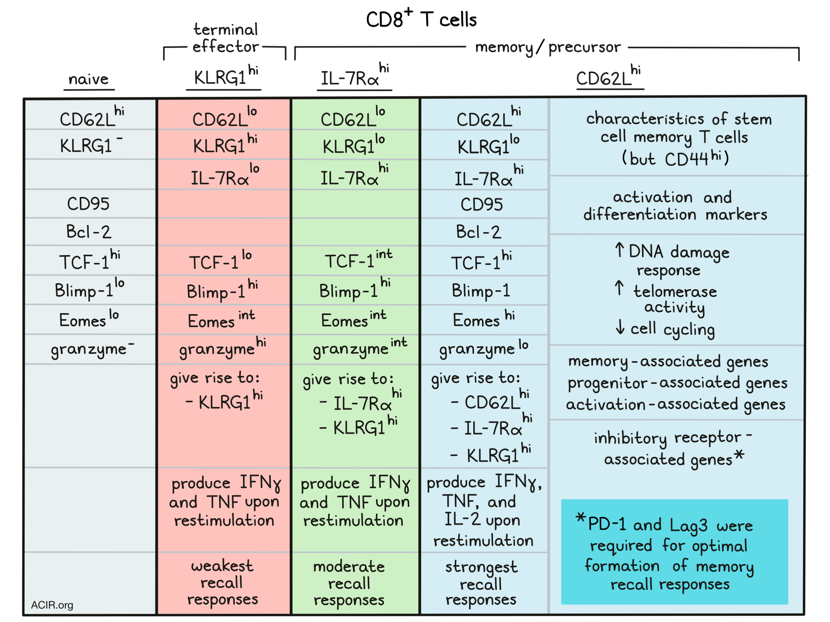
At day 8 post-infection (pi), the researchers identified a small CD62LhiKLRG1loIL-7Rαhi subpopulation (CD62Lhi) and compared this subset to naive CD8+ T cells and to the effector subsets CD62LloKLRG1loIL-7Rαhi (IL-7Rαhi) and CD62LloKLRG1hiIL-7Rαlo (KLRG1hi). The CD62Lhi subset expressed CD95, Bcl-2, and TCF-1 at similar levels as naive CD8+ T cells, but also expressed activation and differentiation markers. The CD62Lhi subset expressed higher levels of Eomes than the other subsets. It also had higher expression of Blimp-1 than naive cells, but lower expression than the IL-7Rαhi and KLRG1hi subsets. The CD62Lhi subset expressed characteristics of stem cell memory T cells (Tscm), but did express granzymes and CD44, in contrast to Tscm. The CD62Lhi, KLRG1hi, and IL-7Rαhi subsets all expressed IFNγ and TNF upon restimulation, while only the CD62Lhi subset produced IL-2.
Next, the researchers purified the three subpopulations and performed bulk RNAseq. Principal Component Analysis revealed a clear separation of these populations from naive T cells. The CD62Lhi cells also had a distinct transcriptional profile from IL-7Rαhi and KLRG1hi cells, expressing memory-associated and progenitor-associated genes. Additionally, this subset, like the others, expressed inhibitory receptor-associated genes, including Pdcd1, Lag3, Ctla4, Cd5, and Tigit, and activation-associated genes.
These data suggested that the small CD62Lhi subset was distinct from the larger KLRG1hi and IL-7Rαhi subsets. To assess whether this phenotype was caused by incomplete activation, the researchers performed proliferative history tracking. They showed that the CD62Lhi cells had undergone at least eight divisions at the peak of expansion, but cell cycle proliferation dynamics tests revealed that the CD62Lhi cells cycled more slowly than the other two subsets, accounting for the differences in relative proportions of each subset.
Based on the expression patterns, the researchers hypothesized that the CD62Lhi subset was a progenitor population during clonal expansion. Since progenitor cells correlate with genomic integrity maintenance in multiple biological systems, they measured markers of surveillance of DNA damage. Phosphorylation of γH2AX, an event occurring in response to DNA damage, was elevated in the CD62Lhi subset as compared with the other two subsets, and this effect was even more pronounced after cells were exposed to ionizing irradiation. Finally, the CD62Lhi subset also had higher telomerase activity, suggesting this subset had genome-protective characteristics. This was confirmed using a genome integrity disruption assay using doxorubicin treatment during the expansion phase.
To further dive into these subsets’ developmental origins, the researchers performed a pseudotime analysis on scRNAseq data. This showed that the KLRG1hi cells were more differentiated than CD62Lhi and IL-7Rαhi cells, but the difference between these last two was not obvious. To perform in vivo lineage tracing, the three subsets were isolated at d6 pi, and adoptively transferred into congenically disparate recipient mice at d4 pi. Four days later, the CD62Lhi subset had raised the most antigen-specific progeny cells. KLRG1hi cells gave rise to only KLRG1hi cells, IL-7Rαhi cells gave rise to IL-7Rαhi and KLRG1hi cells, and the CD62Lhi subset produced all three subsets, suggesting self-renewal.
To study recall responses, naive recipient mice received isolated d8 pi cell subsets and were rechallenged 30 days later. The CD62Lhi subset produced robust recall and showed self-renewal. The IL-7Rαhi population displayed less extensive recall, while the KLRG1hi subset contributed least to the recall response. The responses were similar when CD62Lhi and IL-7Rαhi cells were co-transferred, suggesting the effects were not due to differences in antigen exposure or inflammation. When d8 pi subsets were transferred to naive mice and analyzed after 6 months without antigen rechallenge, the mice receiving CD62Lhi cells still had mostly CD62Lhi cells.
These results suggested that the CD62Lhi cells are highly activated, but restricted in proliferation and effector differentiation. Since this could be due to being excluded from antigen stimulation in the expansion phase, the localization in the spleen’s red and the white pulp on d8 was investigated. The CD62Lhi subset was localized mainly in the white pulp, suggesting access to antigens. Out of the three cell populations, this subset also expressed the highest levels of Nur77, an immediate early gene upregulated by TCR stimulation, and had the highest levels of costimulatory molecules and inflammatory cytokine receptor expression, suggesting strong and recent TCR signaling.
The CD62Lhi cells expressed the highest levels of PD-1, Lag3, CTLA-4, and other inhibitory receptors, potentially limiting the proliferation of this subset. Cotransfer of wild-type and PD-1-deficient P14 cells in the mouse model showed a decrease in CD62Lhi cells after intranasal CMV infection, but not intraperitoneal. Hypothesizing that multiple pathways may work together to preserve the recall memory characteristics, the researchers then transferred PD-1 and Lag3 double knockout cells. This resulted in a decreased frequency of IL-7Rαhi and CD62Lhi cells, with the largest difference in the CD62Lhi population. It also impacted memory responses, as rechallenge in naive mice transferred with these cells resulted in a reduced recall response, with fewer CD62Lhi cells.
These results provide further knowledge into the origins of cells giving rise to memory and recall responses, and shed a light on the paradoxical importance of checkpoint molecules in the development of such responses. It will be of interest to investigate whether long-term use of checkpoint inhibitors impacts memory responses in patients receiving cancer immunotherapy.
Write-up by Maartje Wouters, image by Lauren Hitchings


