Weekly Digests
‹ Back to January

Dysfunction: what’s in a name?

January 30, 2019

To gain insight into the mechanisms underlying response and resistance to immunotherapy, Li, van der Leun, Yofe, and Lubling et al. set out to molecularly characterize the expressed gene sets of tumor-infiltrating immune cells in order to quantitatively and qualitatively analyze the heterogeneity within and between cancer patients. The results were recently published in Cell.

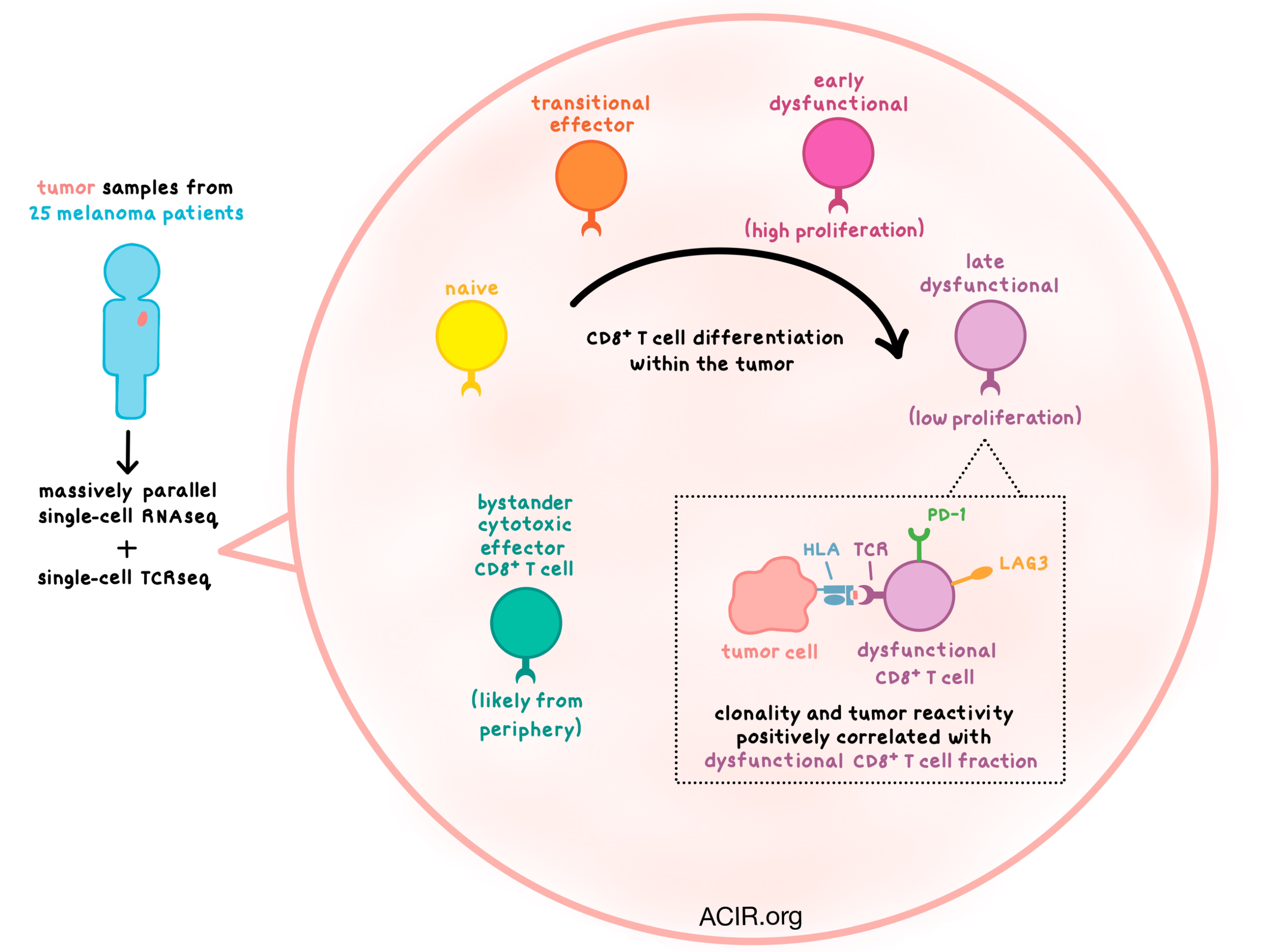
Using massively parallel single-cell RNA sequencing (MARS-seq), the researchers began by examining tumor samples from 25 melanoma patients with various stages of disease and treatment histories. Focusing on T and NK cells first, clustering analysis revealed a number of subsets, including naive T cells; cytotoxic effector (GZMH+), transitional effector (GZMK+), and dysfunctional (PD-1+LAG3+) CD8+ T cells; a small subset of CD4+ and CD8+ memory T cells; Tregs, follicular helper (TFH), and dysfunctional CD4+ T cells; and NK cells. The largest CD8+ T cell cluster was the dysfunctional subset. The observed heterogeneity of molecular phenotypes among T cells was consistent across patients with different disease stages, metastatic sites, and prior treatments, pointing to a universal process that shapes the T cell population within melanoma tumors.

The researchers then used the MARS-seq data to create two transcriptional scores: one for dysfunction (including LAG3, PD-1, and CXCL13) and one for cytotoxicity (including FGFBP2 and CX3CR1). Based on these scores and the clustering analysis, they observed a spectrum of CD8+ T cell states, on a continuum from naive, to transitional, to dysfunctional, indicating that these cells gradually increased their expression of immune checkpoint genes. A similar gradient was observed with Tregs, and interestingly there was some overlap in genes and transcription factors activated in both Tregs and dysfunctional CD8+ T cells. However, the cytotoxic T cells appeared to form a distinct cell population.

Turning their analysis to myeloid cell subsets, the team identified macrophages, monocytes, dendritic cells, plasmacytoid DCs, osteoclast-like cells, B cells, and plasma cells. The data suggested that, similar to T cells, monocytes undergo continuous development and differentiation within the tumor, and that the tumor is not just infiltrated with mature myeloid cells.

Comparing data between patients, the researchers saw that while the myeloid cell distribution was patient-specific, most of the T cell transcriptional states showed up in many patients (albeit with varying frequency). The dysfunctional CD8+ T cell population fraction was particularly variable (ranging from 3.6% to 72.1% of tumor-infiltrating T cells), and it was negatively correlated with naive-like T cell fraction and positively correlated with TFH fraction.

To gain a better understanding of the clonal structure and dynamics of various T cell states, the researchers coupled MARS-seq with single-cell TCRβ sequencing. TCR clonotype distribution varied widely across patients, with some T cell infiltrates exhibiting large TCR diversity but little clonal expansion, and with other T cell infiltrates dominated by a few T cell clones. Clonality positively correlated with the dysfunctional CD8+ T cell fraction, which constituted the most proliferative tumor-infiltrating immune cell subset. More proliferation was observed in the early stages of the dysfunctional program acquisition, but with increased dysfunctionality, proliferation tapered off. Similarly, Tregs were highly proliferative mostly in the early stages of differentiation.

TCRβ genotype provided an opportunity to link clonality and phenotype. Clonal analysis once again showed overlap between transitional and dysfunctional CD8+ T cells, while cytotoxic cells formed a distinct “bystander” subset. To find a potential source of these cytotoxic T cells, the team conducted MARS-seq of peripheral blood, and found that PBMCs contained no dysfunctional cells, but had naive-like, transitional, and cytotoxic CD8+ T cells. This suggests that cytotoxic cells arrive from the periphery rather than differentiate within the tumor.

Asking the question of whether their study results would extend beyond melanoma, the researchers analyzed a dataset of T cells from lung adenocarcinoma samples. They found some differences in the specifics of the dysfunctional gene program between tumor types, but most observations were consistent with melanoma data.

Finally, the team analyzed the relationship between T cell states and tumor reactivity by co-culturing ex vivo-expanded patient T cells with autologous tumor cells. Tumor reactivity, measured by IFNγ and TNFα production, varied significantly between TILs from different patients, with some TILs showing no reactivity. In general, tumor-reactive T cells tended to be in a dysfunctional CD8+ T cell state with a trend toward increasing reactivity with increasing dysfunctionality.

Taken together, the transcription, clonality, and proliferation data suggest a model in which antigen-driven interactions within the tumor microenvironment lead to continuous T cell differentiation toward an early dysfunctional and then highly dysfunctional state. Although the tumor reactivity of the cytotoxic T cells could not be directly evaluated, this model, coupled with the albeit weak association between dysfunctionality and tumor reactivity, may suggest that T cells with a cytotoxic signature are bystanders with no relationship to the tumor. Such observations underscore the power of single-cell analysis in unraveling the complexity of immune cell heterogeneity and its relationship to immune cell functionality.

by Anna Scherer

References:

Li H., van der Leun A.M., Yofe I., Lubling Y., Gelbard-Solodkin D., van Akkooi A.C.J., van den Braber M., Rozeman E.A., Haanen J.B.A.G., Blank C.U., Horlings H.M., David E., Baran Y., Bercovich A., Lifshitz A., Schumacher T.N., Tanay A., Amit I. Dysfunctional CD8 T Cells Form a Proliferative, Dynamically Regulated Compartment within Human Melanoma. Cell. 2018 Dec 20.

In the Spotlight...

Approaches to treat immune hot, altered and cold tumours with combination immunotherapies

In this review, Galon and Bruni classify complex tumor microenvironments into four categories – hot, altered-excluded, altered-immunosuppressed, or cold – based on their cytotoxic T cell landscapes, immune contexture, and immunoscore. By taking the key features of the cancer-immune interactions into account, they propose rational combination treatments for each tumor category, drawing from both conventional therapies (chemotherapy, radiation) and immunotherapies (checkpoint blockades, adoptive cell transfer, vaccination, etc.).

PI3Kα/δ inhibition promotes anti-tumor immunity through direct enhancement of effector CD8+ T-cell activity

Carnevalli and Sinclair et al. explored the effect of a dual PI3Kα/δ inhibitor (typically used to directly target tumor cells) on immune cells in the TME and found that it showed strong antitumor efficacy on an intermittent dosing schedule in various murine tumor models. PI3Kα/δ inhibition induced dynamic suppression of Tregs, promoted an inflammatory signature, and increased infiltration by antitumor immune cells, including CD8+ T cells. Independent from the effect of Treg suppression, PI3Kα/δ inhibition directly increased the survival and activation of CD8+ T cells, likely via induction of IL-2 autocrine signaling in weakly activated T cells.

IL-15 is a component of the inflammatory milieu in the tumor microenvironment promoting antitumor responses

Santana Carrero et al. show that the soluble IL-15/IL-15Rα complex (sIL-15) was abundant in early tumors and was required in combination with type I IFN for lymphocyte infiltration, but declined as tumors matured. sIL-15 was produced primarily by myeloid cells and sometimes by tumor cells or non-hematopoietic cell types. In established tumors, sIL-15 was low, but IL-15-expressing myeloid cells were abundant, and intratumoral STING agonism increased sIL-15 release. STING agonism also increased cytolytic cell proliferation in secondary lymphoid organs and mediated regression of injected and distal tumors, which was dependent on IL-15.

Differential control of human Treg and effector T cells in tumor immunity by Fc-engineered anti-CTLA-4 antibody

Ha et al. studied the potential complementary or opposing outcomes of anti-CTLA-4 antibody (mAb) treatment on CTLA-4+ suppressive CD4+ Tregs and newly antigen-activated CD8+ T cells using ADCC/ADCP-active, FcR affinity-enhanced or -silent mAbs in vitro with human PBMCs and in a mouse model. Concurrent FcR-enhanced mAb and tumor vaccine treatment effectively reduced CD45RA-FoxP3+ Tregs but also reduced activated CD8+ T cells, while delayed antibody treatment spared CD8+ T cell generation, translating to improved tumor control in mice. Timing of interventions may also be relevant to other Treg-depleting approaches.

From Pluripotent Stem Cells to T cells

In this review, Montel-Hagen and Crooks discuss past and present research supporting the goal of developing off-the-shelf T cell products for cancer immunotherapy from pluripotent stem cells (PSCs). PSCs can be genetically modified with prearranged TCR or CAR constructs, have the unique capacity for unlimited self-renewal, and can be differentiated into functional tumor-specific T cells.

A non-functional neoepitope specific CD8+ T-cell response induced by tumor derived antigen exposure in vivo

Vormehr and Reinhard et al. used a matrix of 2474 peptides representing all point mutations in CT26 murine colon carcinoma to screen for neoantigen-specific CD8+ T cells following treatment with a TLR7 agonist, MHCII neoepitope vaccination, or anti-PD-L1. From the neoepitopes, only mutated Smc3-specific CD8+ T cells were detected, which failed to lyse CT26 cells or control tumor growth, and engineered Smc3-TCR T cells did not recognize CT26 cells. Analysis of MHCI-bound peptides revealed variable and low expression of Smc3-MHCI complexes on CT26 cells, indicating that the Smc3 neoantigen may elicit specific yet non-functional CD8+ T cells.

Deep learning using tumor HLA peptide mass spectrometry datasets improves neoantigen identification

To improve HLA-I neoepitope prediction, Bulik-Sullivan et al. developed a computational model called EDGE and trained it on a large set of HLA-I peptide mass spectrometry, HLA type, and RNAseq data from human tumor and normal tissue samples. EDGE identified predictive models for 53 alleles, learned whether genes were prone to presentation, and predicted the stability of HLA-peptide complexes. EDGE achieved a positive predictive value that was folds higher than that of current state-of-the-art models and was able to reliably identify neoantigens and neoantigen-specific T cells using routine clinical specimens.

Low and variable tumor reactivity of the intratumoral TCR repertoire in human cancers

To determine the extent to which human intratumoral TCR repertoires are tumor reactive, Scheper and Kelderman et al. performed an unbiased functional analysis of TILs by isolating paired α/β TCRs, transducing them into donor cells, and testing for tumor reactivity. Intrinsic tumor reactivity was low or absent in ovarian and colorectal cancer samples, indicating that the vast majority of infiltrating T cells – even those expressing PD-1 – were functionally bystanders. The researchers suggest that incorporating and expanding on the tumor recognition potential of T cells could guide and enhance immunotherapy.

Everything New this Week In...

Close Modal

Small change for you. Big change for us!

This Thanksgiving season, show your support for cancer research by donating your change.

In less than a minute, link your credit card with our partner RoundUp App.

Every purchase you make with that card will be rounded up and the change will be donated to ACIR.

All transactions are securely made through Stripe.