While conventional type 1 DCs (DC1s) are recognized as key drivers of CD8+ T cell antitumor immunity, a variety of antigen-presenting cell (APC) subsets reside within the tumor microenvironment, and their roles remain incompletely understood. Recently reported in Immunity, Duong et al. interrogated the specific contribution of a novel DC subset in mediating tumor regression.
Duong et al. began by exploring two tumor lines expressing the same model antigen (SIY), but one (MC38-SIY) progressed while the other (MC57-SIY) spontaneously regressed. These tumors had different DC profiles; the regressing MC57-SIY tumors had higher proportions of CD103+ DC1s and fewer CD11b+ DC2s. However, spontaneous rejection of MC57-SIY tumors was not dependent on DC1s. Compared to wild-type mice, Batf3-/- mice (lacking DC1s) implanted with MC57-SIY had equivalent SIY-specific TIL levels and similarly rejected their tumors (dependent on CD8+ T cells). MC38-SIY, on the other hand, had fewer SIY-specific TILs and grew more rapidly in Batf3-/- versus wild-type mice, showing a clear importance of DC1s in this model. These findings demonstrated that CD8+ T cell immunity, not facilitated by DC1s, rejected MC57-SIY tumors.
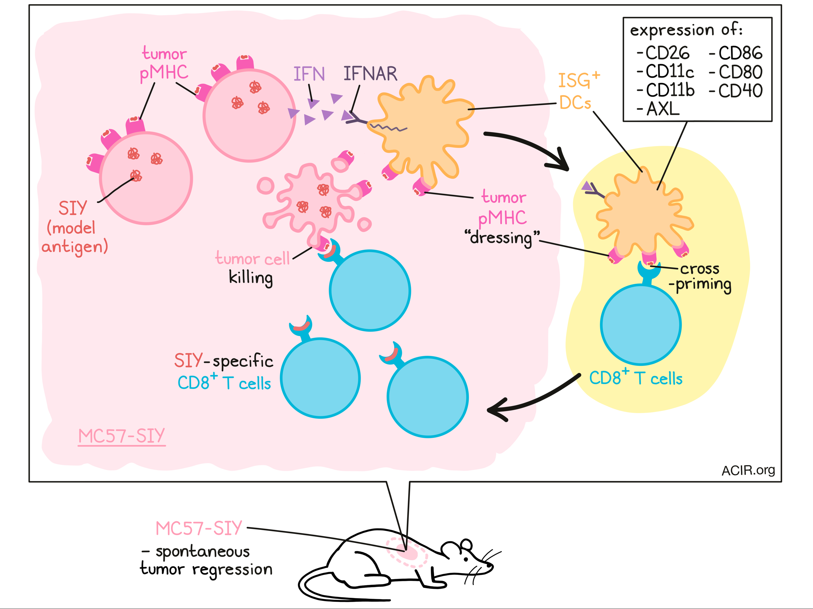
With DC1s found to be non-essential in MC57-SIY tumor rejection, the team next questioned which APC subsets could be responsible. They sorted several APC types from MC57-SIY tumors, and observed that only CD11c+ DCs, not monocytes or macrophages, could induce SIY-specific T cell proliferation ex vivo. Likewise, depletion of CD11c+ cells or all cDCs (through Zbtb46) in vivo led to MC57-SIY tumor progression, confirming that some DC subset(s) must constitute the APCs of interest. To investigate, the authors conducted scRNAseq on immune cells from MC57-SIY tumors. Although several DC clusters were identified, one stood out, which shared certain characteristics with DC2s, but otherwise had a novel gene signature enriched in interferon-stimulated genes (ISGs). Testing the importance of this cluster, termed ISG+ DCs, in tumor rejection, the authors injected MC57-SIY tumors into Ifnar1-/- mice. Without IFN-I signaling, tumor control was lost and SIY-specific T cell responses were reduced compared to wild-type mice. This effect was recapitulated with specific ablation of IFN signaling in CD11c+ DCs. Thus, the authors concluded that IFN-I signaling, likely involving ISG+ DCs, may mediate MC57-SIY tumor regression.
Next, the team characterized ISG+ DCs using targets identified through sequencing (AXL and CD11b) as surface markers for flow cytometry. AXL+CD11b+ DCs were found in MC57-SIY tumors in wild-type, Batf3-/-, and RAG2-/- mice. These DCs more closely resembled DC2s than DC1s (expressing CD11b, but lacking CD103, CD24, and CCR7), and displayed a conventional DC phenotype (CD26). ISG+ DCs were more mature (CD86, CD80, CD40 expression) than DC1s or DC2s/moDCs. Lineage tracing studies confirmed that ISG+ DCs are conventional DCs, arising from the same pathway as DC1s and DC2s. Additionally, the ISG+ DC gene signature was detected in human tumors, suggesting that this DC subset may be present across species.
The researchers next questioned whether ISG+ DCs can prime CD8+ T cells. In ex vivo coculture, tumor-derived ISG+ DCs expanded antigen-specific CD8+ T cells equivalently to DC1s, which means ISG+ DCs must also engage in some mode of cross-presentation. Based on the observation that knockout of the B2M gene in the MC57-SIY tumor cell line abrogated tumor control in Batf3-/- mice, the team hypothesized that ISG+ DCs could be “dressing” themselves with tumor peptide–MHC complexes (pMHC), and tested this idea in two settings. First, they implanted MC57-SIY tumors into B2M-/- bone marrow chimeric mice. Second, they implanted MC57-SIY tumors (H-Kb) into BALB/c mice (H-Kd). In both models, the only source of H-Kb pMHC would be from the tumor itself. Interestingly, H-Kb pMHC were detected on ISG+ DCs, significantly more than on DC1s or DC2s/moDCs, and could induce antigen-specific CD8+ T cell responses ex vivo and in vivo. Altogether, these results suggested that ISG+ DCs can dress themselves with tumor pMHC to present antigen to CD8+ T cells.
Next, Duong et al. assessed the functionality of CD8+ T cell priming by ISG+ DCs. Batf3-/- mice (lacking DC1s) were implanted with MC57-SIY tumor cells, to presumably prompt SIY-specific CD8+ T cell responses through ISG+ DCs, as the authors had previously concluded. Then, six days later, MC38-SIY (progressor) tumors were implanted on the opposing flank. Compared to mice previously injected with PBS, the prior injection of MC57-SIY significantly decreased MC38-SIY tumor growth. In fact, a portion of mice experienced complete regressions, indicating that ISG+ DCs could elicit functional CD8+ T cell responses capable of regressing an otherwise progressively growing tumor.
The team now considered the importance of IFN signaling in ISG+ DC function. Because ISG+ DCs express a variety of interferon-stimulated genes, they might acquire this state following IFN signaling within the tumor microenvironment, potentially driven by tumor cells. Supporting this idea, MC57-SIY cells expressed higher levels of IFNAR-related transcripts than MC38-SIY cells, and bone marrow-derived DCs cultured in MC57-SIY conditioned media upregulated IFN signaling. Testing the importance of tumor IFN signaling in vivo, the researchers implanted Irf3-/- or wild-type MC57-SIY cells into Batf3-/- mice. Unlike the wild-type MC57-SIY cells, which were spontaneously rejected, the Irf3-/- MC57-SIY tumors progressed, and SIY-specific CD8+ T cell responses were dramatically reduced. Without IFN signaling, the normally heightened ISG+ DC maturation also decreased. Thus, tumor-derived IFN appears to play a key role in ISG+ DC maturation, enabling effective induction of CD8+ T cell responses and tumor rejection.
Finally, the researchers questioned whether the importance of IFN signaling for ISG+ DCs could inform a therapeutic strategy. MC38-SIY tumors were implanted into Batf3-/- mice (where they would typically progress rapidly) with or without recombinant IFN-β protein. The addition of IFN-β boosted SIY-specific CD8+ T cell immunity, which was lost when B2M-/- MC38-SIY cells were used instead, again showing the importance of pMHC dressing.
Overall, the researchers demonstrated the importance of a non-DC1 DC subset in eliciting functional, tumor-specific CD8+ T cell responses, and how it could be improved by supplementing IFN-β signaling. A screening of multiple murine and human cancer cell lines found that IFN signaling (Ifnb1 expression) was variable and generally low, suggesting a broad applicability for this approach. Further understanding the diversity and roles of APC subsets can support immunotherapy research, from cancer vaccine technologies to checkpoint blockade therapy.
Write-up by Alex Najibi, image by Lauren Hitchings
Meet the researcher
This week, first author Ellen Duong answered our questions.

What prompted you to tackle this research question?
Dendritic cells (DCs) are critical activators of antitumor T cell responses. As a bulk population, DCs are highly heterogeneous and consist of several phenotypically distinct and functionally specialized subsets. It is increasingly appreciated that under inflammatory contexts, these DC subsets can assume distinct activation states with differential impacts on their function. In this work, we aimed to characterize the DC states in tumors that are associated with productive antitumor T cell responses and identify the signals that drive them. By studying these states, we hope to uncover new insights into how we can effectively harness the DC compartment to drive antitumor T cell immunity.
What was the most surprising finding of this study for you?
It is well established that cross-presenting DC1 are required for the generation of cytotoxic T cell responses. Thus, our observation that DC1 can be dispensable for antitumor immunity against a spontaneously regressing tumor was quite surprising. Through functional assays and transcriptional profiling approaches, we identified a DC cluster expressing interferon-stimulated genes (ISG+ DC) in regressor tumors. Like DC1, ISG+ DC could activate cytotoxic T cells, and we demonstrated that they were the predominant stimulatory antigen-presenting cells in mice lacking DC1. In contrast to cross-presenting DC1, ISG+ DC activated cytotoxic T cells by MHC-I dressing with tumor-derived peptide–MHC-I complexes. This was another surprising finding. and presents an opportunity to overcome the requirement for cross-presentation to drive antitumor cytotoxic T cell responses.
What was the coolest thing you’ve learned (about) recently outside of work?
The incubation temperature for turtle eggs dictates their gender! Cooler incubation temperatures give rise to males, whereas warmer temperatures produce females. For the longest time, I assumed my red-eared slider (Thaddeus) was male, but I learned from a veterinarian visit that Thaddeus is actually female! This makes sense in retrospect, as I originally got her as a hatchling during the summertime.


