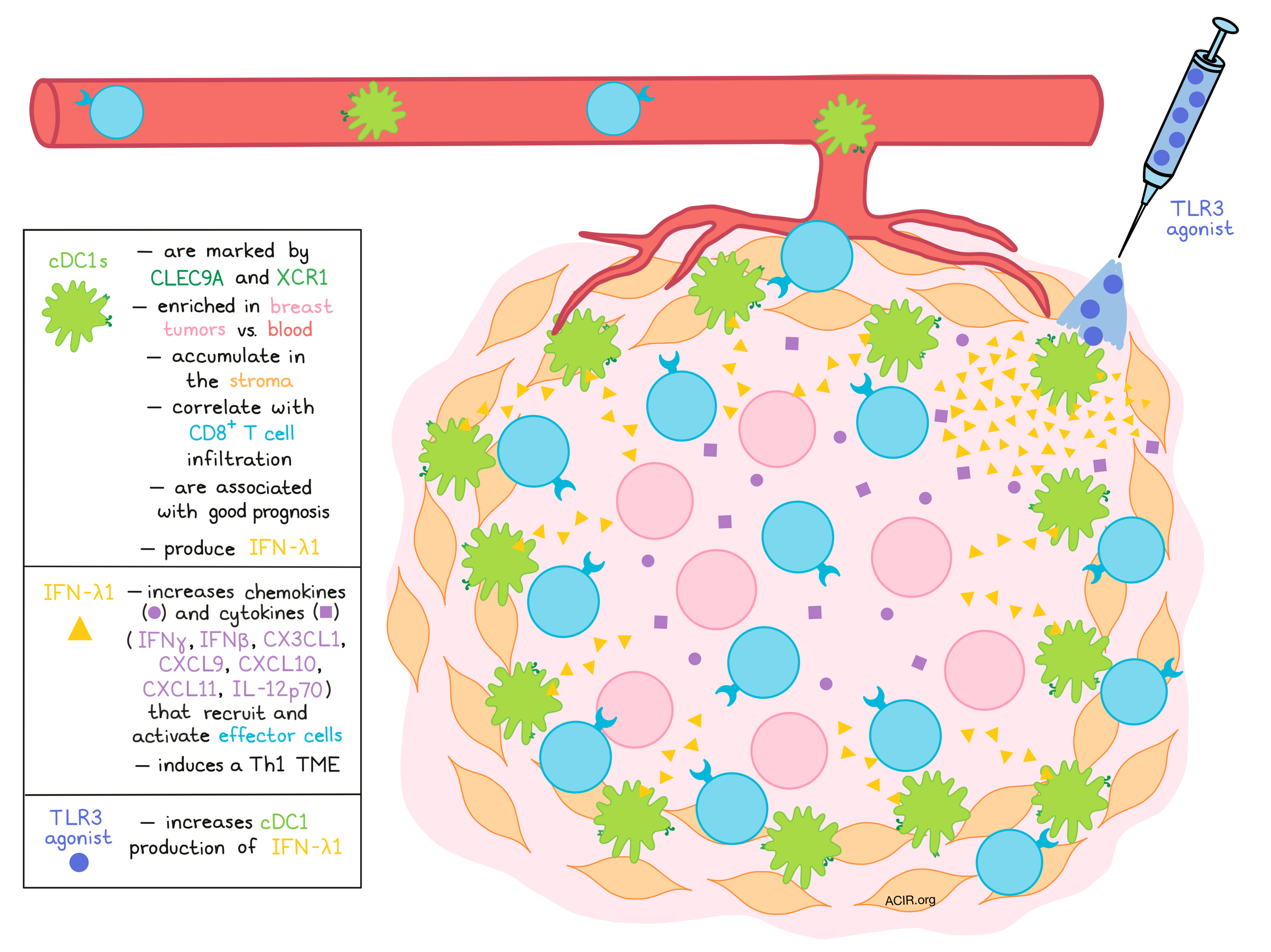
Dendritic cells play a critical role in kickstarting immune responses, and cDC1s, which cross-present antigens and activate CD8+ T cells, are of particular interest for inducing responses to immunotherapy. Previous research has shown tumor infiltration by cDC1s to be associated with favorable patient prognoses and improved clinical responses to anti-PD-1. In a paper recently published in Science Immunology, Hubert et al. analyzed primary breast tumor samples and public datasets to further investigate the role of cDC1s in patient outcomes, and to explore possible strategies that could take advantage of cDC1s to improve outcomes in the future.
Performing flow cytometry analysis of fresh primary breast tumors as well as PBMCs, Hubert et al. identified four DC compartments within tumors: plasmacytoid DCs (pDCs), cDC1s, cDC2s, and Langerhans cells (LCs). Tumor-associated cDC1s were detectable across different subtypes of breast cancer, and could be identified based on their specific expression of CLEC9A and XCR1. cDC1s also showed the highest levels of BTLA and low levels of DC-LAMP, suggesting moderate maturation. Unlike other DC subsets, the portion of cDC1s within DCs was higher in tumors than it was in blood, indicating recruitment or accumulation of cDC1s in breast tumor tissues – a phenomenon that was most evident in the triple-negative breast cancer subtype.
Using available transcriptomic datasets for breast cancer to evaluate the prognostic value of cDC1s, the researchers identified a strong association between cDC1 infiltration and a good prognosis. The presence of cDC2s and LCs also positively correlated with patient survival, but to a lesser extent.
Investigating the precise localization of cDC1s by in situ hybridization with CLEC9A and CD8 probes, the researchers imaged and analyzed 8 breast tumor samples and found that cDC1s were predominantly located in the stroma, and not the tumor bed. Here, the researchers identified a strong correlation between cDC1s and CD8+ T cell infiltration, which was supported by imaging showing close contact between the two cell types and was validated by transcriptomic data from TCGA. The correlation to CD8+ T cell infiltration was shared with cDC2s and pDCs, but not LCs, despite the correlation of LCs with improved prognosis.
To investigate the contributions of individual DC subsets, Hubert et al. used DC infiltration scores to define groups of tumors enriched for one particular subset, then used pairwise comparisons across all the sets to identify enriched gene signature pathways for each subset. In tumors that were mostly infiltrated by cDC1s, type I/III IFN and type II IFN signatures were strongly enriched, as were genes involved in antigen processing and presentation, and genes involved in cross-talk between DCs and NK cells. Tumors that were most infiltrated by cDC2s had the lowest IFN signatures, and were enriched for genes involved in immunosuppressive pathways. Tumors highly infiltrated by pDCs, LCs, or cDC2s also showed an enriched epithelial-to-mesenchymal transition (EMT) signature, indicating a more mesenchymal phenotype.
Following up on the observations that a type I/III gene signature was enriched in cDC1-exclusive tumors, the researchers investigated whether IFN-III was produced by the cDC1s in human tumors. TCGA data showed that IFN-III was present and was more abundant in tumors than in normal adjacent tissue. In breast tumor samples, IFN-λ1 was found to be the most abundant subtype of IFN-III, and further, correlated with the presence of cDC1s. Using intracytoplasmic flow cytometry analysis, the researchers found that in a third of human breast tumors, cDC1s exclusively were capable of spontaneously producing IFN-λ1 in the absence of ex vivo stimulation. Together, this suggested to the researchers that IFN-λ1 production was a specific feature of cDC1s in human tumors, and that it likely plays a key role in antitumor immunity.
To further explore the role of IFN-λ1, Hubert and colleagues analyzed IFNL1 and IFNLR1 (the specific chain of its heterodimeric receptor) expression using public transcriptomic datasets, and identified that high expression of these genes correlated with greater relapse-free survival. Next, using over 100 breast tumor samples, they found that IFN-λ1 was strongly correlated with CXCR3 ligands, CX3CL1 chemokines, TNFα, and IL-12p40, which are involved in the recruitment and activation of CD8+ T cells. To test whether IFN-λ1 might induce production of these cytokines and chemokines, the researchers treated human breast tumor samples with IFN-λ1 and saw that it increased levels of IFN-β, CX3CL1 and two CXCR3 ligands (CXCL10 and CXCL11), CX3CL1, IL-12p70, and IFNγ, suggesting that IFN-λ1 induces a T helper 1 (Th1) immune soluble microenvironment in human breast tumors.
Exploring how cDC1s and the production of IFN-λ1 might be utilized therapeutically to promote antitumor immune responses, Hubert et al. interrogated TLR3 stimulation as a strategy to induce the production of IFN-λ1 by cDC1s in breast tumors. They treated breast tumor cell suspensions and patient PBMCs with TLR3 agonist polyI:C plus resiquimod/R848 to stimulate other DC subsets through TLR7/8. In PBMCs, this induced TNFα production by all DC subsets, IFNα production by pDCs, and IFN-λ1 production by both cDC1s and pDCs. Similar results were observed in tumor cell suspensions, except that tumor-associated pDCs were unable to produce IFNα or IFN-λ1. Since tumor-associated cDC1s efficiently produced IFN-λ1 in response to TLR stimulation in 11 of 12 tumor samples, the researchers next looked at the impact of just TLR3 stimulation on the tumor microenvironment as a whole. Using fresh tumor thick sections, they found that IFNγ, CXCL9, CXCL10, and CX3CL1 were markedly increased. Together, this suggested that cDC1 stimulation via TLR3 agonism could induce a microenvironment that favors the recruitment and activation of cytotoxic effector cells.
Overall, Hubert et al. showed that IFN-λ1 was selectively produced by cDC1s in human tumors and was associated with the induction of immune-stimulating chemokines and cytokines, as well as with favorable outcomes in breast cancer. They also uncovered evidence that TLR3 activation of intratumoral cDC1s could be used in a therapeutic setting to promote the production of IFN-λ1 and induce a favorable immune microenvironment within tumors – a strategy that warrants further investigation.
by Lauren Hitchings
Meet the researcher
This week, Jenny Valladeau answered our 3 questions.

What prompted you to tackle this research question?
It was shown previously in our lab by Nathalie Bendriss-Vermare and colleagues that a population of DC called pDC was inhibited in the tumoral context. At the same time, it was shown that a population of DC termed cDC1 seemed to be more efficient at cross-presenting tumoral antigens, so it was logical to analyze whether those cDC1 infiltrate human breast tumors and if they are inhibited. The objective was clearly to propose novel immunotherapies by reactivating or targeting those very particular antigen-presenting cells.
What was the most surprising finding of this study for you?
Many studies have described that type I IFNs are very important cytokines in the antitumor immune response. When we analyzed the soluble cytokine milieu of more than 100 human breast tumors, the most surprising thing was finding out that most of the time there was no type I IFN, but rather type III, which is not a well known cytokine.
What was the coolest thing you’ve learned (about) recently outside of work?
In the current conditions of lockdown, I was surprised to see how much human beings need social interaction! Indeed, although our basic needs such as access to food are met, we have experienced how much stress confinement can cause. I was already interested in Ethology, but I was happy to go back to it to better understand what man has to gain from being a gregarious animal like herrings or penguins!


