Tumor neoantigen-specific T cells generated by vaccines can synergize with immune checkpoint blockade (ICB) for antitumor efficacy, but not much is known about the dynamics of these cells during therapy. Therefore, Liu, Chen, and Zhang et al. assessed the tumor microenvironment (TME) and lymphatic organs in response to this treatment strategy in a mouse model, using scRNAseq and TCRseq to define the phenotype and dynamics of therapy-generated neoantigen-specific T cells. Their results were recently published in Nature Cancer.
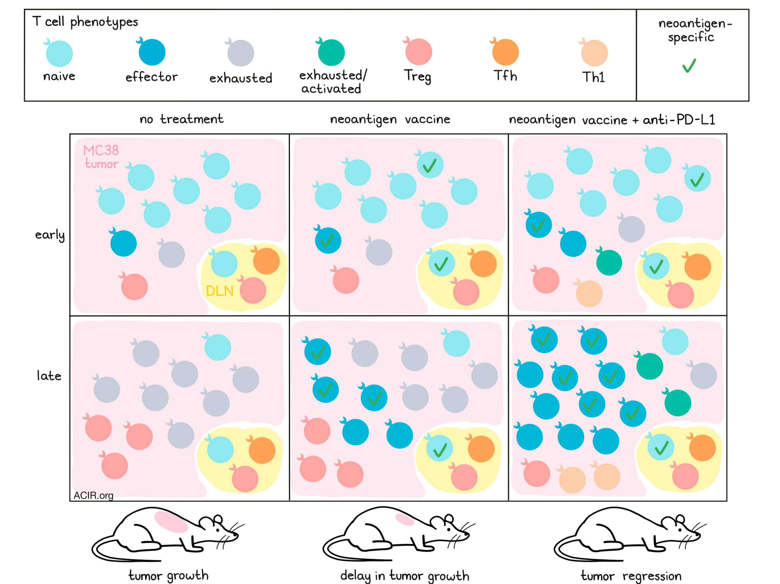
The researchers made use of the colorectal carcinoma model MC38, which has well defined MHC-I-restricted immunogenic neoantigens. The 9-mer, mutated, ADP-dependent glucokinase (ADPGK) peptide (ASMTNMELM) was used as the neoantigen for the vaccine, which was adjuvanted with two toll-like receptor agonists. Two doses of the vaccine resulted in a delay in tumor growth, but no regression. In the draining lymph nodes (DLN), tetramer-positive neoantigen-specific T cells could be detected after vaccination; however, only low levels of these cells were found in the tumor and these had an exhaustion phenotype (PD-1, TIM-3, TOX). PD-L1 expression was detected on myeloid cell populations, with higher levels in the tumor than in the DLN.
While monotherapy with anti-PD-L1 or the vaccine resulted in delayed tumor growth, the combination treatment resulted in complete tumor regression and improved survival in all animals. To establish the dynamic changes of T cell populations in response to this treatment regimen, scRNAseq with targeted TCR capture (scTCRseq) was performed on T cells from the tumor, DLN, and spleen.
Unsupervised graph-based clustering identified 23 clusters, which were distinguished based on differentially expressed genes. Lymphoid tissues were enriched for naive, memory, regulatory, and follicular helper T cells. There were two Treg clusters in tumors, which expressed higher levels of inhibitory receptors, transcription factors, and Gzmb than lymphoid Tregs. Tumoral CD8+ T cells expressed high levels of activation and exhaustion markers (Ifng, Tnfrsf9, Fasl, Tnf, Pdcd1, Lag3, and Havcr3), and low levels of naive and stem cell markers (Ccr7, Tcf7, and Sell). There was also a resident memory T cell cluster, an effector T cell cluster, and an exhaustion cluster (Pdcd1, Lag3, Havcr3, Tigit, Ctla4). Another cluster was specific to the ICB treatment group, which had an exhaustion phenotype, but expressed higher levels of granzymes than the exhaustion cluster.
To study T cell status dynamics, T cells obtained at day 10 (early-stage; prior to any therapy) and day 20 (late-stage; untreated or following therapy) of tumor progression were compared. Early-stage T cells in the tumor were primarily of a naive phenotype, which was minimally present at the late stage, regardless of treatment. On the other hand, cells with an exhaustion phenotype and Tregs were predominantly found at the late stage. Vaccine monotherapy reduced the dysfunctional CD8+ T cell percentage, increased the proportion of effector T cells, and increased the Treg ratio. Combination therapy increased CD8+ effector cells and reduced dysfunctional CD8+ T cells and Tregs; Th1-like cells were the dominant CD4+ T cell population. Flow cytometry confirmed that late-stage tumors had reduced levels of naive CCR7+ T cells and that combination therapy reduced the number of Tregs, exhausted PD-1+TIM-3+CD8+ T cells, and TOX+CD8+ T cells, and increased the number of CD4+ Th1 T cells.
To characterize neoantigen-specific T cells, the researchers vaccinated naive mice and sorted tetramer+ T cells for TCRseq. Using the iSMART algorithm, TCRs were clustered according to their similarity. Comparison between TCR groups obtained from treated and untreated mice recovered 20 vaccine-enriched groups. These enriched TCR groups were then used to track neoantigen-specific T cells in the scRNAseq data. Specific TCRs from multiple groups were enriched in the combination treatment group, and these TCRs were mainly found in the effector and naive-like clusters, and least in the exhausted cluster. Combination treatment enriched for neoantigen-specific T cells, which were mainly of an effector T cell phenotype. These data were confirmed using tetramer staining, and ELIspot analyses showed that these cells had higher IFNγ production in response to tumor cells, consistent with enrichment of the Ifng pathway in the effector T cell cluster. Furthermore, in the MC38 C57/Bl6 model, T cells specific to other MC38 epitopes were found after combination treatment, suggesting treatment resulted in epitope spreading.
To assess whether effector T cells induced by combination therapy were derived from pre-existing tumoral T cells or infiltrated from lymphoid tissue, mice were treated with the S1P inhibitor FTY720, which blocks T cell evasion from lymph nodes. Confirming that specific T cells infiltrated from lymphoid tissue, this treatment eliminated the efficacy of the combination therapy and reduced the percentage of IFNγ-producing CD8+ and tetramer+ T cells.
The researchers then determined whether DLN-derived neoantigen-specific T cells were able to mediate durable antitumor effects in the presence of anti-PD-L1. Lymphocytes from vaccinated, naive mice were collected from the DLN 6 days after vaccination, and adoptively transferred to MC38-bearing Rag1-/- mice, which were then treated with anti-PD-L1. This therapy controlled tumor growth and increased the percentage of antigen-specific T cells in the spleen and tumor.
Finally, the researchers explored whether a mouse-derived gene signature of neoantigen-specific T cells based on this data could also be found in human cancers. The five most discriminative, differentially expressed genes from the effector T cell cluster that are also found in the human genome were used to define the Cancer Antigen Specific T cell (CAST) score. In the TCGA database, the CAST score was predictive of improved overall survival in melanoma, head and neck, cervical, and pancreatic cancer in patients with a high CD8+ T cell infiltration; no effect was observed for patients with low CD8+ T cell infiltration. In a basal cell carcinoma dataset of samples obtained before and after anti-PD-1 treatment, CAST correlated with CD8+ T cell clonal frequency, and in 3 out of 4 patients, the CAST score increased after PD-1 blockade.
These data confirm that the assessed characteristics found in this mouse study might be translatable to the situation in patients, and shines a light on the dynamics and phenotypes of neoantigen-specific T cells in response to vaccination and checkpoint blockade therapy.
Write-up by Maartje Wouters, image by Lauren Hitchings.


