Weekly Digests
‹ Back to August

The cell types behind the synergy in dual checkpoint blockade

August 8, 2018

Dual checkpoint blockade targeting PD-1 and CTLA-4 is one of the most effective immunotherapeutic strategies in the clinic, but while the mechanisms of action of each monotherapy have been studied in detail, the mechanism behind the synergy of this dynamic duo is not fully understood. In a recent study, published in Cancer Immunology Research, Beavis et al. dug deeper into which immune cell subsets might make the effect of dual checkpoint blockade greater than the sum of its parts.

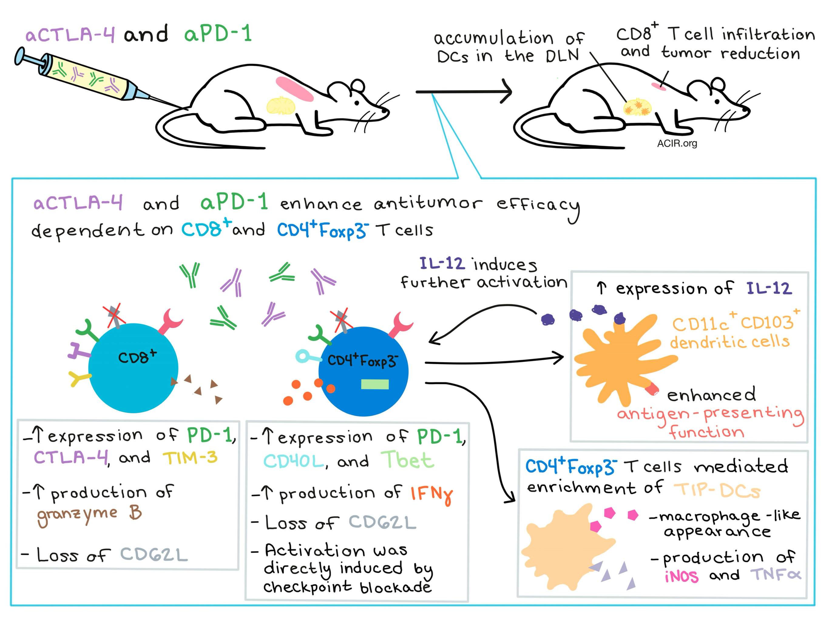
Beavis et al. began by using two murine tumor models expressing low levels of the foreign antigen OVA to confirm that combination treatment with both anti-PD-1 mAb and anti-CTLA-4 mAb significantly reduced the growth of established tumors and enhanced survival in mice compared to either monotherapy. To determine which T cell subsets were involved in the antitumor effect, the team performed a series of cellular depletion studies. Interestingly, depletion of CD8+ T cells had no effect on tumor growth in the absence of therapy, but reduced the efficacy of dual checkpoint blockade. Meanwhile, depletion of CD4+ T cells decreased tumor growth in the absence of therapy – likely due to the depletion of Foxp3+ Tregs – and dual checkpoint therapy did not further improve this effect, suggesting that CD4+ T cells play a necessary role in the response to dual checkpoint blockade.

In order to distinguish between the effect of CD4+ helper T cells (CD4+Foxp3- T cells) and Tregs (CD4+Foxp3+ T cells), the researchers utilized DEREG mice, in which Tregs can be specifically depleted with administration of diphtheria toxin. Treg depletion alone reduced tumor growth, but the effects of dual checkpoint blockade were still observable, while depleting the remaining CD4+Foxp3- T cells then attenuated the effect of the dual checkpoint blockade, suggesting that CD4+Foxp3- T cells play an essential role in dual checkpoint efficacy.

Next, Beavis et al. isolated CD8+ T cells and CD4+Foxp3- T cells from the tumors and draining lymph nodes (DLNs) of treated mice and analyzed their phenotypes. Compared to no treatment or checkpoint monotherapy, dual checkpoint blockade was found to increase the portion of CD8+ T cells expressing PD-1, CTLA-4, TIM-3, and granzyme B. In CD4+Foxp3- cells, it increased expression of PD-1, CD40L, Tbet, and IFNγ, indicating an enhanced state of activation. In both CD8+ and CD4+Foxp3- T cell subsets, the presence of IFNγ was found to be critical for activation, and combination therapy induced the loss of expression of CD62L, indicating a transition to an effector cell phenotype. Another series of immune subset depletion studies in mice, as well as ex vivo cytokine analysis, revealed that combination therapy directly activated CD4+Foxp3- T cells, as activation (measured by increased Tbet and IFNγ production and loss of CD62L) was independent from the presence of CD8+ T cells and Tregs.

Beavis et al. next explored the myeloid compartment to better understand the role of antigen-presenting cells (APCs) in dual checkpoint blockade. Interestingly, in treated tumors, CD4+Foxp3- T cells (independent of CD8+ or CD4+FoxP3+ T cells) mediated enrichment of a “TIP-DC” subset, which was primarily defined by production of TNFα and iNOS and macrophage-like appearance and qualities. Whether these TIP-DCs contribute to the observed antitumor efficacy was not determined. The team also observed that IL-12 production, a marker of myeloid cell activation, was predominantly associated with CD11c+CD103+ dendritic cells (DCs), and was upregulated by this subset in mice treated with dual checkpoint blockade. Treatment also led to an accumulation of CD103+ DCs in DLNs, concurrent with a reduction in tumor volume, possibly representing DC trafficking from the tumor to the DLN following antigen uptake. CD103+ DCs in treated mice also showed evidence of enhanced antigen-presenting function, dependent on CD4+Foxp3- T cells.

Interestingly, the increased production of IL-12 by activated CD103+ DCs was found to be essential for the upregulation of Tbet and IFNγ production by CD4+Foxp3- T cells and for the overall antitumor efficacy of dual checkpoint blockade. Together, this suggests an interplay in which CD4+Foxp3- T cells are directly activated by dual checkpoint blockade, triggering the activation of CD103+ DCs and upregulation of IL-12, which in turn induces further activation of CD4+Foxp3- T cells and CD8+ T cells, potentially contributing to memory cell formation. This cycle of activation allows CD4+Foxp3- T cells to exert their antitumor efficacy to the fullest extent, accounting, at least in part, for the enhanced antitumor effect observed in dual checkpoint blockade.

by Lauren Hitchings

References:

Beavis P.A., Henderson M.A., Giuffrida L., Davenport A.J., Petley E.V., House I.G., Lai J., Sek K., Milenkovski N., John L.B., Mardiana S., Slaney C.Y., Trapani J.A., Loi S., Kershaw M.H., Haynes N.M., Darcy P.K. Dual PD-1 and CTLA-4 checkpoint blockade promotes antitumor immune responses through CD4+Foxp3- cell-mediated modulation of CD103+ dendritic cells. Cancer Immunol Res. 2018 Jul 17.

In the Spotlight...

Wnt3a Neutralization Enhances T-cell Responses through Indirect Mechanisms and Restrains Tumor Growth

Following the observation that Wnt3a in MC38 tumors was primarily produced by stromal cells, Pacella et al. examined the effects of neutralization of Wnt3a on T cell effector functions. Administration of Wnt3a-neutralizing antibody in tumor-bearing mice reduced MC38 tumor growth and led to preferential expansion of tumor antigen-specific CD8+ effector memory T cells with increased expression of Tbet and IFNγ, and decreased expression of Tcf1. The effect was not the result of the hypothesized interruption of B-catenin signaling in CD8+ T cells but rather the result of restored dendritic cell maturation and activity following Wnt3a neutralization.

Activation of NKT cells in an anti-PD-1-resistant tumor model enhances anti-tumor immunity by reinvigorating exhausted CD8 T cells

Bae et al. demonstrate that in mice with tumors resistant to anti-PD-1, treatment with antigen-presenting cells (APCs: B cells and monocytes) loaded with α-galactosylceramide leads to improved tumor control via IL-2 production by invariant natural killer T cells and IL-12 production by CD11c+ dendritic cells and CD11b+ myeloid cells. The two cytokines restore function to exhausted CD8+ T cells and reduce their PD-1 expression, leading to delayed tumor growth. Addition of anti-PD-1 synergizes to induce complete response and improve survival in mice.

Enhancing T cell therapy through TCR-signaling-responsive nanoparticle drug delivery

Tang and Zheng et al. developed a drug delivery system comprising a protein of interest (e.g., cytokine) cross-linked with a reversible disulfide linker, forming a nanogel that is anchored to the T cell surface via CD45, which allows for prolonged surface retention. Upon TCR stimulation, T cells increase cell surface reduction activity, which triggers the release of the protein via cross-link cleavage. In vivo, adoptive transfer of T cells with IL-15-loaded nanogels significantly slowed tumor growth, improved survival, and lessened toxicity (due to tumor-targeted release) in mice with melanoma, compared to transfer of T cells combined with free IL-15.

TIGIT immune checkpoint blockade restores CD8+ T cell immunity against multiple myeloma

Guillerey and Harjunpaa et al. show that TIGIT is the dominant immune checkpoint in multiple myeloma (MM) in both mice and humans, and that the percentage of TIGIT+CD8+ T cells correlate with MM burden in mice. In human MM, TIGIT+CD8+ T cells are exhausted and have impaired cytokine production, proliferation, and cytotoxicity. Blockade of the TIGIT pathway in vivo in mice, either via knockout or antibody, reduced tumor burden and improved survival in a CD8+ T cell-dependent manner, and anti-TIGIT antibody also increased CD8+ T cell effector function.

IL-35 hinders endogenous anti-tumor T cell immunity and responsiveness to immunotherapy in pancreatic cancer

Mirlekar et al. found that in the context of pancreatic ductal adenocarcinoma (PDA), IL-35 is produced primarily by Bregs and CD4+ T cells (both FoxP3+ and FoxP3-). In mice lacking IL-35, PDA tumor growth was reduced, infiltration by activated cytotoxic CD8+ T cells and effector CD4+ T cells (including those expressing IFNγ and TNFα) was increased, and Treg frequency was decreased. While PDA is typically resistant to anti-PD-1 immunotherapy, mice lacking IL-35 responded to anti-PD-1 (accompanied by enhanced CD8+ T cell infiltration), suggesting that targeting IL-35 could help overcome resistance to immunotherapy in PDA.

Initial phase I/IIa trial results of an autologous tumor lysate, particle-loaded, dendritic cell (TLPLDC) vaccine in patients with solid tumors

In an ongoing, open label, phase I/IIa clinical trial, patients with measurable or fully resected solid tumors were treated with a personalized tumor lysate dendritic cell vaccine that was prepared using a novel, scalable, and rapid method requiring only 1 mg of tumor and 120 mL of patient’s blood. The vaccine was well-tolerated. Among 31 patients with residual disease, 2 had complete response (both had failed prior therapy and received no concurrent therapy), 4 had partial response, and 6 had stable disease. In the adjuvant arm, 6 of 13 patients remained disease-free at a median follow-up of 22.5 months.

Everything New this Week In...

Close Modal

Small change for you. Big change for us!

This Thanksgiving season, show your support for cancer research by donating your change.

In less than a minute, link your credit card with our partner RoundUp App.

Every purchase you make with that card will be rounded up and the change will be donated to ACIR.

All transactions are securely made through Stripe.