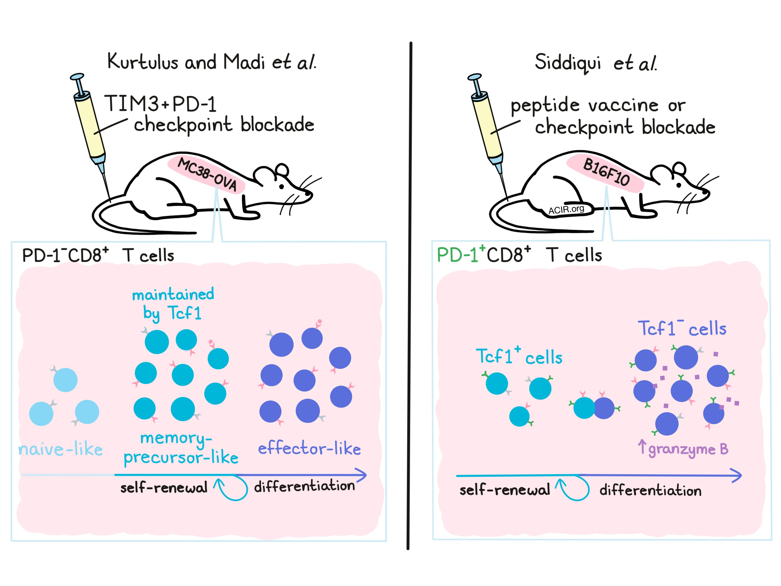
Two articles, recently published back to back in Cell, identify important roles for T cells expressing Tcf1 (encoded by Tcf7), a transcription factor known to play roles in maintaining a relatively undifferentiated T cell state, promoting self-renewal and T cell memory. Kurtulus and Madi et al., working primarily with the MC38-OVA colon carcinoma model, focused their investigation on PD-1-CD8+ T cells, while Siddiqui et al., working with the B16F10 melanoma model, focused on PD-1+CD8+ T cells. Both teams defined a critical role for Tcf1+ T cells as a self-renewing precursor population critical to response to immunotherapy.
Kurtulus and Madi et al. began their investigation by analyzing bulk and single-cell RNA profiles of TILs to explore the dynamics of effector CD8+ T cell responses to checkpoint blockade. They found that following treatment with combination TIM-3 and PD-1 blockade in a murine OVA-MC38 colon carcinoma model, a variety of transcriptional changes occured, primarily within PD-1-TIM-3-CD8+ T cells (hereafter referred to as just PD1-CD8+ T cells). PD1-CD8+ T cells showed increased effector profiles and increased frequency following checkpoint blockade due to increased expansion in response to tumor-antigen stimulation. Adoptive transfer of this cell subset into MC38-OVA tumor-bearing RAG-/- mice showed that the transferred cells were able to maintain a population of PD-1-CD8+ T cells, while also producing a pool of PD-1+TIM3+CD8+ T cells. This suggested that PD-1-CD8+ TILs act as precursors with the potential to both self-renew and give rise to more differentiated cells.
Based on transcriptional programs, cell surface markers, and previously defined CD8+ T cell signatures, the researchers were able to identify three distinct subsets within PD-1-CD8+ T cells defined by expression of CD62L, Slamf7, and CXCR5: naive-like, memory-precursor-like, and effector-like. Effector-like and memory-precursor-like subsets both expanded in response to checkpoint blockade, contained tumor-antigen specific cells, and exhibited cytotoxic activity, though the memory-precursor-like cells exhibited higher polyfunctionality. Shifts in the proportions of subsets with checkpoint blockade and overlap in their gene expression profiles indicated that PD-1-CD8+ T cells likely follow a differentiation trajectory from naive-like, to memory-precursor-like, to effector-like. Hypothesizing that the memory-precursor-like PD-1-CD8+ T cell subset was largely responsible for maintaining long-lasting antitumor responses, the researchers identified high expression of the transcription factor Tcf1 in this subset. Targeted genetic knockout of Tcf7 in CD8+ T cells dramatically reduced the number and polyfunctionality of memory-precursor-like T cells and reduced the efficacy of checkpoint blockade. Finally, the gene signatures of the memory-precursor-like and effector-like subsets shared features with human TIL phenotypes that have previously been associated with better prognosis and response to checkpoint blockade in cancer patients, suggesting that these findings may be broadly applicable and relevant in the clinic.
In a separate study, Siddiqui et al., also identified an important role for Tcf1 (Tcf7), though their work largely focused on the PD-1+CD8+ T cell compartment. Siddiqui et al. first observed that adoptively transferred tumor-specific T cells lacking Tcf7 were less effective in controlling tumor growth in vivo than their Tcf7-expressing counterparts. The researchers found that most TILs from palpable untreated tumors were Tcf1+ and PD-1+. Therapeutic peptide vaccination expanded the Tcf1+PD-1+ TIL subset, concurrent with an expansion of Tcf1-PD-1+ TILs, which were more differentiated and frequently co-expressed granzyme B. When Tcf1+ cells were selectively ablated from vaccinated mice, Tcf1- T cells also decreased and tumors progressed, suggesting that the Tcf1+ subset sustains the Tcf1- subset in the context of therapeutic vaccination. Further, based on selective Tcf1 deletion and blockade of lymphocyte trafficking, the source of Tcf1- cells was found to be intratumoral Tcf1+ cells, suggesting that Tcf1+ cells are capable of expanding and differentiating on site, mediating the proliferative response to immunotherapy. Similar results were observed in the context of immune checkpoint blockade, suggesting that checkpoint blockade may rely not on the reversal of exhaustion in existing T cells, but on the expansion and differentiation of Tcf1+PD-1+ memory-like T cells within the tumor.
Like Kurtulus and Madi et al., Siddiqui et al. turned to clinical data to determine whether the subset they had identified was relevant in human patients. In peripheral blood from melan A peptide-vaccinated melanoma patients, the researchers were able to identify non-naive, tumor antigen-specific TCF1+PD-1+CD8+ T cells and TCF1-PD-1+CD8+ T cells with the same antigen specificity. Using publicly available single-cell RNAseq data for TILs from human melanoma, they confirmed that their subset of interest was present and had an expression profile similar to that observed in mice. In patients treated with checkpoint blockade, the fraction of TCF1+PD1+ cells was higher among CD8+ TILs than in patients who were not treated with checkpoint blockade. Using The Cancer Genome Atlas, Siddiqui et al. found that the combined expression of TCF7 and PDCD1 predicted improved patient survival.
Together, these studies by Kurtulus and Madi et al. and Siddiqui et al. suggest that immunotherapy acts on the less differentiated Tcf1+CD8+ T cells, which produce an expanded pool of more differentiated Tcf1-CD8+ T cells. While Kurtulus and Madi et al. interrogated PD-1- T cells and Siddiqui et al. focused on PD-1+ T cells, both identified the self-renewing and memory-stimulating effects of Tcf1 expression as critical to effective immunotherapy. Finally, given the potential of Tcf1+ memory or stem-like T cells to both self-renew and differentiate into Tcf1- effector-like cells upon treatment with checkpoint blockade, our understanding that checkpoint blockade reinvigorates existing exhausted cells may be replaced by a model in which checkpoint blockade depends on newly differentiated Tcf1- effector CD8+ T cells.
by Lauren Hitchings


