A wide variety of factors are known to contribute to the success or failure of cancer immunotherapy. In an effort to identify specific factors, including cell states, Sade-Feldman and Yizhak et al. profiled the transcriptomes of immune cells isolated from 48 tumor samples from 32 patients with melanoma who had been treated with checkpoint blockade therapy (anti-PD-1, anti-CTLA-4, or combination) with known clinical outcomes. The results of these analyses and related functional studies were recently published in Cell.
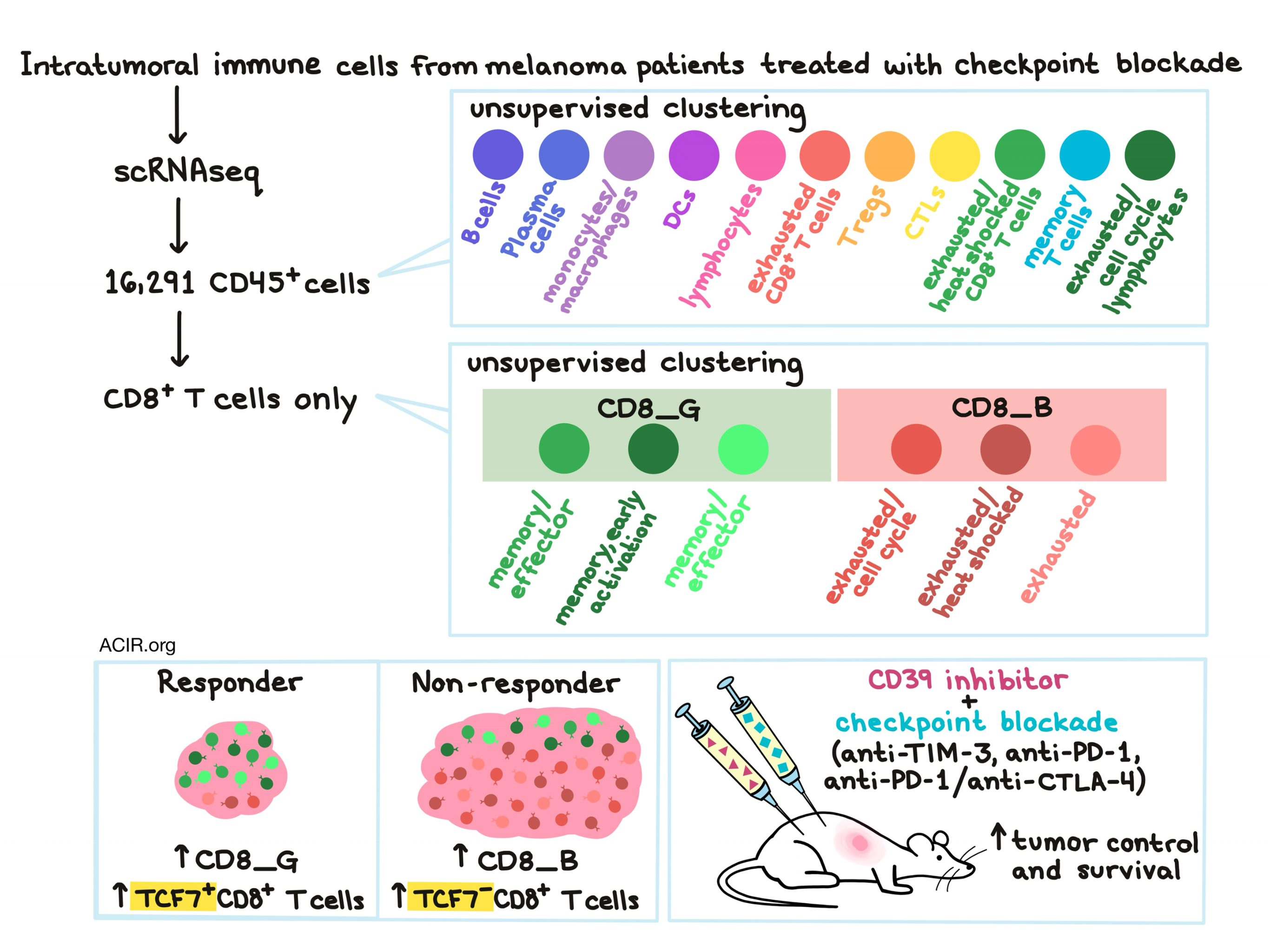
To begin their analysis, Sade-Feldman and Yizhak et al. categorized patients and individual lesions as responders (complete response, partial response) or non-responders (progressive disease, stable disease). They then performed single-cell RNA sequencing (scRNAseq) on CD45+ immune cells, identifying 16,291 cells suitable for analysis. Unsupervised clustering of the cells identified 11 distinct clusters. In responding lesions, one cluster of B cells and one cluster of T cells were significantly enriched, while in non-responders a cluster of monocytes/macrophages, a cluster of dendritic cells, a cluster of T cells, and a mixed cluster of T and NK cells were enriched. Signatures of lymphocyte activation and exhaustion were identified in both responding and non-responding lesions both before and after therapy. All individual patients showed changes in their immune profiles between baseline and post-treatment samples, though interestingly, none of the observed changes were shared between patients.
Based on their high frequency in tumors and their known role in mediating checkpoint blockade, Sade-Feldman and Yizhak et al. focused their analysis on CD8+ T cells. Clustering of CD8+ T cells identified two distinct CD8+ T cell states: CD8_G had increased expression of genes linked to memory, activation, and cell survival and reduced expression of co-inhibitory molecules, while CD8_B was enriched for genes linked to exhaustion. While CD8_G and CD8_B cells coexisted in all lesions, CD8_G cells were enriched in responders and CD8_B cells were enriched in non-responders, and the CD8_G/CD8_B ratio could predict the regression or progression of tumors.
In an even finer analysis of clustering, CD8_G and CD8_B could be further subcategorized. Within CD8_G cells, two clusters were marked by memory and effector functions, while a third was marked by memory and early activation. Within CD8_B cells, one cluster was marked by exhaustion and cell cycle genes, another was marked by exhaustion and heat shock genes, and a third was marked by exhaustion alone.
Looking into individual markers, the researchers noted that the transcription factor TCF7 was a top marker associated with responding lesions and that it was often expressed in CD8_G cells. TCF7 has previously been identified for its key role in differentiation, self-renewal, persistence of memory CD8+ T cells, and reinvigoration of exhausted CD8+ T cells in response to checkpoint blockade. In the current dataset, more TCF7+CD8+ T cells were found in responding samples and more TCF7-CD8+ T cells were found in non-responding samples, and a higher TCF7+CD8+/TCF7-CD8+ ratio corresponded with longer survival, indicating that TCF7 expression in CD8+ T cells was associated with response to checkpoint blockade and survival.
Another marker noted by researchers in this study was CD39, which was identified as an exhaustion marker that was co-expressed with TIM-3. CD39+TIM3+CD8+ T cells showed reduced expression of TNFα and IFNγ and reduced antitumor efficacy ex vivo compared to CD39-TIM3-CD8+ T cells (which also expressed TCF7). In a mouse model of melanoma, dual blockade of CD39 and TIM3 reduced tumor size and enhanced survival compared to either monotherapy. The addition of a CD39 inhibitor to either anti-PD-1 or anti-PD-1/anti-CTLA-4 treatments also showed a synergistic effect evidenced by further reduction of tumor growth and enhanced survival. The effects of CD39 inhibition were dependent on CD8+ T cells and led to increased IFNγ production and proliferation in response to TCR stimulation.
To better understand the underlying transcriptional regulation behind the observed CD8+ T cell states, Sade-Feldman and Yizhak et al. turned to analysis of the epigenetic landscape. Patterns of open chromatin (identified by ATAC-seq) aligned with previously identified upregulated transcripts and showed that key regulatory elements and transcription factors including TCF7 and BATF regulate memory-like and exhaustion-like programs, respectively, in CD8+ T cells in human melanoma.
Finally, looking at the TCR clonal landscape, the researchers found that non-responders were enriched for persistent TCRs (observed both before and after therapy), but that those T cells did not fall into memory or effector clusters. They also observed a rise in TCR clones (typically singlets) in effector clusters that did not exist prior to therapy, indicating that lymphoid-derived T cells may play a role in effective immunotherapy. Enriched TCRs (defined as being detected in multiple T cells) were more likely to be associated with exhausted T cell phenotypes, which may suggest persistent exposure to tumor antigen. Further, identical TCRs could be found in exhausted and memory states, suggesting that T cells can transition between states.
Overall, Sade-Feldman and Yizhak et al. identified markers, transcription factors, and immune cell states – particularly CD8+ T cell states – associated with response or lack of response to immunotherapy in melanoma and identified TCF7 expression as potentially predictive of outcome. With more information about the immune contexture within melanoma and other types of tumors, researchers may be able to more effectively predict response to therapies and treat cancer.
by Lauren Hitchings


