Weekly Digests
‹ Back to November

What’s keeping natural killer cells in check?

November 15, 2017

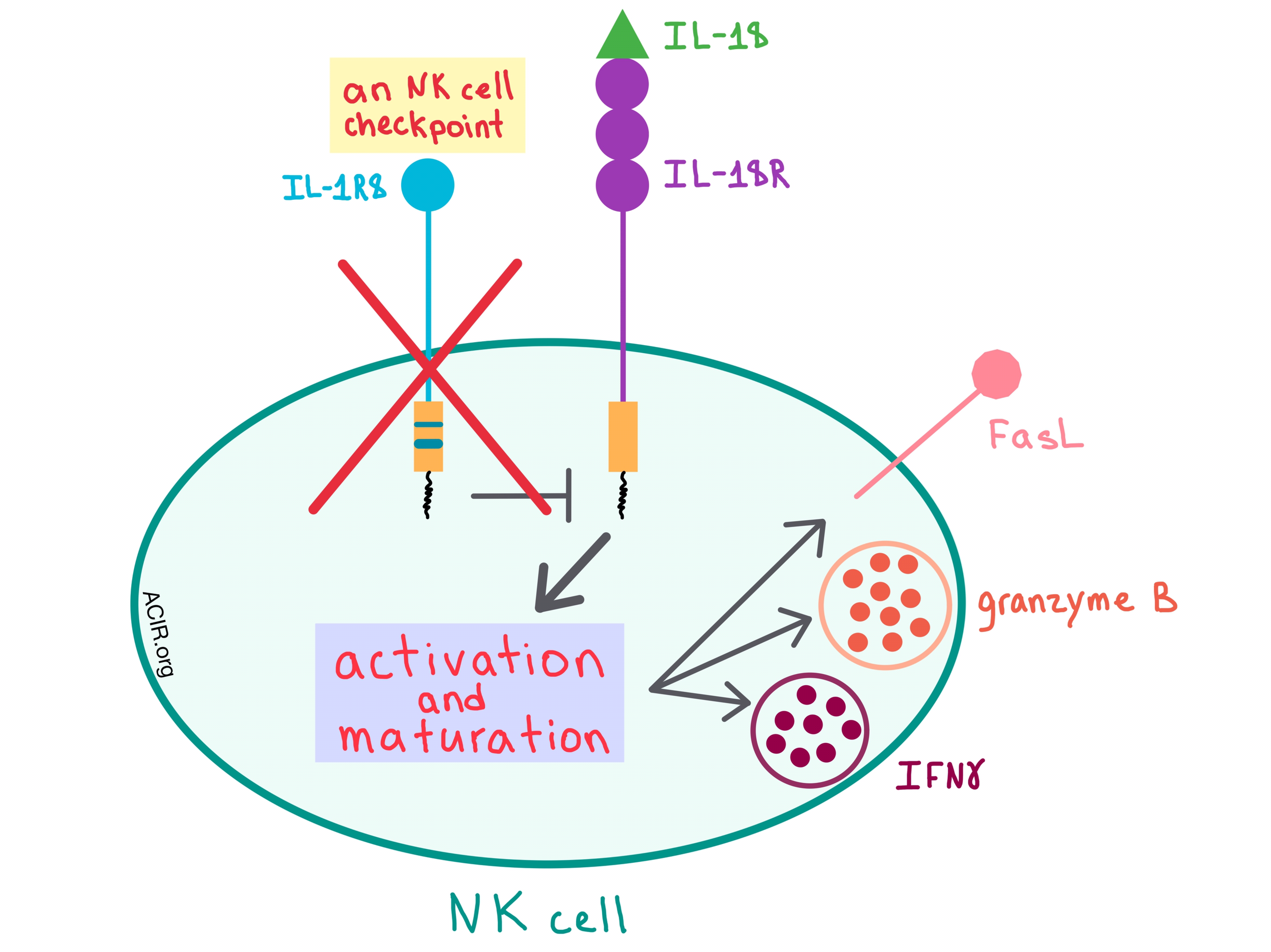
In a recent paper published in Nature, Molgora et al. demonstrated that interleukin-1 receptor 8 (IL-1R8), which is a negative regulator of the IL-1 receptor (ILR) and Toll-like receptor (TLR) pathways, acts as a checkpoint for natural killer (NK) cell maturation and function, and removing this checkpoint could affect tumorigenesis and metastasis.

Although many cells express IL-1R8, Molgora et al. found that among both human and mouse immune cells, it is particularly abundant on NK cells. As human NK cells mature, the mRNA levels and protein expression of IL-1R8 increases, and this pattern is recapitulated in mouse NK cells, thus allowing the researchers to examine the effects of this receptor in mouse models.

To understand how IL-1R8 acts on NK cells, Molgora et al. utilized IL-1R8-deficient (Il1r8-/-) mice, and found that the knockout of IL-1R8 resulted in increased NK cell frequency and count and a more mature NK cell phenotype. NK cells within the peripheral blood of these mice had an upregulated expression of activating receptors, and, when stimulated ex vivo with IL-18, the cells increased the production of IFNγ and granzyme B and expression of FasL, indicating that IL-1R8 influences NK cell function.

Since IL-18 is involved in NK cell differentiation and function, the researchers sought to further elucidate its relationship with the IL-1R8 pathways. Removing the effect of IL-18 either genetically or via an antibody blockade in Il1r8-/- mice abrogated enhanced NK cell maturation and function, suggesting that IL-1R8 regulates the IL-18 signaling pathways in NK cells. To better understand the mechanism of action of IL-1R8, the researchers performed RNA sequencing, which revealed that the transcriptional changes in NK cells due to IL-1R8 deficiency involved activation pathways (such as MAPK), adhesion molecules (such as ICAM-1), and increased production of some chemokines (such as CCL4).

To confirm that IL-1R8 also acts as a checkpoint in humans, Molgora et al. treated normal human NK cells with a combination of IL-18 and IL-12, and observed an inverse correlation between IL-1R8 levels and IFNγ production. In addition, partial silencing of IL-1R8 with small interfering RNA resulted in increased IFNγ production, thus indicating that, like in mouse cells, IL-1R8 acts as a negative regulator of NK cell activation in humans.

Having confirmed that the relationship between IL-1R8 and NK cell maturation and function is similar between humans and mice, the researchers went on to test the effect of IL-1R8-mediated regulation of NK cells in a tumor setting in several mouse models. Because the liver has a high concentration of NK cells, the team started with a model of hepatocellular chemical carcinogenesis. Compared with Il1r8+/+ mice, fewer tumor lesions that were also smaller in size developed in the Il1r8-/- mice, and within these lesions, there were more NK cells, higher levels of antitumor cytokines (eg, IFNγ), and lower levels of pro-inflammatory cytokines. Consistent with other studies suggesting that NK cells may have a beneficial role in preventing blood-borne metastases, similar results were observed in a model of sarcoma spontaneous lung metastasis, where Il1r8-/- mice had fewer metastases, although the primary tumors were unaffected. In both mouse models, the protection against cancer development or spread was NK cell-dependent, as depletion of the NK cells (but not CD4+ or CD8+ T cells) abrogated the protective effect. Il1r8-/- mice were also protected against MC38 colon carcinoma liver metastasis, and genetically knocking out IL-18 negated this protection, once again confirming that IL-1R8 acts through the IL-18/IL-18R axis. Further, adoptive transfer of Il1r8-/- NK cells into tumor-bearing Il1r8+/+ mice reduced the number and volume of liver and lung metastases, while adoptively transferred Il1r8+/+ NK cells had no effect.

As the effects of an immune checkpoint blockade can often go both ways, IL-1R8-deficient mice experience deregulated activation by ILR or TLR ligands, which leads to increased inflammation and, in turn, the development of certain cancers and autoimmune disease. The differential effect of IL-1R8 deficiency on carcinogenesis may reflect the fundamental differences in tissues at different anatomic sites, including the level of NK cell infiltration. Therefore, NK cells have the potential to affect solid tumors and metastases if the appropriate checkpoints, such as IL-1R8, are blocked and the tissue-specific immunological landscape is taken into consideration.

by Anna Scherer

References:

Molgora M., Bonavita E., Ponzetta A., Riva F., Barbagallo M., Jaillon S., Popović B., Bernardini G., Magrini E., Gianni F., Zelenay S., Jonjić S., Santoni A., Garlanda C., Mantovani A. IL-1R8 is a checkpoint in NK cells regulating anti-tumour and anti-viral activity. Nature. 2017 Nov 2.

In the Spotlight...

Neutralization of IL-8 decreases tumor PMN-MDSCs and reduces mesenchymalization of claudin-low triple-negative breast cancer

A clinical-stage monoclonal antibody HuMax-IL8 targeting IL-8 reverted the tumor from mesenchymal to a more epithelial state, reduced the recruitment of polymorphonuclear myeloid-derived suppressor cells to the tumor, made tumor cells more susceptible to lysis by NK and T cells, and reduced tumor volume in a claudin-low triple-negative breast cancer (TNBC) xenograft model. Combining the antibody with docetaxel enhanced its effect, suggesting that HuMax-IL8 could have a role in combination therapies for TNBC and other cancers.

Intratumoral CD8+ T-Cell Apoptosis is a Major Component of T Cell Dysfunction and Impedes Anti-Tumor Immunity

Horton et al. found that tumor-infiltrating PD-1+LAG-3+4-1BB+ CD8+ lymphocytes (TILs) within progressing tumors were stuck in a vicious cycle of antigen-stimulated proliferation and enhanced apoptosis (possibly due to high levels of DNA damage), leading to limited accumulation and poor tumor control. Preventing apoptosis by either overexpression of the anti-apoptotic molecule Bcl-xL or combination of agonist 4-1BB antibody with a blockade of either CTLA-4 or PD-L1 led to increased CD8+ TIL accumulation and improved tumor control.

Combination of CD40 agonism and CSF-1R blockade reconditions tumor-associated macrophages and drives potent antitumor immunity

Using a combination of anti-CSF-1R and agonist anti-CD40 antibodies, Wiehagen et al. shifted the tumor microenvironment toward a more proinflammatory state, and improved tumor control and survival by reducing tumor-associated macrophages and Tregs, increasing the frequency of newly-differentiated proinflammatory macrophages, increasing the number of activated dendritic cells in the draining lymph nodes, and enhancing CD8+ T cell and CD4+ helper T cell effector function and infiltration within the tumor.

The immunopeptidomic landscape of ovarian carcinomas

In a comprehensive study utilizing 42 epithelial ovarian carcinoma (EOC) bulk tumors and more than 100 benign tissue samples (5 tissue types, including ovary/fallopian tube), Schuster et al. identified over 50,000 HLA-presented peptides, including multiple HLA class I and class II peptide epitopes found exclusively on EOC tissue in >10% of patients. Priming of CD8+ T cells from healthy volunteers demonstrated the immunogenic potential of a number of these EOC-specific epitopes.

SLAMF7-CAR T-cells eliminate myeloma and confer selective fratricide of SLAMF7+ normal lymphocytes

SLAMF7 is highly expressed in multiple myeloma (MM), but also in subsets of normal T, B, and NK cells. Gogishvili et al. engineered SLAMF7-CAR T cells from both healthy donors and MM patients that effectively targeted newly diagnosed and relapsed/refractory myeloma in vitro and demonstrated complete resolution of MM manifestations after a single dose in a murine xenograft model (although MM eventually reemerged from anatomical sanctuaries). Subsets of normal B, T, and NK cells that were SLAMF7-/low were spared from fratricide and remained functional.

A Discrete Subset of Monocyte-Derived Cells among Typical Conventional Type 2 Dendritic Cells Can Efficiently Cross-Present

Sheng et al. discovered a distinct subset of antigen-presenting cells (APCs) that shares the migratory nature and markers (CD11chiMHCIIhiCD11b+CD24+) of conventional cDC2 dendritic cells and the monocytic origin, phagocytic properties, and markers (F4/80hiCD64+CD169+MERTK+) of tissue-resident macrophages. In metastatic melanoma and colorectal cancer mouse models, these “hybrid” APCs were enriched in the tumor microenvironment, and were capable of cross-presenting tumor-derived antigens.

Everything New this Week In...

Close Modal

Small change for you. Big change for us!

This Thanksgiving season, show your support for cancer research by donating your change.

In less than a minute, link your credit card with our partner RoundUp App.

Every purchase you make with that card will be rounded up and the change will be donated to ACIR.

All transactions are securely made through Stripe.