Immune checkpoint blockade is a powerful treatment for many cancers, and a number of factors are known to predict or contribute to responses. However, some cancers that seem set up for success, like high-grade serous epithelial ovarian cancer (HGSOC), still fail to respond to therapy. In a recent study published in Cancer Cell, Duraiswamy et al. investigated why many HGSOCs fail to respond to PD-1 blockade despite the presence of TILs, including intraepithelial (ie)TILs that specifically infiltrate tumor islets and are associated with tumor reactivity and longer survival.
To begin, Duraiswamy et al. looked at TILs from 74 samples of chemotherapy-naive, advanced HGSOCs using multispectral immunofluorescence microscopy (mIF) and found that markers of TCR activation and tumor specificity were almost exclusive to tumors harboring ieTIL. When purified, these ieTILs exhibited effector memory or terminally differentiated phenotypes and evidence of recent TCR engagement. Further, TILs that were clonally expanded or produced granzyme B, IFNγ, or IL-2 were increased in tumor islets versus adjacent stroma, suggesting that tumor islets serve as hubs of antitumor immunity.
Next, investigating PD-1 expression on TILs from ovarian cancer, the researchers showed that TILs – especially TILs from within tumor islets, terminally exhausted CD8+ TILs, and tumor antigen-specific TILs – were enriched for PD-1+ cells. A fraction of these PD-1+ TILs also expressed markers associated with memory, polyfunctional effector functions, and proliferation. In tumor digest cultures, PD-1 axis-targeting antibodies enhanced the polyfunctionality of these TILs in response to tumor-associated peptides. Looking at the effects of anti-PD-1 in situ on antigen-specific TIL clonotypes using tetramer-sorted cells, the researchers noted variable proliferation, with some TILs proliferating and others becoming depleted. When TILs that emerged following treatment with anti-PD-1 were transferred into mice bearing patient-derived tumors, they more effectively rejected tumors compared to TILs not exposed to anti-PD-1. Further, immunodominant clones that rejected tumors could be traced to islet compartments of the original autologous tumors.
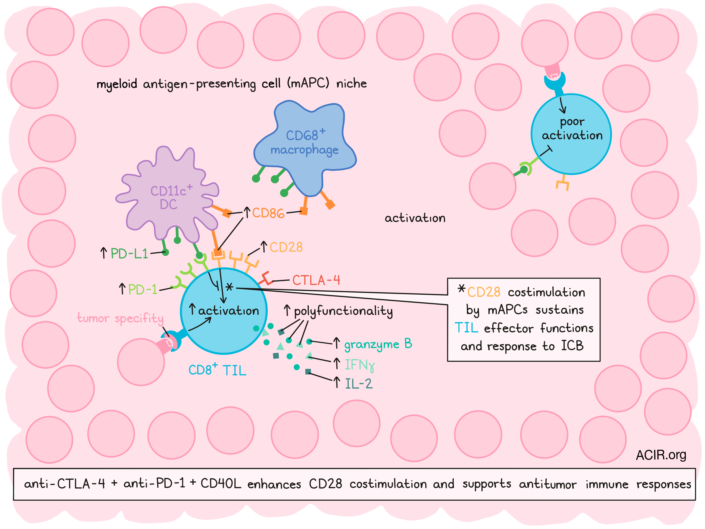
Looking more specifically at PD-1+CD8+ TILs and tumor islets, Duraiswamy et al. found that ieCD8+ TILs correlated with the presence of iePD-L1+ myeloid cells, but not with PD-L1+ tumor cells. The iePD-1+CD8+ TILs also physically clustered with iePD-L1+CD11c+ DCs and iePD-L1+CD68+ macrophages, and were observed in simultaneous contact with myeloid cells and tumor cells. Frequent interactions between TILs and myeloid cells, evidence of intimate membrane interfaces, and higher TIL activation and polyfunctionality within myeloid niches suggested that these cells formed functional immune synapses. DCs and macrophages from within niches also expressed increased PD-L1, suggestive of reciprocal activation. Ultimately, higher frequencies of TIL–APC clusters within tumors were associated with higher frequencies of polyfunctional ieCD8+PD-1+ TILs expressing activation and effector markers, and with longer survival.
Given the observed interactions between TILs and myeloid cells, the researchers hypothesized that polyfunctional PD-1+CD8+ TILs embedded in mAPC niches receive costimulatory signaling. FACs analysis confirmed increased CD28 expression on both PD-1+ and PD-1- CD8+ TILs, and increased CD28 expression on a per cell basis among PD-1+ CD8+ TILs, especially those that had recently been TCR-activated. Within the same tumors, a higher portion of APCs expressed PD-L1 and CD86.
Analyzing the molecular states of TILs, the researchers then performed single-cell RNA and TCR sequencing on 23,000 CD8+ TIL from 17 ovarian tumor digest cultures stimulated with TAA peptides. Unsupervised clustering revealed 7 clusters, each of which was assigned an exhaustion (TEX) and costimulation (CD28cost) score. Cells with high scores in both exhaustion and costimulation were the most clonally expanded, and showed a gene expression profile that indicated antigen experience. Within clonally expanded TIL, individual clones exhibited similar exhaustion scores, but a wide range of costimulation scores. Clonally expanded cells with high scores in both exhaustion and costimulation displayed enhanced polyfunctionality, effector fitness, and genes for effector machinery components compared to cells with high scores in exhaustion and low scores in costimulation.
Duraiswamy et al. hypothesized that the functional state of CD8+ TILs could be dependent on CD28 signaling, which is affected by the availability of local CD28 signals and by PD-1. In line with this, depletion of APCs and disruption of CD28 signaling each abrogated both T cell activation and the effects of anti-PD-1. In patient data, pre-treatment upregulation of activated T cell and mAPC signatures correlated with response to anti-PD-1. Upon treatment, a brief increase in CD8+ TILs expressing CD28 was observed in responding TMEs. In the setting of PD-1 blockade in culture, forced expression of CD28 ligands (CD80/CD86) restored cytolytic function in previously exhausted T cells.
CTLA-4, which attenuates CD28 signaling, was also found to be a hallmark of tumor-reactive CD8+ TILs, with most TAA-specific TIL expressing CTLA-4. When anti-CTLA-4 was added to tumor digest cultures with anti-PD-1 and TAA peptides, increases in T-bet, granzyme B, IFNγ, and proliferation were observed; these effects were dependent on local mAPCs and CD28. Similar results were observed in vivo, where the combination of anti-PD-1 and anti-CTLA-4 increased expansion of antigen-specific cytotoxic TIL and improved survival over either monotherapy.
In search of predictors of response, the researchers found that ovarian digest cultures that responded to immune checkpoint blockade (ICB) had higher clonal expansion of cells with high exhaustion and costimulation signatures at baseline. A gene signature of the top 5 genes enriched in patients with positive outcomes following pembrolizumab treatment was also identified in data from TCGA for ICB-responding patients with melanoma and other disease, and correlated with objective responses. A high frequency of TILs with high exhaustion and costimulation scores, as well as a high frequency of TILs in proximity to CD11c+ cells were also associated with responses to therapy.
Finally, investigating possible interventions to enhance CD28 costimulation and expand the portion of responders, the researchers turned their attention towards myeloid cell CD40 activation to enhance the availability of CD28 ligands on APCs. The addition of CD40L to dual ICB and peptide stimulation induced polyfunctional TIL activation in more tumor-bearing mice, and this triple combination most effectively cleared tumors. Baseline activation states of both TILs and myeloid cells could be used to predict responses to ICB, but CD40L overcame poor baseline activation of myeloid cells.
Based on their research, Duraiswamy et al. showed that in certain tumor settings, myeloid cells provide CD28 costimulation to CD8+PD-1+ TILs within mAPC niches, guiding the phenotype and function of these cells and their contribution to antitumor immunity in the setting of ICB. Interventions that target this costimulation, like ICB and CD40L, could potentially increase the portion of patients who benefit from ICB.
By Lauren Hitchings


