Weekly Digests
‹ Back to November

Macrophages swallow tumor cells to protect cancer from immune response

November 7, 2018

In a recent paper published in Cell, Su, Zhao, and Xing et al. explored the effect of antibody-dependent cellular phagocytosis (ADCP) on tumors and unexpectedly discovered that following monoclonal antibody treatment, ADCP in macrophages led to suppression of natural killer (NK) and T cells as well as poor antitumor response in breast cancers and lymphomas.

To begin, the researchers fluorescently labeled HER2+ breast cancer cell lines (SKBR3 and BT-474) and monocyte-derived human macrophages with different dyes and co-cultured them either in the presence or absence of trastuzumab (a humanized anti-HER2 monoclonal antibody used in the treatment of breast and gastric cancers). Trastuzumab induced phagocytosis of HER2+ cancer cells by macrophages via Fc gamma receptors (FcγRs). In a series of experiments, the team was surprised to find that ADCP in macrophages inhibited the trastuzumab antibody-dependent cellular cytotoxicity (ADCC) by autologous NK cells and suppressed the proliferation and cytotoxicity of tumor-specific T cells. Similar results were observed with Raji CD20+ lymphoma cells and rituximab (an anti-CD20 monoclonal antibody used in the treatment of leukemia and lymphoma).

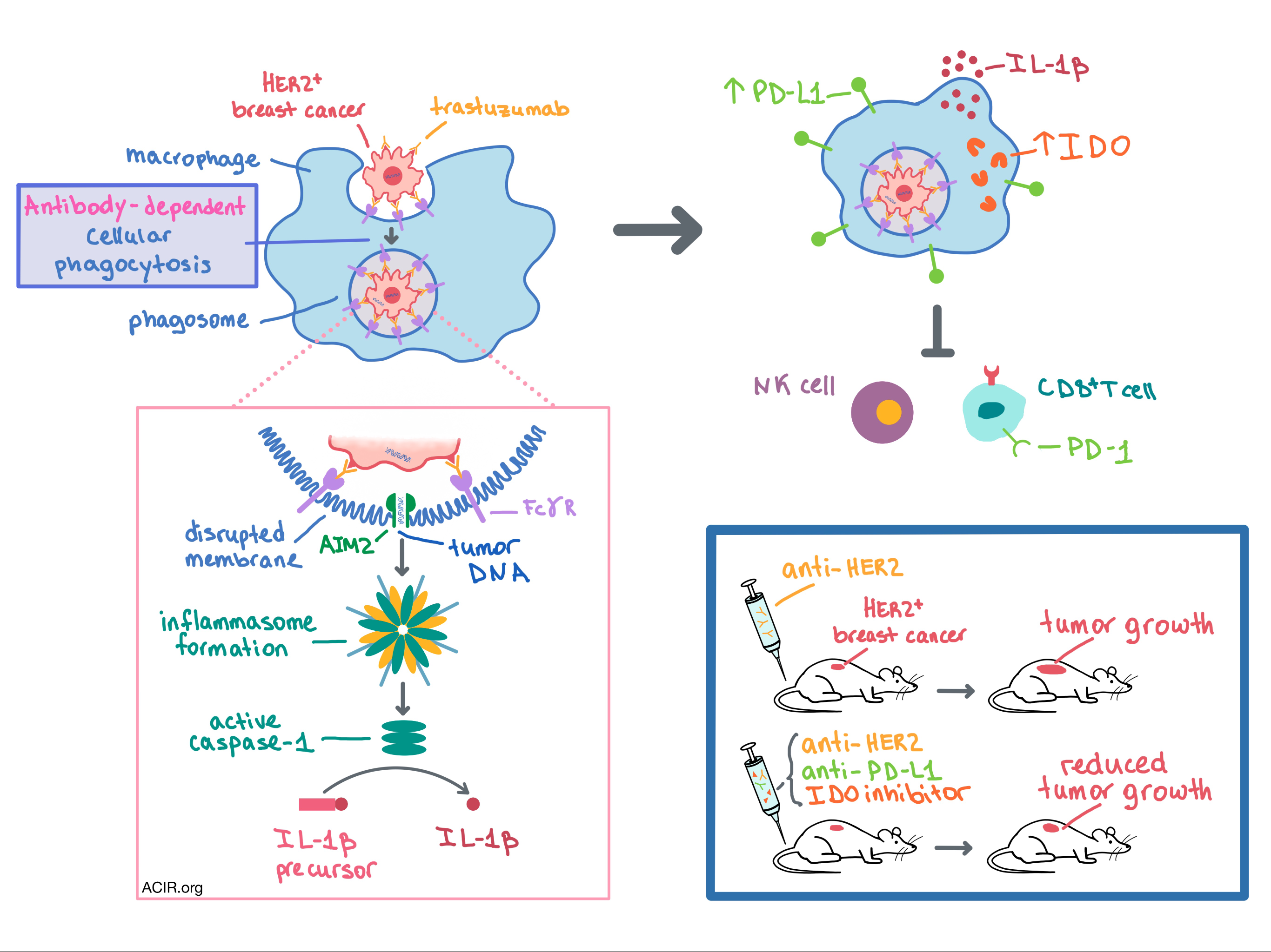
In order to explain the surprisingly immunosuppressive effect of ADCP in macrophages, the researchers dove deep into mechanistic studies. In their numerous experiments, the team utilized gene expression analysis, qRT-PCR, transcriptomic profiling, Western blot, immunohistochemistry, and confocal microscopy. Ultimately, the following mechanism emerged. After antibodies recognize and bind their target on the tumor cell, FcγRs on the surface of macrophages bind the Fc antibody fragments, initiating ADCP. Once the tumor cell-containing phagosome is inside the macrophage, FcγR signaling recruits AIM2 (a cytosolic double-stranded DNA sensor) to the membrane of the phagosome. The phagosomal membrane is disrupted, and AIM2 detects tumor DNA (both mitochondrial and genomic), leading to inflammasome formation. The inflammasome activates the caspase-1 enzyme, which cleaves the IL-1β precursor to create its bioactive, mature form. The active IL-1β is released, leading to the overexpression of PD-L1 and IDO in the macrophages that phagocytosed antibody-coated tumor cells as well as in the adjacent bystander macrophages. PD-L1 and IDO expression in macrophages then mediates immunosuppression of NK and T cells. The mechanism by which IL-1β, usually a proinflammatory cytokine, leads to the upregulation of PD-L1 and IDO in macrophages remains unclear.

Having observed that macrophage ADCP was responsible for the suppression of NK and T cells in vitro, the researchers sought to explore the effects of trastuzumab in vivo. To do this, Su, Zhao, and Xing et al. expressed human HER2 in the mouse E0771 breast cancer cell line and injected these cells into transgenic mice that are immune tolerant to human HER2. In one set of these mice, macrophages were genetically ablated. Mice were then treated with 4D5, a mouse anti-human HER2 antibody that is the parental drug to trastuzumab. In mice without macrophages, the treatment led to decreased tumor growth and increased infiltration of NK and CD8+ T cells in the tumor compared to mice with intact macrophages. In addition, the treatment increased the expression of PD-L1 and IDO in tumor-associated macrophages (TAMs). While adding anti-PD-L1 or an IDO inhibitor to 4D5 reduced tumor growth in mice with intact macrophages, combination of all three therapies synergized to boost the therapeutic effect even further. Mechanistically, the triple therapy dramatically increased infiltration of NK and CD8+ T cells in the tumor. These beneficial effects were not observed in mice without macrophages. The involvement of the AIM2/IL-1β pathway in suppression of the anti-HER2 antibody’s therapeutic effect was observed in vivo via knockout and blockade experiments.

The researchers then sought to confirm the clinical relevance of their pre-clinical observations. To that end, they analyzed resected primary tumors from HER2+ breast cancer patients who had received neoadjuvant trastuzumab prior to surgery. In non-responders, macrophages were more abundant while NK and CD8+ T cell infiltration was reduced compared with responders. Levels of PD-L1 and IDO in the pre-treatment tumor biopsies were low; however, PD-L1 and IDO expression increased significantly in TAMs, but not in tumor cells, following trastuzumab treatment. Higher post-treatment number of PD-L1+IDO+ TAMs was associated with poor response to trastuzumab.

Overall, Su, Zhao, and Xing et al. demonstrate, and mechanistically explain, that trastuzumab therapy in humans and mice with HER2+ breast cancer leads to upregulation of PD-L1 and IDO in TAMs via ADCP, which in turn inhibits the antitumor response of NK and CD8+ T cells. These results provide the rationale for combining immune checkpoint blockade with therapeutic antibody treatment to improve anti-cancer response.

by Anna Scherer

References:

Su S., Zhao J., Xing Y., Zhang X., Liu J., Ouyang Q., Chen J., Su F., Liu Q., Song E. Immune Checkpoint Inhibition Overcomes ADCP-Induced Immunosuppression by Macrophages. Cell. 2018 Oct 4.

In the Spotlight...

Neoadjuvant immune checkpoint blockade in high-risk resectable melanoma

In this phase II clinical trial, patients with high-risk resectable melanoma were treated with neoadjuvant checkpoint blockade. In the 12 patients treated with nivolumab, a 25% ORR (25% pathologic CR) was observed. In the 11 patients treated with nivolumab/ipilimumab, a 73% ORR (45% pCR) was observed and all patients in this arm were surviving at 15 months. Response to nivolumab alone appeared dependent on a pre-existing, antigen-experienced, clonal, and diverse T cell infiltrate. The trial closed early because neoadjuvant nivolumab treatment delayed surgical resection in two patients and toxicity was high in combination-treated patients.

Neoadjuvant versus adjuvant ipilimumab plus nivolumab in macroscopic stage III melanoma

In this phase 1b clinical trial, 20 patients with palpable stage III melanoma were divided evenly into groups to receive four doses of combination nivolumab and ipilimumab as adjuvant therapy (4 doses after surgical resection) or neoadjuvant therapy (2 before, 2 after), though high toxicity reduced the median number of doses to 2. The neoadjuvant arm had better pathological responses and fewer relapses, with no responders relapsing within the follow-up period. Neoadjuvant treatment induced higher expansion of tumor-resident T cell clones in peripheral blood. Low T cell infiltration, low clonality, low IFNγ signature, and low PD-L1 associated with relapse.

Enhanced detection of neoantigen-reactive T cells targeting unique and shared oncogenes for personalized cancer immunotherapy

To more completely identify neoantigen-reactive T cells in epithelial cancer TIL samples, Yossef et al. sorted cells expressing either or both of PD-1 and a co-stimulatory molecule (CD134 or CD137) into microwells at low density (3 cells per well to reduce overgrowth by non-exhausted cells) and following expansion screened for neoantigen reactivity across the entire mutanome. The method was at least two-fold more sensitive than previous TIL fragment culture approaches, facilitated rapid TCR α/β cloning, and resulted in identification of MHC class II TCRs for two highly relevant oncogenes (KRASG12V and TP53G245S).

CD84 regulates PD-1/PD-L1 expression and function in chronic lymphocytic leukemia

Lewinsky et al. showed that CD84 (which is self-associative and upregulated in chronic lymphocytic leukemia (CLL) and the TME) mediated the upregulation of PD-L1 on CLL and other cells within the TME via a cellular pathway (distinct from IFNγ-induced activation) involving Akt/mTOR, S6, and STAT3. Activation of CD84 on T cells directly contributed to upregulation of exhaustion markers and decreased the functionality of T cells. Blocking CD84 reduced expression of PD-L1 on CLL cells and resulted in increased lysis of CLL by patient-derived T cells.

Enhanced Anti-lymphoma Activity of CAR19-iNKT Cells Underpinned by Dual CD19 and CD1d Targeting

Rotolo et al. presented an optimized protocol for the generation of CAR19 invariant NKT (iNKT) cells (which naturally target CD1d), showing higher expansion, cytotoxicity, and polyfunctionality compared to standard CAR T cells. CAR19-iNKT cells were more effective at eliminating CD19+CD1d+ lymphomas, including intracranial disease (possibly due to enhanced chemokine receptor expression); eradicating relapsed disease; and extending tumor-free and overall survival in mice. CD19-iNKT activity was enhanced by ɑGalCeramide or CD1d upregulation. Because iNKT cells do not incite GVHD, an off-the-shelf product could be developed.

Bifunctional PD-1 x αCD3 x αCD33 fusion protein reverses adaptive immune escape in acute myeloid leukemia

Herrmann et al. developed a checkpoint inhibitory T cell-engaging (CiTE) antibody consisting of an αCD33 single-chain variable fragment (scFv), an αCD3ε scFv, and the extracellular domain of PD-1 (PD-1ex), which has a low affinity to PD-L1 compared with αPD-L1 scFv. The CiTE antibody selectively bound acute myeloid leukemia (AML) cells and T cells, efficiently activated T cells, and led to AML cell lysis in vitro and complete tumor eradication in a murine xenograft model. Locally restricted checkpoint blockade and the preferential targeting of CD33+PD-L1+ but not other PD-L1+ cells by CITE reduced on-target, off-leukemia side effects in mice.

Pan-tumor genomic biomarkers for PD-1 checkpoint blockade-based immunotherapy

Cristescu et al. evaluated the independent and combined application of two potential biomarkers of anti-PD-1 therapy response – tumor mutational burden (TMB) and T cell-inflamed gene expression profile (GEP) – using discovery and validation cohorts from HNSCC and pan-tumor trials. TMB, with a cutoff of ~80-200 mutations depending on tumor type, and GEP independently separated responders from non-responders. Both overall response rate and progression free survival were highest in TMBhi/GEPhi patients. The frequency of TMBhi/GEPhi varied across tumor types and reflected the observed checkpoint therapy responsiveness.

Tumor-Derived cGAMP Triggers a STING-Mediated Interferon Response in Non-tumor Cells to Activate the NK Cell Response

Marcus et al. analyzed the role of the cGAS-cGAMP-STING pathway in activating NK cells in the setting of tumors resistant to T cell response and found that NK cell killing of tumor cells was dependent on tumor expression of cGAS and host expression of STING but not cGAS. Mechanistically this suggests that constitutively activated cGAS in the tumor synthesizes cGAMP, which is taken up by non-cancerous host cells where it activates STING and leads to type I IFN production. In patients with melanoma, tumor cGAS expression positively correlated with expression of immune activation genes, NK cell infiltration, and improved survival.

Everything New this Week In...

Close Modal

Small change for you. Big change for us!

This Thanksgiving season, show your support for cancer research by donating your change.

In less than a minute, link your credit card with our partner RoundUp App.

Every purchase you make with that card will be rounded up and the change will be donated to ACIR.

All transactions are securely made through Stripe.