Weekly Digests
‹ Back to November

Stress and metabolism in the cancer-exposed T cell

November 14, 2018

Tumors create a hostile microenvironment that disrupts T cell metabolism and effector function and allows for tumor progression, but the specific pathways between the environmental stressors and the disruption of T cell metabolic function are not fully understood. In a letter recently published in Nature, Song et al. investigated this mechanism in the context of ovarian cancer, elucidating the intracellular pathways that lead to metabolic dysfunction in T cells and identifying potential points of intervention.

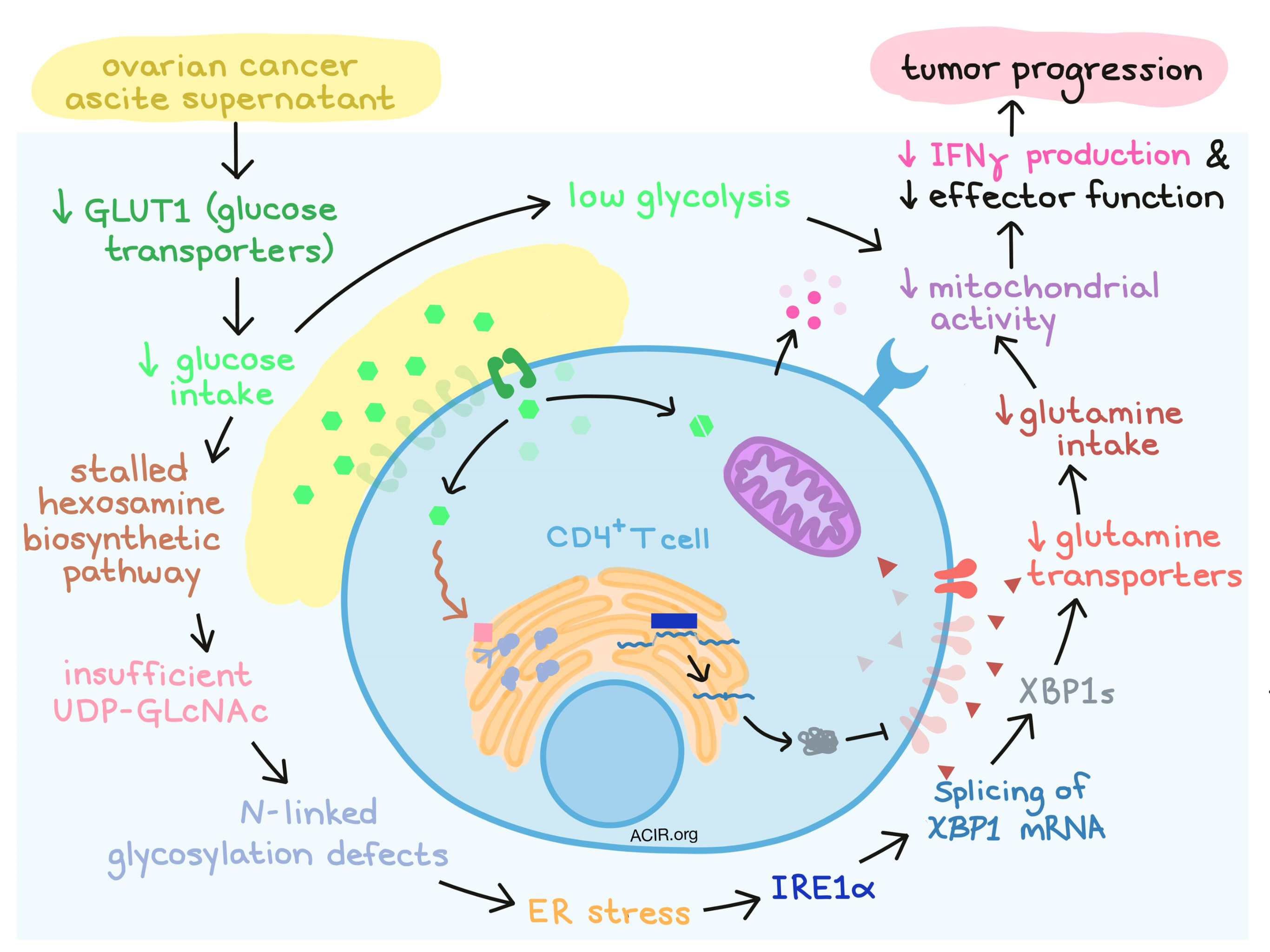
Due to its known role in endoplasmic reticulum (ER) stress, protein folding pathways, and enhanced tumorigenic capacity, Song et al. considered the IRE1α-XBP1 pathway the prime suspect in their investigation. Under conditions of stress in the endoplasmic reticulum (ER), the IRE1α enzyme excises a fragment from XBP1 mRNA, and the spliced version of the mRNA encodes the functionally active form of the XBP1 protein, known as XBP1s. When the researchers isolated CD4+ and CD8+ T cells from patient-derived ovarian cancer ascites, they observed an increase in XBP1 splicing. XBP1 levels also correlated with expression of genes that indicated activation of the unfolded protein response (UPR), and therefore with ER stress. This signature was associated with reduced T cell infiltration, indicating that this pattern may influence T cell functionality.

To better understand the role of IRE1α-XBP1, Song et al. isolated CD4+ T cells from healthy women and found that they increased expression of XBP1 in a dose dependent manner upon exposure to the supernatants of ascites derived from ovarian cancer patients. T cells treated with the ER stressor tunicamycin also showed evidence of strong XBP1 activation that could be abrogated by administering an IRE1α inhibitor, indicating that ER stress induces IRE1α-XBP1.

Working backwards to identify the source of ER stress, the researchers noticed that exposure to ascites fluid suppressed the major glucose transporter GLUT1 in CD4+ T cells, which would compromise glucose uptake and cause internal glucose deprivation. A lack of internal glucose would stall the hexosamine biosynthetic pathway used to make uridine diphosphate N-acetylglucosamine (UDP-GLcNAc), which is required for N-linked protein glycosylation. In line with this established mechanism, ascites-exposed CD4+ T cells showed N-linked protein glycosylation defects associated with ER stress. Supplementing GLcNAc, a direct substrate for N-linked protein glycosylation, minimized ER stress and halted downstream XBP1 activation, validating the link between limited glucose uptake and upregulated IRE1α-XBP1.

Next, Song et al. investigated what happens downstream of XBP1 activation and noticed that the glycolytic capacity and oxygen consumption were low in ascite-exposed CD4+ T cells, implicating mitochondrial dysfunction. Blockade of IRE1α in wild-type cells exposed to ascites fluid abrogated this effect on mitochondrial respiration. Engineered XBP1-deficient CD4+ T cells had superior mitochondrial respiration compared to WT under conditions of glucose withdrawal, indicating a direct role for IRE1α-XBP1 in dampening mitochondrial activity. Mechanistically, the researchers found that XBP1s mediated the activation of an ER-associated degradation system that controls the abundance of glutamine carriers, likely through post-translational mechanisms. A lower number of glutamine carriers limits influx of glutamine, which is needed to sustain mitochondrial respiration when glucose is in limited supply. Treating T cells rendered metabolically dysfunctional by ascites exposure with a glutamine analogue, or forcing overexpression of glutamine transporters, restored some respiratory capacity to the T cells.

The researchers then performed transcriptional analysis on wild-type and XBP1-deficient T cells from mice bearing ovarian tumors and found that within ascites, XBP1-deficient T cells showed global transcriptional reprogramming, upregulation of genes related to activation and mediation of antitumor response, and improved effector capacity compared to wild-type T cells. Mice engineered with XBP1-deficient T cells and challenged with ovarian cancer had a higher portion of CD4+ T cells producing IFNγ and a higher portion of CD8+ T cells with increased IFNγ and perforin production. Further, these mice showed superior antitumor immunity, delayed malignancy, and longer overall survival.

In order to show that their findings in mice were relevant to humans, Song et al. evaluated healthy human CD4+ T cells exposed ex vivo to ascite supernatants derived from ovarian cancer patients and found that IFNγ production, indicative of effector function, decreased. Administration of an IRE1α inhibitor partially alleviated this effect and restored some mitochondrial respiration.

Overall, Song et al. elucidate the pathway by which the ovarian cancer microenvironment reduces mitochondrial activity in CD4+ T cells, allowing for tumor immune escape. What they describe can be considered an “immunometabolic checkpoint” controlling antitumor immunity. In outlining this mechanism, the researchers note several points at which intervention could be applied to restore T cell functionality. Controlling ER stress or inhibiting IRE1α-XBP1 might limit metabolic stress and enhance the anticancer immune response, but further research will be needed to understand the potential benefits and consequences of these strategies.

by Lauren Hitchings

References:

Song M., Sandoval T.A., Chae C.S., Chopra S., Tan C., Rutkowski M.R., Raundhal M., Chaurio R.A., Payne K.K., Konrad C., Bettigole S.E., Shin H.R., Crowley M.J.P., Cerliani J.P., Kossenkov A.V., Motorykin I., Zhang S., Manfredi G., Zamarin D., Holcomb K., Rodriguez P.C., Rabinovich G.A., Conejo-Garcia J.R., Glimcher L.H., Cubillos-Ruiz J.R. IRE1α-XBP1 controls T cell function in ovarian cancer by regulating mitochondrial activity. Nature. 2018 Oct.

In the Spotlight...

WDFY4 is required for cross-presentation in response to viral and tumor antigens

Using cDC1 from murine bone marrow and whole cells as antigens, Theisen et al. conducted a CRISPR screen to identify genes involved in cross-presentation and uncovered WDFY4. cDC1s from WDFY4-/- mice were unable to cross-present whole cell antigens but were fully capable of presenting soluble antigens, cytoplasmic antigens, or MHC class II epitopes and demonstrated all other relevant DC functions (phagocytosis, upregulation of co-stimulatory molecules, production of IL-12). The membrane protein WDFY4 appears to be important in proper intracellular trafficking of endocytosed whole cell antigens, such as virally infected or tumor cells.

Blocking immunoinhibitory receptor LILRB2 reprograms tumor-associated myeloid cells and promotes antitumor immunity

Myeloid cells respond to multiple environmental cues which modulate their phenotype and Chen et al. demonstrate that LILRB2, a myeloid signaling receptor which naturally binds HLA class I, serves as a checkpoint, directing myeloid cells toward an anti-inflammatory phenotype. Antibody blockade of LILRB2 during macrophage maturation enhanced NF-κB/STAT1 activation, resulting in multiple gene expression changes including reduction of PD-L1 expression and decreased STAT6 activation, shifting cells away from the M2 phenotype and enhancing antitumor efficacy in humanized or Lilrb2-transgenic mice when combined with anti-PD-1/anti-PD-L1.

Entinostat converts immune-resistant breast and pancreatic cancers into checkpoint-responsive tumors by reprogramming tumor-infiltrating MDSCs

Mice with breast or pancreatic tumors that were treated with entinostat (ENT, a histone deacetylase inhibitor) in combination with checkpoint inhibition (ICI: anti-PD-L1, anti-CTLA-4, or both) experienced improved survival compared with ENT alone. ENT shifted the MDSC TME population toward G-MDSCs, which were rendered less immunosuppressive due to reduced expression of arginase-1 and PD-L1. Mechanistically, ENT inhibited the VEGF, ErbB, and TOR pathways, and although each checkpoint had differential effects, the impact converged on decreased STAT3 phosphorylation, resulting in increased infiltration of CD8+ effector T cells.

LILRB4 signalling in leukaemia cells mediates T cell suppression and tumour infiltration

In a search by Deng et al. for co-inhibitory or costimulatory domains related to survival in AML, leukocyte immunoglobulin and co-inhibitory receptor B4 (LILRB4), a molecule known to inhibit T cell activity, emerged. LILRB4 expression is restricted to monocytes and subsets of AML. In vitro experiments with LILRB4-/- AML cells or antibody blockade of LILRB4 demonstrated enhanced T cell proliferation and cytotoxicity and decreased AML migration. Multiple in vivo models confirmed improved tumor control. Mechanistically, LILRB4 signaling in AML cells occured through SHP2/NFκB and upregulated uPAR/ARG1 to suppress T cells.

Phage-Based Anti-HER2 Vaccination Can Circumvent Immune Tolerance Against Breast Cancer

Encouraged by predicted structural differences between HER2 and its oncogenic splice variant (Δ16HER2), Bartolacci and Andreani et al. developed DNA vaccines against both isoforms. Both vaccines induced a specific antibody response and protected mice from transplantable, Δ16HER2-expressing breast cancers, but failed to elicit a response in Δ16HER2 transgenic mice that develop spontaneous tumors. Bacteriophages expressing the entire Δ16HER2 extracellular domain or specific epitopes from the novel splice junction broke immune tolerance, delayed tumor growth, and reduced the number and size of tumors in transgenic mice.

The Effectiveness of Checkpoint Inhibitor Combinations and Administration Timing Can Be Measured by Granzyme B PET Imaging

Utilizing the previously developed granzyme B PET imaging agent (GZP, a small peptide which binds only to the active, secreted form of granzyme B), Larimer et al. demonstrated that in mice with colon carcinoma (CT26 or MC38) treated with anti-PD-1, anti-PD-1 + anti-CTLA-4, or anti-PD-1 + anti-TIM-3, low GZP signal was highly correlated with non-response on an individual level. The average GZP tumor-to-background ratio in a given treatment group directly correlated with percent survival and provided a basis for early comparison of the relative success of the immunotherapy regimens.

Human breast tumor-infiltrating CD8+ T cells retain polyfunctionality despite PD-1 expression

Egelston et al. analyzed PD-1 and other phenotypic markers in human breast cancer (mostly ER+/HER2-) and melanoma and observed that CD8+ TILs from both tumor types were primarily CCR7-/CD45RA- effector memory cells displaying a seemingly exhausted Eomes+/TIGIT+/2B4+ and terminally differentiated CD127-/KLRG1- profile. However, breast cancer PD-1+CD8+ TILs showed enhanced IFNγ, TNFα, and IL-2 production and stronger cytolytic capability in conjunction with a bispecific antibody compared to same cells derived from melanoma. These results reveal a more nuanced relationship between PD-1 expression and T cell exhaustion.

A Novel Anti-LILRB4 CAR-T Cell for the Treatment of Monocytic AML

John, Chen, and Deng et al. capitalized on the widespread expression of LILRB4 on the M5 subset of AML, including CD34+ leukemic stem cells, and lack of expression on most normal tissues (except for monocytes), to develop a CAR T cell targeting LILRB4. LILRB4 CAR T cells were highly cytotoxic to M5 AML in vitro and in vivo, extending survival in a xenograft model. No effect of the CAR T cells was observed on hematopoietic cell populations in ex vivo human CD34+ hematopoietic colony forming assays or in a human-reconstituted murine model despite in vitro cytotoxicity to primary monocytes.

Everything New this Week In...

Close Modal

Small change for you. Big change for us!

This Thanksgiving season, show your support for cancer research by donating your change.

In less than a minute, link your credit card with our partner RoundUp App.

Every purchase you make with that card will be rounded up and the change will be donated to ACIR.

All transactions are securely made through Stripe.