Tumor-infiltrating lymphocytes (TILs) can exhibit a variety of functional and transcriptional states, but the connection between phenotype and antigen specificity remains unclear, and decoding this relationship could provide insight into patient response to immune checkpoint blockade (ICB) therapy. With this goal in mind, Oliveira et al. and Caushi & Zhang et al. examined TILs in melanoma and lung cancer, respectively, through state-of-the-art single-cell techniques, recently published in Nature.
To begin, Oliveira and the team collected CD8+ TILs from four melanoma patients and performed single-cell RNA, TCR, and CITEseq to interrogate gene expression, TCR clonotypes and surface proteins, respectively. Clustering cells by gene and protein expression, distinct TIL phenotypes arose at varying levels of exhaustion. Interestingly, cells with the same TCR clonotype typically resided at the same exhaustion level, falling primarily in one of two groupings: exhausted (TEx; terminally exhausted, progenitor exhausted, and proliferating T cells), which contained the most highly expanded clonotypes, or non-exhausted memory (TNExM; memory, effector memory, γδ-like, and NK-like T cells) T cells.
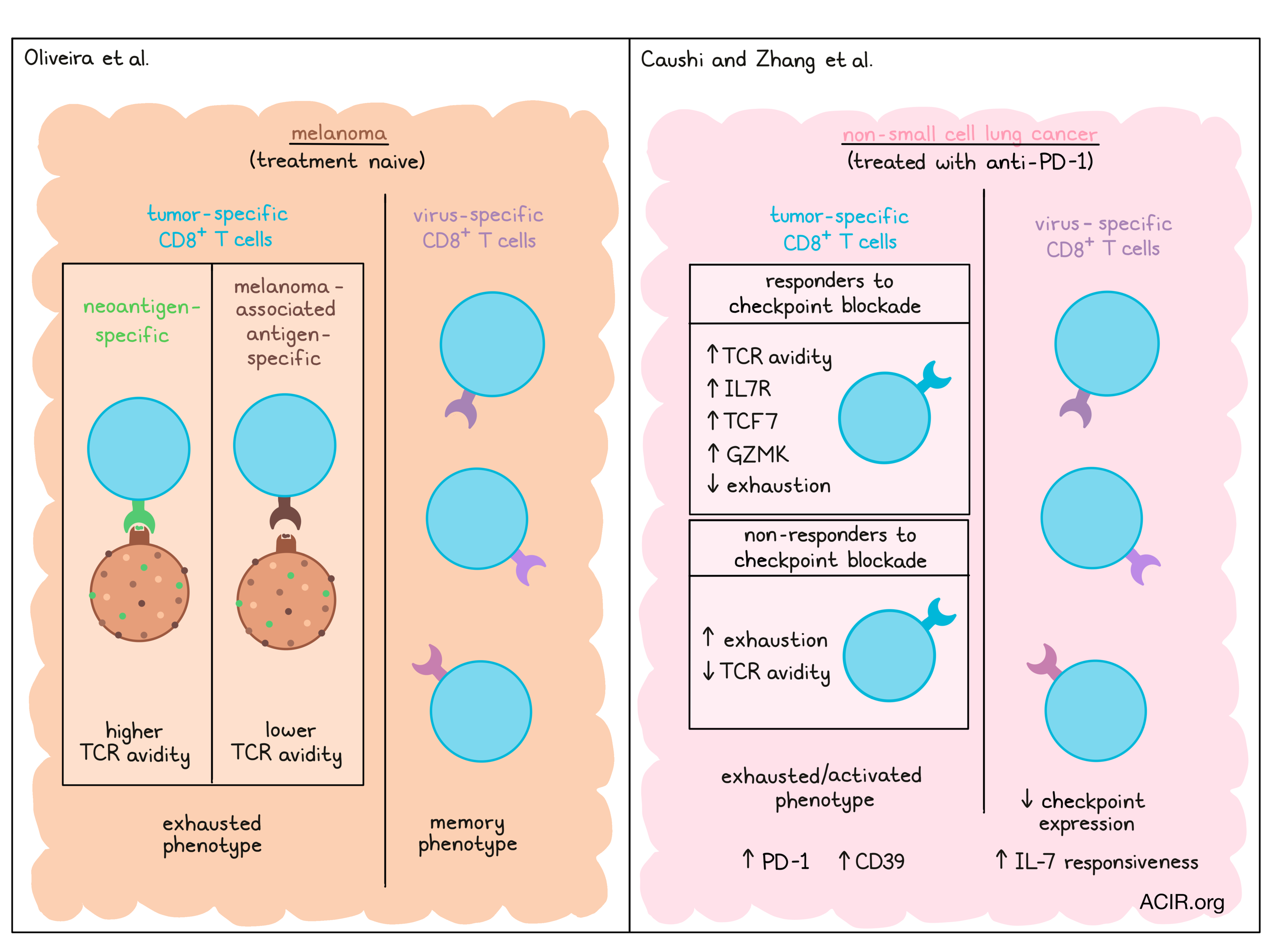
The authors questioned whether TCR antigen specificity could relate to TEx or TNExM dominance within TCR clonotypes. To screen for antigen recognition (CD137 upregulation), they transduced TEx- or TNExM-associated paired α/β TCRs into healthy donor T cells and cocultured them with patient-derived melanoma cells (to detect tumor specificity), virus-infected cell lines (for viral specificity), or autologous PBMCs (as controls). Strikingly, 83% of the analyzed TEx TCRs displayed melanoma reactivity, compared to only 10% of the TNExM TCRs. On the other hand, TNExM TCRs were enriched in viral recognition and matched several known TCR clonotypes for Epstein-Barr virus (EBV), influenza, and yellow fever. These findings were validated in a separate patient cohort, altogether suggesting that tumor antigen recognition may drive the exhausted phenotype.
Next, the researchers delved deeper into characterizing the melanoma-specific TILs. These cells partitioned into several clusters, all of which highly expressed PD-1 and CD39, but the majority displayed terminally exhausted or acutely activated profiles. To determine the specific antigen targets, the team co-cultured cells expressing the identified TCRs with cell lines expressing shared melanoma-associated antigens (MAAs), personal neoantigens, or shared viral antigens. Matching the prior findings, both MAA and neoantigen-specific TCRs were largely TEx cells; these melanoma-specific T cells upregulated exhaustion and reduced memory gene expression compared to the virus-specific TCRs, which had a TNExM phenotype. Although their transcriptional signatures were similar, MAA-specific TCRs had lower avidity than neoantigen-specific TCRs for their cognate antigen. Furthermore, TCR avidity was inversely related to antigen expression levels on the melanoma cells, and positively correlated with experimentally measured peptide–HLA affinity, but not peptide–HLA stability. Thus, although tumor-specific T cells may have had different TCR avidities based on the antigen type, they still acquired an exhausted state.
Finally, the authors considered how their findings with TILs could relate to T cells in circulation. In blood, T cells with MAA and neoantigen-specific TCRs were rare and displayed an exhausted phenotype. TNExM TCRs, which tended to be non-melanoma-specific, were much more abundant than TEx TCRs. In a separate cohort of ICB-treated patients with melanoma, higher TEx levels in the blood correlated with disease progression and worse survival outcomes. Overall, the team found that T cell features are linked between tumors and blood, and could hint towards immunotherapy response.
While Oliveira et al. studied TILs in melanoma, Caushi and Zhang et al. explored the phenotype and antigen specificity of TILs from non-small cell lung cancer (NSCLC). The researchers sequenced RNA and TCRs of T cells from healthy lungs and tumor tissue of patients with NSCLC who had received neoadjuvant anti-PD-1 therapy, finding multiple clusters of T cells. Broadly, gene expression could differentiate between TILs and healthy lung T cells, but not between TILs in patients who did or did not respond to ICB.
To probe more deeply into antigen specificity, the authors used the mutation-associated neoantigen functional expansion of specific T cells (MANAFEST) assay, previously developed for antigen-specific T cell identification and TCR sequencing [1]. TCRs specific to both viral and neoantigen epitopes were found, but neoantigen-specific T cell abundance did not relate to ICB response. However, phenotypic differences were observed in neoantigen-specific T cells between ICB responding and non-responding patients. Neoantigen-specific CD8+ T cells from responding patients increased expression of memory- and effector-associated genes such as IL7R, TCF7, and GZMK, and decreased features of exhaustion and checkpoints (e.g., TOX2, CTLA4), relative to non-responding patients’ neoantigen-specific CD8+ T cells. Additionally, responding patients displayed a higher avidity of neoantigen-specific TCRs. Thus, although the broad TIL sequencing did not distinguish responders from non-responders, characterization of neoantigen-specific T cells uncovered critical differences.
Next, the authors compared the transcriptional profiles of neoantigen and virus-specific (EBV or influenza) CD8+ T cells. EBV-specific CD8+ T cells tended to share an effector T cell phenotype, relative to influenza and neoantigen-specific CD8+ T cells, which displayed tissue-resident memory profiles (expressing HOBIT and CD103). Although both EBV- and neoantigen-specific T cells expressed genes indicating CTL activation (e.g., IFNG), gene expression for cytolytic function (e.g., GZMK, EOMES) was reduced in neoantigen-specific T cells. Broadly, the neoantigen-specific T cells shared features of immune checkpoints and exhaustion, with increases in genes encoding PD-1, CTLA-4, TIM3, and CD39 compared to virus-specific T cells, and were also less responsive to IL-7 signaling than influenza-specific cells.
To conclude, Caushi and Zhang et al. profiled blood T cells longitudinally in a patient with complete pathological response to anti-PD-1 therapy. Two weeks after initiating ICB, the majority of neoantigen-specific T cell clones displayed a tissue-resident memory phenotype, but by four weeks, had diverged into either effector or memory profiles. The transition from tissue-resident memory to effector cells coincided with increases in genes involved in T cell activation, homing, and IFNγ signaling, providing insight into the changes in T cell phenotype that can occur after ICB therapy.
In summary, recent advances in immune-profiling technologies have allowed unparalleled resolution at the single-cell scale. Applying these techniques to study CD8+ T cells within tumors, thought to be the main players in ICB response, could have important implications in understanding the response to fruitful ICB treatment, improving that response, or even predicting patients who could benefit.
Write-up by Alex Najibi, image by Lauren Hitchings
[1] Danilova, L., et al. The Mutation-Associated Neoantigen Functional Expansion of Specific T Cells (MANAFEST) Assay: A Sensitive Platform for Monitoring Antitumor Immunity. Cancer Immunology Research 2018.
Meet the researcher
This week, co-first authors on “Transcriptional programs of neoantigen-specific TIL in anti-PD-1-treated lung cancers” Jiajia Zhang and Justina X. Caushi and first author on “Phenotype, specificity and avidity of antitumour CD8+ T cells in melanoma” Giacomo Oliveira answered our questions.
What prompted you to tackle this research question?
JZ: Mutation-associated neoantigen (MANA)-specific T cells play a key role in tumor control and response to immune checkpoint inhibition (ICI). However, the majority of tumor-infiltrating lymphocytes (TILs) are not specific for the tumor. To better understand the mechanisms underlying T cell dysfunction in the context of tumor killing, we need to study the specific T cells that recognize tumor antigens. In our lab, we have the capacity to identify those MANA-specific T cells from bystander T cells using the FEST (Functional Expansion of Specific T cells assay). This platform provides tremendous opportunity to study the biology of tumor-specific T cells and differential response to immunotherapy.
JC: I was very fortunate to join the lab as a graduate student at the time of completion of the first-in-human clinical trial of neoadjuvant anti-PD-1 (nivolumab) in resectable non-small cell lung cancer. We had an opportunity to then analyze these irreplaceable patient samples to understand just what the T cells were doing in the tumor microenvironment and in the periphery during and post anti-PD-1 administration. We were interested to see if mutation-associated neoantigen (MANA)-specific T cell phenotype correlated with clinical response to help identify mechanisms by which we can overcome resistance to PD-1 blockade. By being able to do transcriptomic analysis at the single cell level, we wanted to understand how anti-PD-1 shaped the T cell response and identify T cell clones that were involved in the antitumor effort. By identifying and transcriptionally profiling these MANA-specific T cell clones, we also wanted to be able to better understand what differentiates these cells from viral-specific T cells and other bystander T cells in the tumor microenvironment.
GO: A major issue in the field of cancer immunotherapy is to understand the phenotype and properties of the T cells that are able to recognize and kill the tumor. Unfortunately, within the tumor microenvironment, the ability of T cells to react against the tumor cells can be variable, since there are also many T cells that lack antitumor function. Up to now, many studies have characterized different types of tumor-infiltrating lymphocytes, but without focusing on the T cells with actual potential to kill the tumor. To tackle this issue and unambiguously describe the properties of antitumor CD8+ T cells, we coupled single-cell profiling of tumor infiltrating lymphocytes with the in vitro analysis of the molecules that drive the ability of the T cells to recognize and kill a target: the T cell receptors. This allowed us to measure in vitro the potential of each TCR against the patient’s tumor cells, in order to separate the tumor-specific cells from the non-tumor-reactive ones. By understanding the properties of true antitumor T cells, it would be possible to foster and elicit their reactivity with novel and more effective immune therapies.
What was the most surprising finding of this study for you?
JZ: I am most amazed that MANA-specific CD8+ TIL expressed a very unique phenotype that was distinct from T cells recognizing viral antigens, such as flu and EBV. MANA-specific T cells presented with a partially activated cytolytic program with co-expression of multiple immune checkpoints and upregulated transcriptional regulators of T cell dysfunction. In addition, MANA-specific CD8+ T cells had hallmark transcriptional programs of tissue-resident memory cells. Particularly for non-responders to immune checkpoint blockade, MANA-specific T cells lacked key mediators of long-term survival and had higher co-expression of immune checkpoints. These findings provide important insights for overcoming resistance to PD-1 blockade.
JC: We were able to identify T cell clones that recognize mutation-associated neoantigens in patients regardless of whether or not they experienced major pathologic response (MPR) in the clinic. We observed that the transcriptomic profiles of these cells were different in MPR vs. non-MPR patients. What was even more impressive to me, however, was that we were able to demonstrate the difference in ligand-dependent signaling in T-cell receptors (TCRs). We found that the MANA-specific TCR’s from patients that experienced MPR had significantly higher functional avidity than MANA-specific TCRs from nonMPR patients. This led us to hypothesize that lack of a clinical response may be due to the tumor-specific T cells exhibiting low activity, owing to poor avidity of their TCR for their cognate peptide-MHC.
GO: One of the most surprising findings was that in all the patients there are CD8+ T cells with the potential to exhibit strong antitumor activity. In all the analyzed cases, the immune system was able to generate a response specific for a spectrum of different tumor antigens. Unfortunately, the vast majority of such T cells responses were characterized by the acquisition of a dysfunctional cell state, and the recognition of tumor antigens shaped the antitumor T cells towards an exhausted phenotype. Thus, we could conclude that in patient with melanoma, there is not a defect in the amount, but rather in the quality of the antitumor response. Within the tumor microenvironment, high antitumor potential and T cell dysfunction are very often associated. Therefore, the disentanglement of these two aspects of the T cell biology through novel cancer immunotherapies might result in generation of more potent antitumor responses.
What was the coolest thing you’ve learned (about) recently outside of work?
JZ: I am a dog person, and recently, I just had my first puppy named Coco. She has been a huge blessing and has brought so much joy and laughter to my life. She is a great emotional support for me in the United States since all my family is in China. I love to take her to trails and jog with her every day, because she has a ton of puppy energy. I hope to take her back to meet my other family members when the pandemic is over and do a family trip to the beach (Coco is a beach girl)!
JC: As an adventurous traveler, I enjoy exploring new places and more often than not, outside of the United States. However, due to the pandemic travel restrictions these past couple of summers, I decided to explore our nation’s picturesque national parks instead. Hiking Angel’s Landing at Zion National Park, the trails of Glacier National Park, and camping along the Appalachian trail have been some of the most incredible adventures to date. I’ve found that you don’t need to fly far to be a wanderer, and to appreciate what you already have close to home.
GO: Since I am Italian, I truly love soccer, and recently I followed with passion the fantastic victory of the Italian team at the European championship (UEFA EURO 2020). Under the guidance of coach Roberto Mancini, the Azzurri (that’s how they are called because of their light blue jersey) defeated all the rivals, and against all the odds, they won the final game against England, right in London, at the Wembley stadium. I think that with this sportive exploit, the Italian team really inspired all the people around the world, showing that starting from the bottom, a group of very young players can reach the top of Europe with talent, with dedication, with a little bit of luck, but most importantly with cohesion. I have learned a lot by watching the Azzurri defeating one after another the most favorite teams, and I bet that all the opponents learned a lot too…