Weekly Digests
‹ Back to June

The many faces of an exhausted T cell

June 6, 2018

Chronic diseases, such as HIV and cancer, are typically associated with an “exhausted” class of T cells characterized by poor function in disease control. Based on the hypothesis that a better understanding of the phenotypic variation among exhausted T cells may more effectively guide immunotherapy approaches, Bengsch et al. set out to systematically categorize exhausted T cell types in different disease settings using a systems immunology approach. The results, published in Immunity, revealed numerous epigenetically defined exhausted T cell subsets with defining features of both disease and anatomical location.

Bengsch et al. began by analyzing gene expression changes in the classically studied chronic lymphocytic choriomeningitis virus (LCMV)-specific exhausted T cells (Tex) compared with naive, effector, and memory T cells after 30 days of infection in order to identify a core exhausted cell gene expression signature. The set of genes with increased or decreased expression, which included CTLA-4, PDCD1, CD38, and ENTPD1 (encoding CD39), was further validated by gene set enrichment analysis. This gene signature was also strongly enriched in tumor-infiltrating lymphocytes (TILs) from melanoma patients and in HIV-specific T cells from HIV progressor patients, suggesting that it may be applicable across disease states.

As epigenetic patterns may be more accurate in describing cell identity than gene expression, Bengsch et al. explored the epigenetic changes in Tex cells in LCMV public data sets using ATAC-seq, which examines the accessibility of open chromatin regions (OCRs). Some of the genes with increased expression and OCR accessibility in Tex cells included inhibitory receptors (Pdcd1, Tigit, Ctla4), ectoenzymes involved in metabolic regulation (Cd38, Entpd1), chemokines and cytokines (Xcl1), and transcription factors (Eomes, Ikzf2, Tox). Genes with decreased expression and OCR accessibility included Ccr7, Il7r, Nt5e, Tcf7, and Lef1. The researchers concluded that this set of genes may serve as a biomarker for exhaustion.

Next, the team sought to apply the population-based epigenomic exhaustion signature to profile Tex cells on a single-cell basis. To do this, they created a mass cytometry panel of 16+ exhaustion-related gene products and other T cell markers of lineage and differentiation state for analysis by cytometry by time of flight (CyTOF). This subset selection was validated via gene set variation analysis of a published CD8+ T cell single-cell transcriptomic dataset from human melanoma tumor-infiltrating lymphocytes (TILs); the validation confirmed that this subset of genes was representative of key features of exhaustion.

After validation, Bengsch et al. applied the CyTOF panel to peripheral blood mononuclear cells (PBMCs) from healthy control subjects, HIV patients on and off antiretroviral therapy, and samples from patients with lung cancer (including PBMCs, macroscopically uninvolved lung tissue, and TILs). They found that PD-1+CD8+ T cells expressed more exhaustion markers than any other CD8+ T cell phenotypes examined. In HIV, molecules predicted to be decreased in Tex correlated with health and mild disease, while molecules predicted to be increased in Tex correlated with advanced disease.

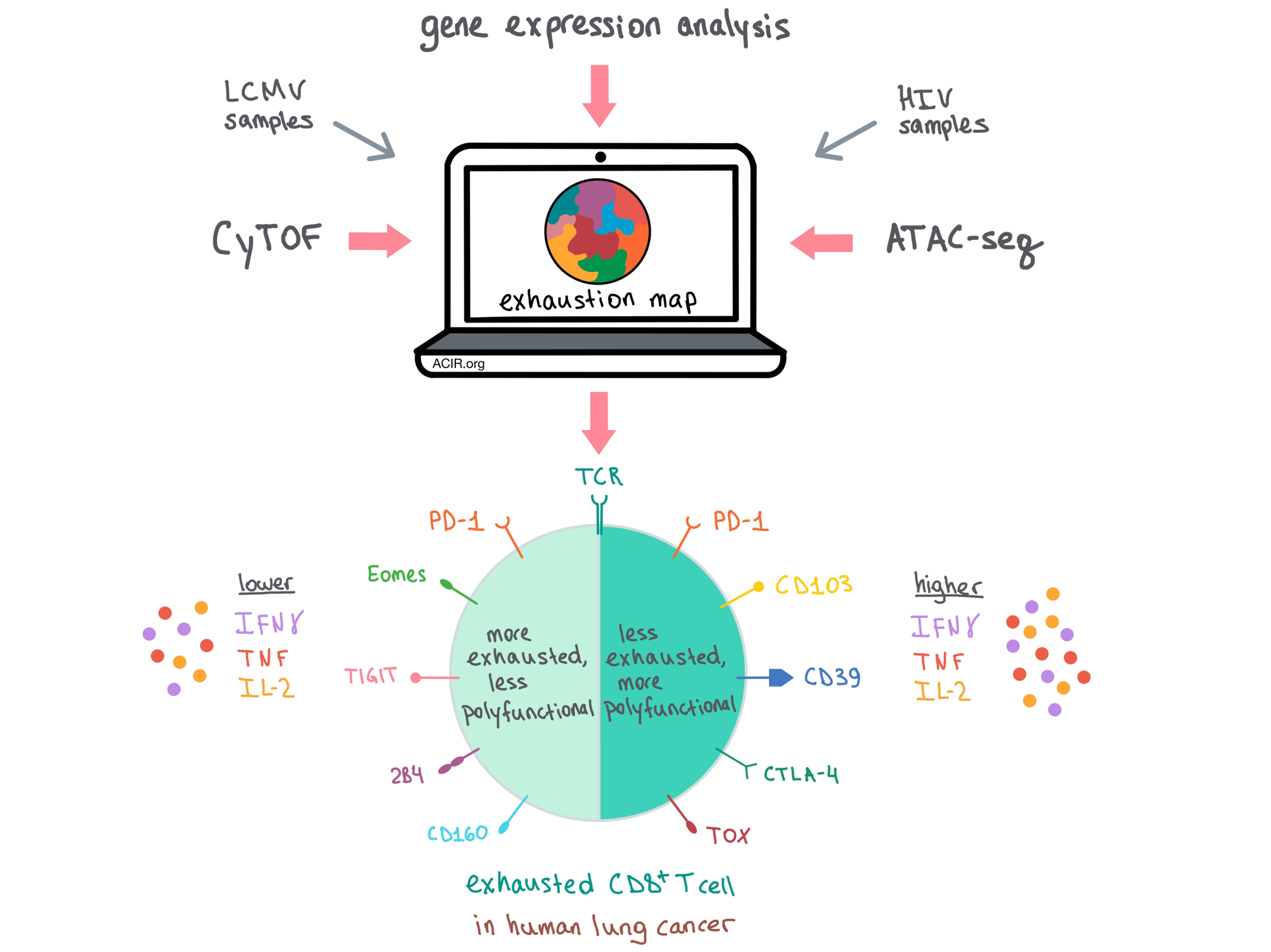
The team then performed a high-dimensional analysis on CyTOF-analyzed non-naive CD8+ T cells to create an “exhaustion map��� and showed that the pattern of marker co-expression differed among multiple Tex cell phenotypes and across patients and diseases. Using a previously developed method, phenograph, the researchers then dug deeper into the data and developed a functional exhaustion score (FES) that increased with hallmarks of exhaustion (e.g., loss of IFNγ, TNF, and IL-2 production) and decreased with the presence of effector or memory functionality to distinctly score each population.

Distinguishing Tex cells from effector T cells using individual markers has been a challenge historically, as some markers of activation also ultimately indicate exhaustion. Using high-dimensional analysis, Bengsch et al. were able to distinguish the two T cell populations by taking into account multiple markers and FES at the same time. Effector T cells had higher expression of CD39, LAG-3, Helios, and CTLA-4, while Tex cells upregulated Eomes, TOX, 2B4, and TIGIT. TCF1, a critical transcription factor in naive and memory cells, was primarily observed in non-exhausted cells, although it was also observed in two exhausted subpopulations.

To find out whether the key features of exhaustion exist across different diseases and tissues, the team analyzed TILs from patients with newly diagnosed lung cancer, and found conserved Tex cell biology across HIV and lung cancer, along with some disease-specific features. Drilling down further into the functionality of these populations in lung cancer, TILs with low IFNγ production co-expressed PD-1 as well as markers of more severe exhaustion, including Eomes, TIGIT, 2B4, and CD160. Meanwhile, the more functional TILs with higher IFNγ production also expressed PD-1, as well as CD103, CD39, CTLA-4, and TOX, but lacked the more severe exhaustion features.

Overall, Bengsch et al. developed a comprehensive systems immunology approach to explore the variation in Tex cells across human diseases, which could provide insights into disease progression and help identify appropriate therapy for patients with specific expression of checkpoint blockade targets.

by Anna Scherer

References:

Bengsch B., Ohtani T., Khan O., Setty M., Manne S., O'Brien S., Gherardini P.F., Herati R.S., Huang A.C., Chang K.M., Newell E.W., Bovenschen N., Pe'er D., Albelda S.M.,Wherry E.J. Epigenomic-Guided Mass Cytometry Profiling Reveals Disease-Specific Features of Exhausted CD8 T Cells. Immunity. 2018 May 15.

In the Spotlight...

Unveiling a CD70-positive subset of cancer-associated fibroblasts marked by pro-migratory activity and thriving regulatory T cell accumulation

Jacobs et al. researched the expression patterns of CD70 by immunohistochemistry in a set of 51 colorectal cancer (CRC) samples and found that CD70 was absent from tumor cells but strongly present in cancer-associated fibroblasts (CAFs), and that high expression of CD70 by CAFs correlated with poor prognosis and survival. Similar to how malignant cells can overexpress CD70 to increase suppressive nTregs, induce T cell apoptosis and exhaustion, and ultimately evade the immune system, CAFs overexpressing CD70 were correlated with nTreg accumulation, IL-2 production, and poor prognosis.

TIGIT and PD-1 dual checkpoint blockade enhances antitumor immunity and survival in GBM

After demonstrating high TIGIT and PD-1 expression on CD4+ and CD8+ cells in brains of glioma patients and confirming these results in a GL261 murine model of glioblastoma, Hung et al. tested TIGIT checkpoint inhibition in combination with anti-PD-1 therapy. Compared to either monotherapy, the combination significantly improved overall survival, elevated TIL CD8+ and CD4+ T cells, and increased the population of IFNγ- and TNFα-producing T cells. Long-term tumor-free murine survivors were immune to rechallenge with GL261-luc cells.

Radiation followed by OX40 stimulation drives local and abscopal antitumor effects in an anti-PD1-resistant lung tumor model

Niknam et al. followed radiation therapy (XRT) with intratumoral OX40 stimulation against a primary anti-PD-1 resistant tumor, resulting in significantly upregulated CD8+ T cells compared to control or OX40 groups alone, and improving primary and secondary tumor response by limiting lung metastases, which raised survival rates in vivo. XRT increased the percentage of splenic OX40+CD4+ T cells, the intensity of OX40 receptor expression, and the level of CD103+ dendritic cells. XRT presumably promoted the release of tumor antigens, which then stimulated OX40 receptor expression on T cells to cause systemic antitumor effects.

Cancer Cell Membrane-Coated Adjuvant Nanoparticles with Mannose Modification for Effective Anticancer Vaccination

Yang et al. developed a novel nanoparticle cancer vaccine by incorporating the TLR7 agonist R837 (as an adjuvant to promote DC activation and maturation) into nanoparticles, coating the nanoparticles in tumor cell membrane (as a source of tumor-specific antigens), and modifying the particles with mannose (to enhance uptake by antigen-presenting cells). In vitro, the vaccine efficiently activated DCs, and in vivo, it induced antigen-specific cytolytic (CD107+) T cells, was prophylactically effective against B16-OVA melanoma tumors, and was therapeutically effective against established tumors, especially when combined with anti-PD-1.

Heat shock protein peptide complex-96 vaccination for newly diagnosed glioblastoma: a phase I, single-arm trial

In a single-arm phase I clinical trial, 20 patients with newly diagnosed glioblastoma received autologous heat shock protein peptide complex 96 (HSPPC-96) vaccine concomitantly with adjuvant temozolomide (TMZ) following the standard regimen of resection, radiotherapy, and TMZ. Treatment was well-tolerated. PFS at 6 months was 89.5%, median PFS was 11 months, and median OS was 31.4 months. Tumor-specific immune response (TSIR) increased ~2-fold after vaccination and was an independent predictor of response, with OS of >40.5 months for patients with above median TSIR versus 14.6 months for patients with below median TSIR.

Tumor-associated calreticulin variants functionally compromise the peptide loading complex and impair its recruitment of MHC-I

Arshad and Cresswell investigated the effects on peptide:MHC I presentation of two naturally occurring frameshift (FS) mutations in calreticulin (CRT), a critical component of the peptide-loading complex (PLC). The researchers found that lack of CRT expression or expression of CRT-FS alone reduces the presence of surface MHC-I. Mechanistically, CRT-FS was still found to associate with MHC-I, but its ability to interact with the PLC and facilitate high-affinity peptide loading on MHC-I was impaired, leading to assembly of low-affinity, unstable MHC-I:peptide complexes that less successfully migrate to the cell surface.

A CS1-NKG2D bispecific antibody collectively activates cytolytic immune cells against multiple myeloma

To target multiple myeloma (MM), Chan et al. developed a bispecific antibody (BiAb) by fusing an anti-CS1 scFv (to target the MM-associated antigen CS1) to an anti-NKG2D scFv (to redirect and activate innate and adaptive immune cell types expressing NKG2D). The CS1-NKG2D BiAb enhanced synapse formation between MM and NK cells, activated NGK2D+ NK cells (as evidenced by downstream phosphorylation of AKT), induced MM-specific INFγ production in IL-2 primed PBMCs, induced CS1-directed cytolysis in vitro, and prolonged survival in an NSG mouse model of human MM.

Everything New this Week In...

Close Modal

Small change for you. Big change for us!

This Thanksgiving season, show your support for cancer research by donating your change.

In less than a minute, link your credit card with our partner RoundUp App.

Every purchase you make with that card will be rounded up and the change will be donated to ACIR.

All transactions are securely made through Stripe.