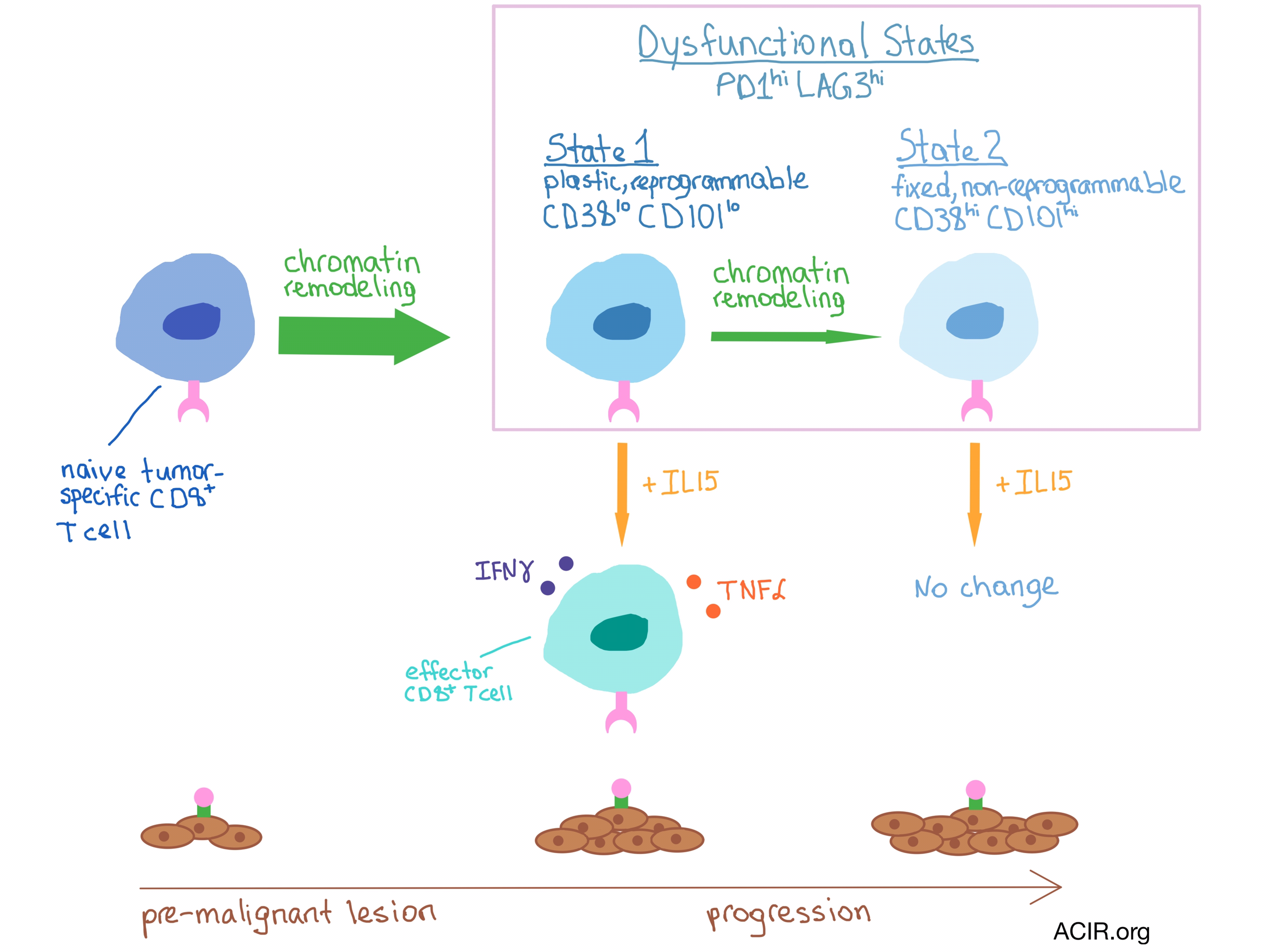
It is well known that tumor-specific CD8+ T cells (TST) found within solid tumors are often dysfunctional and do not affect tumor progression. TST become dysfunctional early on in the tumorigenesis process and exhibit the same characteristics as tumor-reactive tumor-infiltrating lymphocytes (TILs) in late-stage human solid tumors. Philip et al. hypothesized that the dysfunctional state of TST is due to tumor-specific epigenetic changes.
The researchers analyzed the changes in chromatin state of TST during tumor development in mice bearing a tamoxifen-inducible SV40 T antigen (TAG)-expressing liver cancer that progresses from pre-malignant lesions to hepatocellular carcinoma within 60-90 days. Congenically marked naive T cells with specificity for TAG (TCRTAG cells) were adoptively transferred into the mice one day prior to tamoxifen administration. The authors then analyzed the genome-wide chromatin accessibility of liver-infiltrating TCRTAG cells at different time points using ATAC-seq (Assay for Transposase-Accessible Chromatin with high-throughput sequencing) and RNA-seq, and compared it to the epigenetic changes during T cell differentiation in an acute infection model. They found that the TCRTAG cells that encountered the TAG tumor antigen in the pre-malignant liver lesions were rendered dysfunctional, expressed inhibitory receptors PD-1 and LAG3, did not produce IFN-γ or TNFα, and differentiated through two discrete chromatin states.
Plastic, Reprogrammable State 1
- TCRTAG cells underwent massive chromatin remodeling by day 5 (consistent with effector T cell differentiation in the acute infection model).
- In vitro culture with IL-15 could restore the effector function of State 1 TCRTAG cells.
- State 1 TCRTAG cells expressed low levels of CD38 and CD101 cell surface markers.
Fixed, Non-reprogrammable State 2
- TCRTAG cells underwent further, more limited, chromatin remodeling between days 7 and 14.
- In vitro culture with IL-15 could not restore the effector function of State 2 TCRTAG cells.
- State 2 TCRTAG cells expressed high levels of CD38 and CD101.
Consistent with the loss of effector function, the accessibility of transcription factor binding sites in genes encoding inhibitory receptors and negative regulators was increased during progression from the plastic to the fixed dysfunctional state in TCRTAG cells, while the accessibility of co-stimulatory molecule genes was decreased. Very few further chromatin accessibility changes occurred after day 14, even after the cancer progressed to established tumor. Remarkably, the authors found that even memory TCRTAG cells that were adoptively transferred into mice bearing established hepatocellular carcinoma lost their effector function as they progressed through the two dysfunctional states after antigen exposure.
The team focused on two transcription factors they had identified with the global changes in binding site accessibility: NFAT, for which binding site accessibility was increased, and TCF, for which binding sites were closed during T cell dysfunction. Pharmacological modulation of these two transcription factors in mice delayed T cell dysfunction and improved the reprogrammability of TST.
The researchers found that the chromatin accessibility state of patient-derived TILs with high PD-1 expression was most similar to that of the State 2 dysfunctional mouse TST. Human PD-1hi TILs displayed heterogeneous expression of CD38 and CD101 markers, which could potentially be used to identify T cells that are amenable to therapeutic reprogramming, turning dysfunctional TST into an effective antitumor therapy.
by Anna Scherer


