Weekly Digests
‹ Back to October

Form follows function: How dendritic cell distribution within lymph nodes affects T cell response

October 18, 2017

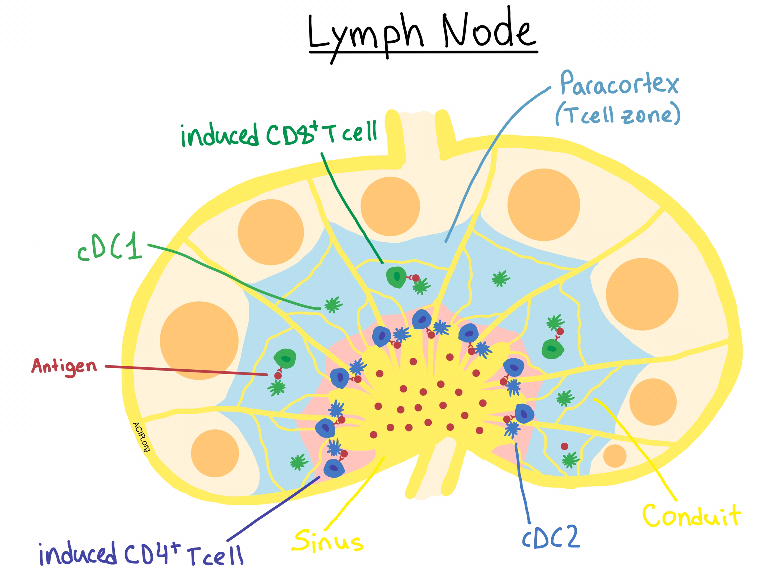
Form follows function, and the fascinating structural microanatomy and related immunological functioning of lymph nodes revealed by Gerner et al. in The Journal of Experimental Medicine highlight their importance for rational vaccine design. Dendritic cells (DCs) within the lymphatic tissues are the key initiators of adaptive immune responses during immunization or in the presence of a growing tumor, but their subsets are known to be asymmetrically distributed within the lymph node (LN). Using several quantitative imaging techniques, including two-photon intravital imaging, multiplex confocal microscopy with 3D reconstruction, and multiplex immunohistochemistry, Gerner et al. explored how antigen dispersal within the LN and the uptake and presentation of antigens by asymmetrically distributed DCs affect the activation of naive T cells after protein immunization.

Two protein antigen models were used for visualization and immunization: EαGFP to image antigen draining, uptake, and presentation by MHC II molecules, and OVA to allow direct assessment of early T cell activation using OVA-specific CD8+ OT-I and CD4+ OT-II T cells. After immunization, the team observed that antigens initially rapidly drained into the lymphatic sinus (LS). Although some smaller antigens traveled into the deep T cell zone via LN conduits soon after immunization, these antigens were rapidly cleared, presumably into the circulation. The majority of antigen appeared to enter the parenchymal LN space independently of LN conduits and via diffusion across the highly permeable LS floor. This diffusion-limited process generated an antigen concentration gradient with maximum antigen concentrations near the LS floor and lower concentrations in the T cell zone deep within the LN cortex. Conjugates of EαGFP to high molecular weight molecules were unable to enter the conduits, demonstrating the size dependence of this clearance mechanism.

To examine antigen uptake and presentation, Gerner et al. focused their attention on two specific subsets of LN-resident DCs: cDC1 cells, which mediate MHC I antigen cross-presentation to CD8+ T cells, and cDC2 cells, which specialize in MHC II antigen presentation to CD4+ T cells. The researchers confirmed their previous findings that LN-resident cDC1 and cDC2 cell populations occupy distinct regions of the lymph node, with the former localized within the deeper paracortical (T cell zone) region, and the latter mostly segregated in the regions near the LS. The antigen gradient determined the uptake by the asymmetrically-distributed DCs, with the centrally-located cDC1s acquiring significantly less antigen than the peripherally-situated cDC2s. The researchers confirmed that it was the location within the LN tissue, and not any cell-intrinsic uptake differences, that accounted for this phenomenon. Interestingly, they observed that the majority of antigen was associated with LN-resident DCs and not migratory DCs, suggesting either limited migratory DC involvement or rapid hand-off of antigen from migratory to LN-resident DCs.

To assess the immunological effects of these anatomical observations, the researchers used OVA immunization and in vivo imaging of CD8+ OT-I and CD4+ OT-II to demonstrate that the asymmetric distribution of cDC1 and cDC2 cells within the draining LN led to OVA-specific CD8+ T cell activation primarily deep within the LN paracortex, and CD4+ T cell priming occurring preferentially, but not exclusively, near the LS. Keeping in mind the antigen gradient across the LN, the researchers further demonstrated (using OVA titration) that effective MHC I presentation and CD8+ T cell activation required higher doses of administered protein than MHC II presentation and CD4+ T cell activation. This dose dependence also significantly affected the number of recruited CD8+ naive T cells, and ultimately, the CD8+ T cell clonal diversity as well as the magnitude of response.

Overall, the results of these imaging experiments demonstrate that lymphoid tissue microanatomy plays a significant role in the regulation of adaptive immune response, and suggest that antigen dosing and design should inform the development of vaccines.

This manuscript is accompanied by two informative videos displaying a 3D reconstruction of the dispersal of antigen within the LN and DC-mediated antigen uptake (Video 1) and of OT-I and OT-II cell clustering around DCs within the LN (Video 2).

by Anna Scherer

References:

Gerner M.Y., Casey K.A., Kastenmuller W., Germain R.N. Dendritic cell and antigen dispersal landscapes regulate T cell immunity. J Exp Med. 2017 Oct 2.

In the Spotlight...

The diverse functions of the PD1 inhibitory pathway

Sharpe and Pauken comprehensively review the great diversity of immune functions impacted by the PD-1 axis, the variety of cell types involved (a range of T cell types as well as non-T cells), and the significant context dependence. A point of emphasis is that PD-1 is not an exhaustion-specific molecule but instead plays a role in a broad swath of T cell biology including controlling early activation, metabolism, memory, proliferation capacity, and homeostatic control.

CD56bright NK cells exhibit potent antitumor responses following IL-15 priming

Wagner et al. used IL-15 or the IL-15R agonist ALT-803 to prime human CD56bright NK cells (from either healthy donors or multiple myeloma [MM] patients) ex vivo, inducing a strong anti-tumor response to hematological tumor targets in vitro and in leukemic NSG mice. CD56bright NK cells isolated from MM patients treated with ALT-803 in a clinical trial demonstrated enhanced, albeit transient, functionality. Anti-tumor effects were attributed to enhanced expression of cytotoxic and adhesion molecules.

Increasing tumor-infiltrating T cells through inhibition of CXCL12 with NOX-A12 synergizes with PD-1 blockade

High concentrations of CXCL12, a chemokine expressed mainly by fibroblasts in the tumor microenvironment, prevent the infiltration of T cells into solid tumors. Zboralski et al. used the RNA aptamer NOX-12 to inhibit CXCL12 in spheroid models in vitro, which increased the number of tumor-infiltrating T and NK cells. The addition of NOX-12 to PD-1 blockade synergistically increased T cell activation in vitro and enhanced antitumor efficacy in vivo.

NY-ESO-1 Vaccination in Combination with Decitabine Induces Antigen-Specific T-Lymphocyte Responses in Patients with Myelodysplastic Syndrome

Decitabine, a front-line chemotherapy for myelodysplastic syndrome (MDS) that induces the expression of the tumor antigen NY-ESO-1, was combined with an NY-ESO-1 vaccine in this Phase I study. In most patients, treatment induced NY-ESO-1-specific responses by CD8+ and CD4+ T cells that could be activated in vitro by autologous myeloid cells expressing NY-ESO-1. Responses were associated with the detectable presence at diagnosis, or later induction of CD141HI conventional dendritic cells.

Loss of MAPK-activated protein kinase 2 enables potent dendritic cell-driven anti-tumour T cell response

Using a melanoma tumor model in a myeloid-lineage (CD11c+) knockout of MAPK-activated protein kinase 2 (MK2), Soukup et al. concluded that MK2 is an intracellular checkpoint that promotes a suppressive dendritic cell (DC) phenotype. Knockout of MK2 reduces the accumulation of myeloid-derived suppressive cells and promotes the expansion of mature CD103+ DCs, thus enhancing CD8+ T cell priming, functionality, and infiltration, and reducing tumor growth in the context of Toll-like receptor agonist and tumor antigen.

Anti-CD137 suppresses tumor growth by blocking reverse signaling by CD137 ligand

Kang et al. uncover the complexity of the CD137/CD137L axis by demonstrating in murine models that although CD137 is a stimulating receptor on T cells, CD137L is an inhibitory receptor in myeloid cells, inhibiting differentiation to anti-tumor CD103+ dendritic cells and M1 macrophages. The authors suggest that the negative signaling by CD137L in myeloid cells is another example of adaptive resistance.

Checkpoint blockade immunotherapy reshapes the high-dimensional phenotypic heterogeneity of murine intratumoural neoantigen-specific CD8+ T cells

Capitalizing on the high-dimensional capability of CyTOF analysis, Fehlings et al. probed the antigen specificity and phenotypic profile of T cells in mice just prior to tumor rejection, revealing distinctions between tumor and peripheral neoantigen-specific T cells; multiple, phenotypically divergent CD8+ T cell populations within tumors; and convergence in phenotype and reduction in markers of dysfunctionality following anti-CTLA-4 therapy.

Tethering IL2 to its receptor IL2Rbeta enhances anti-tumor activity and expansion of natural killer NK92 cells

Jounaidi et al. created a chimeric IL2-IL2Rβ fusion protein (CIRB) that acts like constitutively activated IL2Rβ. NK92 cells expressing CIRB were independent of exogenous IL2, showed signs of increased activation and cytotoxicity compared to parental or IL2-secreting NK92 cells, were resistant to immunosuppressive TGFβ1, survived longer following irradiation, and showed superior persistence and antitumor efficacy in vivo. NK92CIRB cells also expressed higher levels of CD16, making them better effector cells for ADCC.

Cancer immunotherapy with recombinant poliovirus induces IFN-dominant activation of dendritic cells and tumor antigen-specific CTLs

Recombinant poliovirus/rhinovirus PVSRIPO targets CD155, expressed on human cancers and on antigen-presenting cells (APCs); cytopathogenic infection kills cancerous cells, releasing pathogen- and damage-related patterns and tumor antigens, while chronic sublethal infection in APCs causes type I interferon-dominant activation. In mice, intratumoral injection of PVSRIPO functions as a potent immune adjuvant that leads to tumor infiltration by APCs and tumor-specific T cells and improves survival.

Dual Face of Vgamma9Vdelta2-T Cells in Tumor Immunology: Anti- versus Pro-Tumoral Activities

Xiang and Wu review Vγ9Vδ2-T cells in the context of immunotherapy. When activated, Vγ9Vδ2-T cells can directly kill tumor cells in an MHC-independent manner, and can further serve helper functions by priming and modulating immunological responses of other innate and adaptive immune cells. Some evidence, however, suggests that under the influence of the tumor microenvironment, Vγ9Vδ2-T cells can be persuaded into a pro-tumor, Treg-like role.

Everything New this Week In...

Close Modal

Small change for you. Big change for us!

This Thanksgiving season, show your support for cancer research by donating your change.

In less than a minute, link your credit card with our partner RoundUp App.

Every purchase you make with that card will be rounded up and the change will be donated to ACIR.

All transactions are securely made through Stripe.