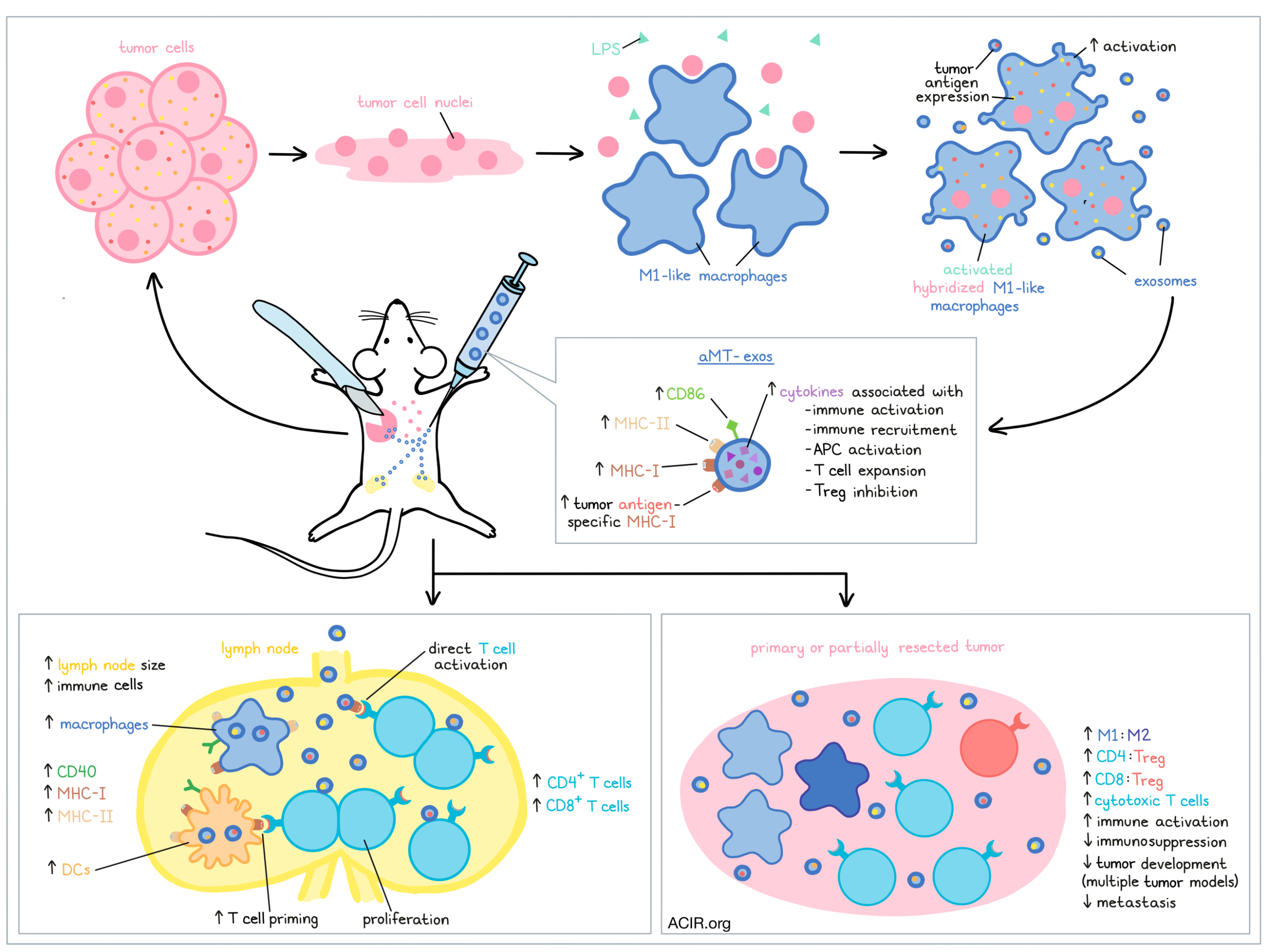
In search of a cancer immunotherapy that increases cancer-specific T cells while modulating the immunosuppressive TME, Wang et al. investigated the potential of using exosomes derived from chimeras of macrophages and tumor cell nuclei. Their strategy was to combine activated macrophages, which produce exosomes capable of activating immunity and reprogramming M2 macrophages, with tumor nuclei, which contribute tumor antigens and tumor homing abilities, to produce exosomes with strong immunotherapeutic potential. Their results were recently published in Science Translational Medicine.
To begin, Wang et al. extracted peritoneal macrophages (M1-like) from mice and treated them with nuclei isolated from E.G7 lymphoma cells expressing OVA. The macrophages internalized the nuclei, and upon stimulation with LPS, the hybridized macrophages increased activation markers and increased expression of OVA peptides on MHC molecules. Exosomes isolated from the supernatants of these cultures (aMT-exos) showed increased MHC-I, MHC-II, and CD86 (attributed to activation) and increased OVA-specific MHC-I (attributed to the incorporation of the tumor nuclei). aMT-exos also contained Increased cytokines and chemokines associated with immune activation, immune cell recruitment, APC activation, T cell expansion, and Treg inhibition. Similar results were observed using different murine tumor lines, including 4T1 TNBC lines and B16 melanoma cells.
Extending to a model for human cells, CD14+ macrophages were isolated from human PBMC samples and were differentiated with GM-CSF into macrophages in vitro. When activated and treated with nuclei from human MDA-MB-231 TNBC, the human macrophages and exosomes expressed tumor components and activation markers, similar to the results seen in murine cells.
Investigating the potential for using aMT-exos for immunotherapy, the researchers subcutaneously administered aMT-exos marked with fluorescent dye into mice bearing solid E.G7 tumors. In vivo imaging showed that aMT-exos accumulated in inguinal LNs and in tumors. Similar results were observed in other tumors with tumor-matched aMT-exos, suggesting that exosomes from hybridized macrophages inherit some extent of the parental tumor’s homing abilities.
Next, Wang et al. examined the effects of aMT-exos in the LNs, and found that inguinal LNs extracted from aMT-exos-treated mice were larger and contained more immune cells, including macrophages and dendritic cells, compared to LNs from control mice. These APCs infiltrated deeply into the paracortical T cell zone, and the proliferative potential and numbers of CD4+ and CD8+ T cells increased, indicating enhanced T cell activation. CD4+ T cells also migrated into germinal centers, indicating potential for activation of a humoral immune response.
Further investigating how aMT-exos contribute to immune activation, the researchers used dye-labeled exosomes and found that APCs and T cells accounted for over 90% of exosome+ cells. Looking at APCs, the researchers found that exosomes were taken up by macrophages and DCs and induced expression of CD40, MHC-I, and MHC-II. In coculture with exosome+ APCs, CD4+ and CD8+ T cells showed increased proliferation and cytolytic activity, suggesting that aMT-exos enhance APC-mediated T cell priming. Looking next at T cells, the researchers found that aMT-exos were present on the surfaces of T cells, and that they induced proliferation of CD4+ and CD8+ T cells, including tumor antigen-specific T cells, fitting with the hypothesis that exosomes could act as miniature APCs to exert direct immunostimulatory effects on T cells. This notion of direct presentation to T cells was further supported by observations of increased antigen-specific T cell proliferation in aMT-exos-treated mice in which DCs, macrophages, or both were ablated. Direct activation was shown to be tumor antigen-specific and promoted increased T cell lysis of target tumor cells. Similar results were observed using human cells.
Turning towards the effects of aMT-exos on the TME, the researchers showed that i.t. Injection of aMT-exos shifted the intratumoral macrophage ratio from M2-dominant to M1-dominant, increased cytotoxic T cells, and increased CD8:Treg and CD4:Treg ratios. It also enhanced expression of immune activation-related genes and pathways, decreased expression of immunosuppression-related genes, and ultimately inhibited tumor development. Macrophage repolarization was confirmed in murine cultures and in a spheroid model of human tumor and immune cells.
To assess the efficacy of aMT-exos as a potential immunotherapy, Wang et al. treated mice with E.G7 tumor bearing mice with either aMT-exos (s.c.), a vaccine consisting of the AS04 adjuvant and tumor exosomes (s.c.), or adoptive T cell transfer (i.v.). Only mice in treatment groups that received aMT-exos survived past day 40, 66% were alive at day 100, and four mice were tumor-free by the end of the experiment. Mice receiving aMT-exos also showed evidence of both antigen-specific T cell activation and TME modulation. Similar benefits were seen in the 4T1 and B16 models.
Looking beyond primary tumors, the researchers investigated the effects of aMT-exos on tumor metastasis. In a mouse model for breast cancer lung tumor metastasis, aMT-exos colocalized with sites of tumor metastasis, where they induced immune effects similar to those observed in primary tumors. Further, fewer metastases were present in the lungs, no metastases were found in other major organs, and mice maintained a steady body weight, suggesting that aMT-exos may limit or prevent metastasis. The addition of anti-PD-1 further increased CD8+ T cells, increased the CD8:Treg ratio. reduced M2 macrophages, and reduced lung metastases compared to aMT-exos alone. Similar results were observed in other tumor metastasis models.
Finally, Want et al. investigated the use of aMT-exos in a personalized partial resection model using tumor cell nuclei from the post-surgical resection to prepare personalized aMT-exos and treat mice bearing partial tumors. When injected into matched mice, personalized aMT-exos infiltrated the residual tumor and LNs. Monotherapy with aMT-exos reduced the recurrence rate at day 14 from 100% in controls to 66.7%, while the combination of aMT-exos and anti-PD-1 reduced recurrence to 16.7%, with 4/6 mice surviving past 100 days. Lung and bone marrow metastases were also eliminated after combination therapy. Similar results were observed in a B16 melanoma post-surgical resection model.
Overall, Wang et al. showed that exosomes derived from hybrids of activated macrophage and nuclei from tumor cells may serve as an effective immunotherapy that enhances immune activation while simultaneously reducing immunosuppression within tumors. Further, aMT-exos have a number of potential clinical advantages, including fast and easy manufacturing, translation across tumor types, introduction of multiple personalized tumor antigens, stability, resistance to reprogramming in the TME, and the potential to be frozen for long-term storage. This strategy warrants further investigation, and an application for a clinical trial for the use of aMT-exos in the post-operative setting is currently underway.
Write-up and image by Lauren Hitchings