Weekly Digests
‹ Back to October

NKILA: A matter of (T cell) life and (activation-induced cell) death

October 3, 2018

Activation-induced cell death (AICD) is part of the immune system’s important checks and balances, but as with multiple other regulatory pathways, tumors can co-opt T cell AICD as a mechanism of immune escape. To better understand how tumors induce AICD and which immune cells are targeted, Huang et al. performed an in-depth discovery and mechanistic analysis and published their results in Nature Immunology.

To begin, Huang et al. evaluated immune cell subsets from breast and lung cancer patients and evaluated their sensitivity to AICD. While tumor infiltrating Tregs and type 2 T helper (TH2) cells were found to be fairly resistant to tumor-induced apoptosis, effector cytotoxic T lymphocytes (CTLs) and type 1 T helper (TH1) cells underwent apoptosis in higher proportions. Confirming this observation in vitro using isolated TILs, CD3 stimulation or exposure to tumor cell antigens induced more apoptosis in tumor-specific CTLs and TH1 cells than in Tregs or TH2 cells. Interestingly, the expression of death receptors including Fas, TNFR2, and TRAILR, was comparable between the different T cell subsets, indicating that despite an abundance of the cognate ligands (FasL, TNF, TRAIL) in these tumors, internal signaling was likely responsible for the differential sensitivity to AICD.

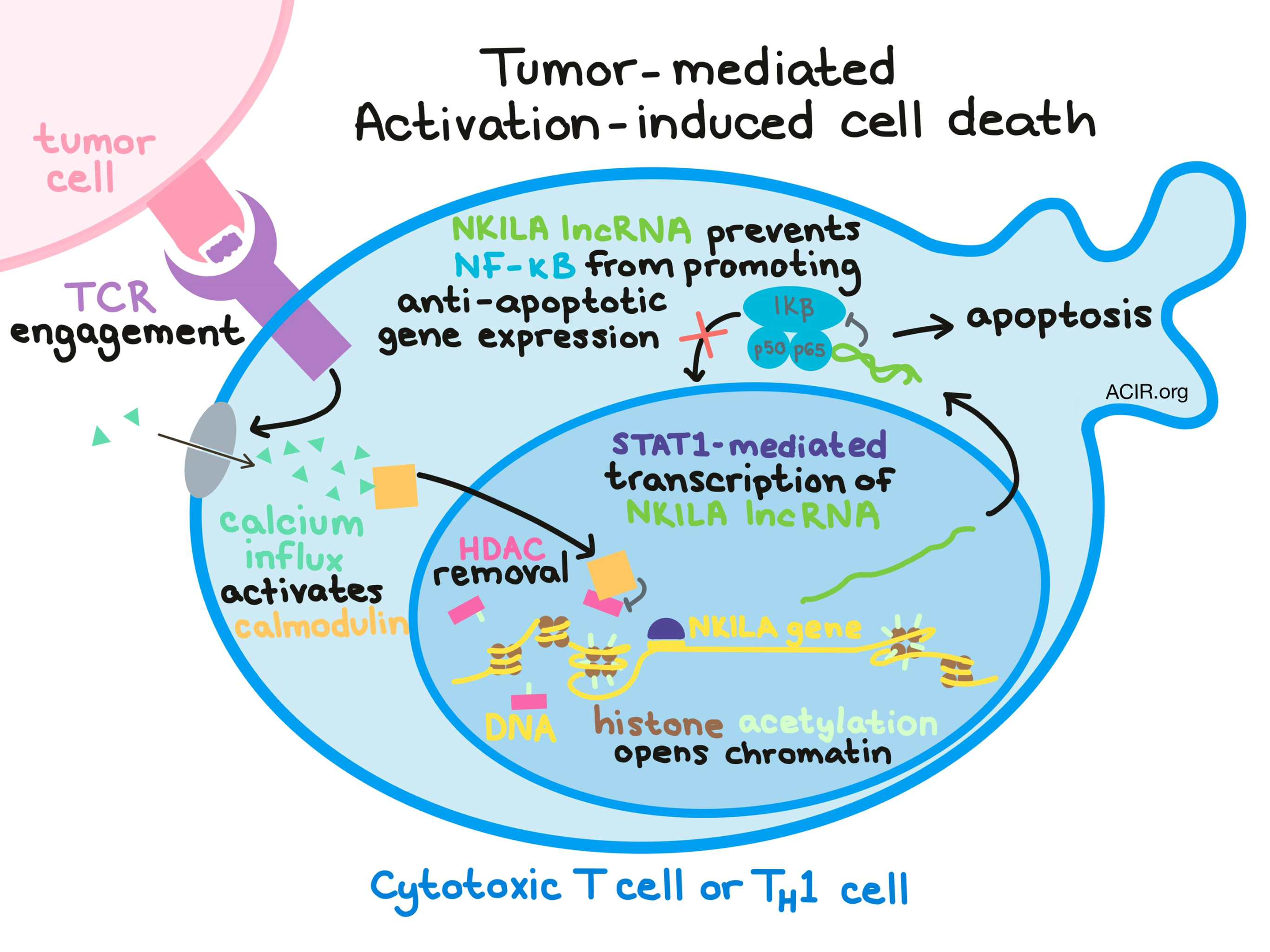
Diving into the mechanism underlying sensitivity to AICD, Huang et al. used gene enrichment analysis and uncovered that rather than being death receptor-dependent, AICD sensitivity was instead dependent on the activity of the NF-κB – a transcription factor that is essential for cytokine production and cell survival. Next, hypothesizing a role for long non-coding RNA (lncRNA), they identified the NKILA lncRNA as an inhibitor of NF-κB activity that functions by binding directly to the p65 protein within the NF-κB-IκBα complex. NKILA lncRNA was found to gradually increase during CTL activation and was most upregulated during the late AICD-sensitive phase. Further, upregulated NKILA was observed in CTLs and TH1 cells, but not in Tregs or TH2 cells, from patient-derived breast and lung cancer samples. In vitro silencing of NKILA with short hairpin RNA (shRNA) in stimulated T cells decreased apoptosis in activated cells, and forcing NKILA overexpression in Tregs or TH2 cells enhanced cells’ sensitivity to tumor-mediated AICD. The NKILA lncRNA-induced sensitivity to AICD was dependent on its ability to inhibit NF-κB.

Once NKILA and NF-κB were confirmed as key players in the mechanism of tumor-mediated AICD, the researchers began to piece together the early events in the tumor-induced mechanism of NKILA upregulation. They found that TCR signaling was critical to induction of AICD and confirmed that TCR signaling leads to an influx of calcium, which results in nuclear translocation of calmodulin – a calcium-modulated protein that mediates multiple cellular events. Next, they demonstrated that calmodulin binds to and inhibits histone deacetylases (HDACs), causing the HDACs to be removed from the NKILA promoter. Upon removal of the HDACs, histone acetyl transferases were recruited to acetylate the histones of the NKILA promoter, thus opening up the chromatin and making it available for transcription. With the chromatin open, STAT1 was able to move in and bind to the promoter at an identified STAT1 binding site, allowing for STAT1-mediated transcription of NKILA. As previously shown, the NKILA lcnRNA then interacts with NF-κB and inhibits its activity, rendering T cells incapable of performing essential cell survival functions and ultimately causing cell death.

To demonstrate the potential clinical relevance of NKILA-induced AICD in cancer, the researchers observed that in patients with breast cancer, high NKILA expression in tumor-specific CTLs from peripheral blood correlated with less infiltration of those tumor-specific CTLs into tumors. Further, patients with a high percentage of NKILAhi tumor-specific CTLs had shorter disease-free and overall survival.

Examining a possible avenue for therapeutic intervention, the Huang et al. transduced CD8+ T cells with NKILA shRNA to silence NKILA expression. When these cells were adoptively transferred into mice bearing patient-derived breast cancer xenografts, the transferred CTLs trafficked to the tumor and underwent less apoptosis, resulting in dramatic tumor cell apoptosis and inhibition of tumor growth compared to mice treated with adoptive transfer of control cells. Further, NKILA silencing upregulated CD107a and perforin, indicating enhanced cytotoxicity of the adoptively transferred cells, and increased the percentages of CD8+CD45RO+ effector T cells infiltrating the tumors.

Overall, Huang et al. identified the T cell subsets that are most susceptible to AICD and presented a thorough understanding of the underlying mechanism that controls this enhanced sensitivity. Further, the researchers presented evidence that this pathway is relevant in the clinical setting and that this mechanism may have the potential to be exploited for immunotherapy.

by Lauren Hitchings

References:

Huang D., Chen J., Yang L., Ouyang Q., Li J., Lao L., Zhao J., Liu J., Lu Y., Xing Y., Chen F., Su F., Yao H., Liu Q., Su S., Song E. NKILA lncRNA promotes tumor immune evasion by sensitizing T cells to activation-induced cell death. Nat Immunol. 2018 Oct.

In the Spotlight...

The anatomical location shapes the immune infiltrate in tumors of same etiology and impacts survival

Comparing the immune landscapes of HPV-induced tumors arising in the cervix (CxCa) or oropharynx (OPSCC), Saskia et al. found that despite similarities in PBMCs, immune infiltrates differed between anatomical locations, with immune profiles closely resembling that of the tissue of origin. CxCa tumors were more infiltrated by CD8+CD103+ and activated CD8+CD103+CD161+ effector T cells, while OPSCC tumors were more infiltrated by B cells, naive CD4+, and CD4+CD161+ effector memory T cells, and had a higher CD4:CD8 ratio. In OPSCC, but not CxCa, infiltration by CD4+ T cells or HPV-specific T cells correlated with survival.

Utilizing TAPBPR to promote exogenous peptide loading onto cell surface MHC I molecules

Ilca et al. determined that exogenous delivery of plasma membrane-targeted TAPBPR (a peptide editor typically expressed intracellularly) or exogenous soluble TAPBPR to target cells in vitro can catalyze the exchange of specific added peptides onto cell surface-expressed MHC-I molecules, allowing for the rapid and efficient (low peptide concentration) loading of high-affinity immunogenic peptides of choice onto MHC-I, bypassing typical intracellular antigen processing. Tumor cells loaded with peptides via TAPBPR were able to engage with TCRs on peptide-specific CD8+ T cells, inducing IFNγ secretion and enhancing T cell-mediated killing of target cells.

Combined analysis of antigen presentation and T cell recognition reveals restricted immune responses in melanoma

To better characterize tumor antigens in cancer and metastases, Kalaora et al. used a combination of whole exome sequencing, neoantigen prediction, HLA-I and -II peptidome analysis, TIL functional analysis, and TCR sequencing. Analyzing 15 tumors from 7 melanoma patients and one melanoma cell line, the researchers identified similarities in tumor-associated antigen (TAA), neoantigen, and TIL TCR profiles between metastases within the same patient. TILs were reactive to a small set of neoantigens and to multiple TAAs. Neoantigen-specific TILs induced killing of up to 90% of matched melanoma cells in vitro and in vivo.

Chemotherapy Combines Effectively with Anti-PD-L1 Treatment and Can Augment Antitumor Responses

Cubas et al. demonstrated in the MC38 murine colon cancer model (which is partially responsive to anti-PD-L1) that combination of anti-PD-L1 and chemotherapy (carboplatin, cisplatin, oxaliplatin, paclitaxel, docetaxel, gemcitabine, or cyclophosphamide) was more effective than either treatment alone. Although most chemotherapeutic agents tested had detrimental effects on T cells in the periphery and draining lymph nodes, effects on T cells in the tumor were limited, and the antitumor response was mostly dependent on TILs present at the time of treatment. Effects on immunosuppressive immune subsets were evident with some drugs.

Recruitment of CCR2+ tumor associated macrophage to sites of liver metastasis confers a poor prognosis in human colorectal cancer

Grossman and Nywening et al. show that in patients with metastatic colorectal cancer, increased preoperative levels of CCR2+ inflammatory monocytes (IMs) were associated with reduced progression-free and overall survival. CCR2+ IMs were recruited to liver metastases via CCL2, which was upregulated in tumors, and once in the tumor, IMs transformed into immunosuppressive tumor-associated macrophages (TAMs). In mice, CCR2 knockout or inhibition reduced TAMs and tumor burden, increased CD8+ and CD4+ effector TILs, and, when combined with chemotherapy, prolonged survival.

A novel bispecific antibody for EGFR-directed blockade of the PD-1/PD-L1 immune checkpoint

Koopmans et al. created PD-L1xEGFR – an IgG1, tetravalent bispecific antibody – and demonstrated in vitro that it targets PD-L1+EGFR+ tumor cells, inhibits tumor cell proliferation, selectively and effectively blocks the PD-1 axis upon concurrent EGFR binding, and enhances the cytotoxic functionality of antigen-experienced T cells. In vivo, PD-L1xEGFR exhibited higher tumor uptake and accumulation than PD-L1xMock antibody. These results suggest that PD-L1xEGFR has the potential to reduce on-target/off-tumor binding that is observed with current PD-L1 antibodies.

Everything New this Week In...

Close Modal

Small change for you. Big change for us!

This Thanksgiving season, show your support for cancer research by donating your change.

In less than a minute, link your credit card with our partner RoundUp App.

Every purchase you make with that card will be rounded up and the change will be donated to ACIR.

All transactions are securely made through Stripe.