Weekly Digests
‹ Back to August

Constitutive IDO1 Expression in Human Tumors is Driven by Cyclooxygenase-2 and Mediates Intrinsic Immune Resistance

August 23, 2017

Cancers are the escape artists of the biological world, each with a unique act by which it eludes or overpowers the immune system of an affected organism. Some human cancers have been found to constitutively express indoleamine 2,3-dioxygenase 1 (IDO1), which rapidly catabolizes tryptophan to kynurenine, creating an immunosuppressive microenvironment that prevents T cell infiltration and proliferation – the classic “cold” tumor. In this study, Hennequart et al. sought to elucidate the mechanism behind constitutive IDO1 expression as an immune escape method of human cancer.

Hennequart et al. performed a series of studies using a BRAF-mutated human melanoma cell line that constitutively expresses IDO1 to study the various pathways involved in this unique model of immune escape. They first confirmed that constitutive IDO1 expression was not caused by the IFNγ signaling pathway that can induce IDO1 expression in almost any tissue, then moved on to instead explore the contributions of cyclooxygenase-2 (COX-2) and prostaglandin E2 (PGE2), the latter of which is known to play a role in normal IDO1 induction in dendritic cells.

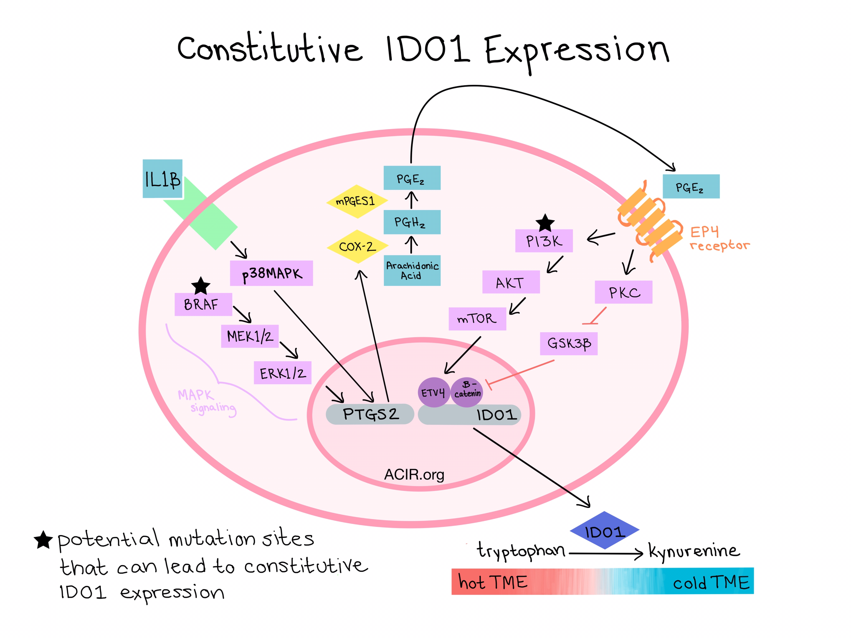
The proposed mechanism underlying constitutive IDO1 expression is as follows:

  • MAPK signaling (BRAF → MEK1/2 → ERK1/2) and/or an autocrine loop of IL1β signaling through the MAPK pathway help to support constitutive expression of COX-2 and mPGES1.
  • COX-2 expression leads to sustained PGE2 production.
  • PGE2 is recognized by EP4 receptors, triggering two separate signaling pathways that induce IDO1 transcription.
    • In the PI3K signaling pathway, PI3K activates AKT, which activates mTOR. mTOR activation likely depends on the transcription factor ETV4, which functions downstream and binds to the IDO1 promoter.
    • In the PKC signaling pathway, PKC deactivates GSK3β. Active GSK3β typically phosphorylates β-catenin, which leads to its degradation. Thus, deactivation of GSK3β leads to an excess of activated β-catenin, which binds to and activates the IDO1 promoter.
  • IDO1 expression leads to degradation of tryptophan into kynurenine.
  • The low tryptophan:kynurenine ratio creates an immunosuppressive microenvironment in which T cells cannot survive or proliferate.

The researchers confirmed that the COX-2/PGE2 pathway is responsible for constitutive IDO1 expression in a wide range of human cancer types including cholangiocarcinoma, head and neck carcinoma, sarcoma, ovarian carcinoma, non-small cell lung carcinoma, and another melanoma. Most of these IDO1-expressing tumor cell lines had mutations in genes from the PI3K or MAPK pathways, or in the receptors that lead to the activation of these pathways. Despite previous reports that STAT3 affects IDO1 production, Hennequart et al. found no direct role of STAT3.

To determine whether they could induce an anti-tumor immune response by targeting the COX-2 pathway to knock down constitutive IDO1 expression, Hennequart et al. used a human ovarian cancer line (SCOV3) that is sensitive to the COX-2 inhibitor, celecoxib. Human tumor xenografts were controlled in mice that received both celecoxib and allogeneic lymphocyte therapies, but not either alone. The same tumors showed reduced IDO1 which likely allowed for improved lymphocyte infiltration and proliferation in tumors that were otherwise “cold” in control mice and mice treated with celecoxib alone. Overall, results from the combination immunotherapy experiments further confirmed the mechanism by which celecoxib induced tumor rejection through repression of IDO1.

These mechanistic studies reveal additional approaches (COX-2 inhibition with the approved drug celecoxib or inhibitors of the PIK3K pathway) to overcome the “non-inflamed” tumor microenvironment characteristic of “cold” tumors caused by constitutive IDO1 expression. Such approaches may be additive or synergistic with the inhibitors of IDO1, such as epacadostat, currently being examined in the the clinic.

by Lauren Hitchings

References:

Hennequart M., Pilotte L., Cane S., Hoffmann D., Stroobant V., De Plaen E., Van den Eynde B.J. Constitutive IDO1 Expression in Human Tumors Is Driven by Cyclooxygenase-2 and Mediates Intrinsic Immune Resistance. Cancer Immunol Res. 2017 Jul 21.

In the Spotlight...

Inducible activation of MyD88 and CD40 in CAR T-cells results in controllable and potent antitumor activity in preclinical solid tumor models

Mata et al. demonstrate that delivering signal 2 to T cells using chemically inducible dimerization of alternative signaling endodomains (MyD88 fused to CD40) improves the activity of a co-delivered HER2-CAR by increasing antigen-driven cytokine production and proliferation capacity, extending persistence of cytotoxicity, and reducing PD-1 upregulation in vitro, culminating in improved tumor control in two solid tumor xenograft models.

GSK 3 inhibition drives maturation of NK cells and enhances their antitumor activity

Inhibition of the tyrosine kinase GSK 3 during IL-15-driven ex vivo expansion of natural killer (NK) cells enhanced the acquisition of a mature NK phenotype, as defined by the increased expression of CD57, T-BET, ZEB2, and BLIMP-1. Cells expanded in this way produced more TNF and IFN-γ in response to target cell recognition, showed enhanced antibody-dependent cellular cytotoxicity, and were more effective against ovarian cancer in murine xenograft models, prompting initiation of a clinical trial.

Notch Shapes the Innate Immunophenotype in Breast Cancer

Shen et al. identified a paracrine feedback loop in which macrophage-derived TGFβ activates Notch in tumor cells which, in turn, primes the tumor cell to respond to TGFβ, and further promotes uPA-dependent production of TGFβ by mature macrophages. These macrophages also produce Notch-dependent cytokines IL1β and CCL2, which promote monocyte recruitment and infiltration, as well as tumor progression.

Identification of essential genes for cancer immunotherapy

Patel et al. employed a “two cell type” CRISPR screen to identify genes expressed in cancer cells that facilitate the effector functions of T cells and thus may promote immune escape by tumors. Both known (HLA-A, B2M) and novel (CD58, APLNR) genes involved in antigen presentation and IFN-γ signaling pathways, among others, were identified and validated.

Using antigen-specific B cells to combine antibody and T-cell based cancer immunotherapy

Wennhold et al. isolated murine antigen-specific B cells, enhanced their antigen presentation capabilities with CD40 stimulation, and showed T cell stimulating properties in vitro and in vivo equivalent to dendritic cells (DC). Combining these DC-like B cells with antigen-specific B cells expressing antibody (following stimulation with CD40, IL-21, and IL-4) significantly enhanced therapeutic efficacy in a pancreatic tumor model. Similar DC-like B cells were isolated from draining lymph nodes or TILs in human cancers.

An unbiased in vivo functional genomics screening approach in mice identifies novel tumor cell-based regulators of immune rejection

Using a genome-wide shRNA library targeting 39,000 transcripts in a murine triple negative breast cancer cell line, Shuptrine et al. employed a novel strategy of differential target gene enrichment or depletion in tumors growing in immune-competent compared to immune-incompetent mice to identify genes involved in immune pathways affecting tumor growth. Both previously known critical regulators (TGFβ, CD47) and novel regulators (Sgpl1, Tex9, Pex 14) were identified and validated.

Immune Protection by a Cytomegalovirus Vaccine Vector Expressing a Single Low-Avidity Epitope

Using murine cytomegalovirus variants containing low and high avidity epitopes expressed by different promoters (strong/early vs weak/late), Borkner et al. demonstrate that low avidity epitopes can generate T-effector memory cells that are as protective as high avidity epitopes (but only when expressed in the strong/early context) and that competition between epitopes is impacted by both avidity and expression. Epitopes low in both parameters are driven to a non-protective T-central memory phenotype.

Normalized Synergy Predicts That CD8 Co-Receptor Contribution to T Cell Receptor (TCR) and pMHC Binding Decreases As TCR Affinity Increases in Human Viral-Specific T Cells

Using polyclonal TCRs specific for a single peptide:MHC complex and a sophisticated two dimensional surface approach to measure peptide/MHC:T cell interactions, Williams et al. demonstrate that peptide/MHC:TCR affinity and on rate positively correlate with T cell function and that the CD8 binding contribution to overall affinity decreases with increasing peptide/MHC:TCR affinity. Some TCRs are independent of CD8 level and would be insensitive to the natural variations in naive cell CD8 expression.

Donor SIRPalpha polymorphism modulates the innate immune response to allogeneic grafts

Using mice deficient in T, B, and NK cells, Dai et al. determined that the ability of myeloid cells to sense and respond to allogeneic grafts is dependent on the differential binding affinity between CD47 on host monocytes and polymorphisms in the extracellular domains of host or donor SIRPα. Dai et al. propose that non-self SIRPα alleles cause innate immune activation by disturbing the balance between activating and inhibitory signals.

Tumor location determines tissue-specific recruitment of tumor-associated macrophages and antibody-dependent immunotherapy response

Using the same melanoma cell line orthotopically implanted in different tissues, Lehmann et al. found that antibody-dependent control of melanoma in the skin required TAMs derived from Ly6Chigh monocytes recruited from blood via the CCL2-CCR2 axis, while control in the lung required CSF-2-dependent, tissue-resident alveolar macrophages.

Everything New this Week In...

Close Modal

Small change for you. Big change for us!

This Thanksgiving season, show your support for cancer research by donating your change.

In less than a minute, link your credit card with our partner RoundUp App.

Every purchase you make with that card will be rounded up and the change will be donated to ACIR.

All transactions are securely made through Stripe.