Weekly Digests
‹ Back to August

Canceling out the consequences with combined PD-L1 and IL-6 blockade

August 22, 2018

Tumors have multiple mechanisms of immune escape, and while certain immunotherapies may help to overcome one pathway, they may have unintended consequences on another. In a study recently published in Cancer Research, Tsukamoto et al. show that both PD-1/PD-L1 blockade and IL-6 blockade have unintended consequences that might hinder their antitumor efficacy, but utilizing both therapies in combination may lead to a synergistic antitumor effect.

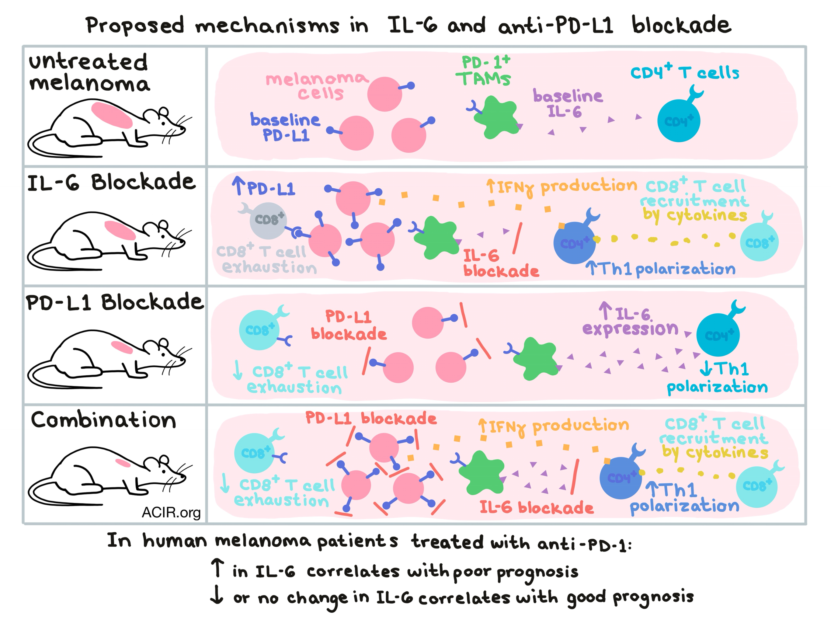
Based on previous research demonstrating that Th1 differentiation of CD4+ T cells can be attenuated by IL-6, Tsukamoto et al. tested the effects of IL-6 blockade in a murine model for melanoma. Following IL-6 blockade, the team observed an increased portion of antigen-specific, IFNγ-producing CD4+ T cells in the draining lymph node, increased CXCR3 expression by CD4+ T cells, recruitment of tumor-specific CD8+ T cells to the tumor site dependent on the presence of CD4+ T cells, decreased Th2 responses, and reduced tumor growth. Together, this data suggests that IL-6 blockade enhanced polarization towards a favorable Th1 phenotype. Despite the overall positive effect of IL-6 blockade, the increase in IFNγ production by Th1 CD4+ T cells was found to increased PD-L1 expression by melanoma cells, which likely puts a damper on the overall antitumor efficacy of IL-6 blockade.

Next, looking at PD-1/PD-L1 blockade as a target, Tsukamoto et al. examined data from melanoma patients treated with nivolumab (anti-PD-1). When the researchers examined levels of IL-6 expression, they found that although baseline levels of IL-6 had little relation to response to therapy, a rise in IL-6 levels upon treatment with anti-PD-1 correlated with shorter progression-free survival (PFS) and poor clinical outcome. Patients whose IL-6 levels remained unchanged or decreased upon treatment with anti-PD-1 were more likely to experience longer PFS and improved clinical outcomes. While only a limited number of patients were utilized in this analysis, the data identifies a rise in IL-6 upon PD-1 blockade as a possible predictive marker of non-response to therapy.

Delving deeper into the connection between PD-1, IL-6, and poor clinical outcomes in melanoma patients, the team turned again to a melanoma mouse model. In mice, anti-PD-L1 antibody increased IL-6 levels in wild-type melanoma-bearing mice, but not in IL-6-deficient mice, indicating that host cells, rather than melanoma cells, were the likely source of the increase in IL-6. The researchers identified a subset of PD-1+ TAMs as the main source of IL-6 in response to PD-L1 blockade and further showed that IL-6 production could be directly controlled by PD-1/PD-L1 ligation in this subset; blockade of the PD-1/PD-L1 axis in TAMs led to increased IL-6 production, while stimulation of the same axis led to decreased IL-6 production.

Knowing that stimulation of the PD-1/PD-L1 axis in TAMs decreased their IL-6 production, the researchers circled back to IL-6’s effect on Th1 polarization. When PD-1-stimulated TAMs (producing less IL-6) were added to a culture of activated CD4+ T cells, more CD4+ T cells produced IFNγ than when control TAMs (producing more IL-6) were added. The addition of an IL-6-blocking antibody to control TAM-treated CD4+ T cells restored differentiation towards a Th1 phenotype. In vivo, depleting TAMs reduced the previously observed increase in IL-6 upon PD-L1 blockade, and increased the IFNγ-producing CD4+ T cells in the tumor. Overall this data suggests that the production of IL-6 by TAMs impairs Th1 differentiation, and that in PD-1+ TAMs, IL-6 production can be upregulated or downregulated upon PD-1/PD-L1 blockade or stimulation, respectively.

Tsukamoto et al. hypothesized that because PD-L1 blockade caused increased IL-6, which had the negative effect of enhancing tumor PD-L1 expression in an IFNγ- and CD4+ T cell-dependent manner, and IL-6 blockade induced upregulation of PD-L1, which had the negative effect of abrogating CD8+ T cell responses, combining the two therapies might cancel out each other’s negative consequences and have a synergistic effect. They found that combining IL-6 blockade and PD-L1 blockade significantly reduced tumor growth in both OVA-expressing MO4 melanoma and CT26 colon carcinoma tumors compared to either monotherapy, though B16F10 melanoma tumors were still refractory to therapy. The synergistic effect of the dual blockade was dependent on CD4+ T cells, which did not increase in numbers, but expressed more IFNγ and CXCR3 upon treatment. Dual blockade also increased intratumoral expression levels of Ccl3/4/5 and Cxcl9/10 – genes for T cell-attracting chemokines.

While anti-PD-1 is emerging as a frontline therapy in cancer, durable effects are still limited to only a subset of patients, and biomarkers of response and additional therapies that improve its efficacy and applications are greatly needed in the clinic. Here, Tsukamoto et al. found that an increase in IL-6 upon anti-PD-1 therapy could serve as an early post-treatment marker of non-response to therapy. Additionally, they show that a blockade of IL-6 could enhance the efficacy of PD-1 checkpoint blockade, though further preclinical and clinical testing will be required to determine whether this hypothesis will hold up in the clinic.

by Lauren Hitchings

References:

Tsukamoto H., Fujieda K., Miyashita A., Fukushima S., Ikeda T., Kubo Y., Senju S., Ihn H., Nishimura Y., Oshiumi H. Combined blockade of IL-6 and PD-1/PD-L1 signaling abrogates mutual regulation of their immunosuppressive effects in the tumor microenvironment. Cancer Res. 2018 Jul 2.

In the Spotlight...

Low-dose cyclophosphamide depletes circulating naive and activated regulatory T cells in malignant pleural mesothelioma patients synergistically treated with dendritic cell-based immunotherapy

In a phase I/II clinical trial, 10 malignant pleural mesothelioma patients received a combination of low-dose cyclophosphamide (CTX) and autologous, tumor lysate-pulsed, dendritic cell vaccine (DC). In the peripheral blood, one dose of CTX increased the proportion of CD8+ T cells, but decreased overall T cells and CD4+ T cells, including all Tregs. However, the percentage of proliferating naive CD45RA+FoxP3med Tregs and their CTLA-4 expression increased. At the end of the combination CTX/DC treatment, almost all cell subsets went back to baseline levels. Unexpectedly, the pretreatment proportion of naive Tregs correlated positively with survival.

Anti-CTLA-4 immunotherapy does not deplete FOXP3+ regulatory T cells (Tregs) in human cancers

Using quantitative immunohistochemistry to monitor the density of T cell subsets (with specificity verified by multiparametric CyTOF analysis), Sharma and Subudhi et al. demonstrate that in human cancers (melanoma, prostate cancer, and bladder cancer), treatment with anti-CTLA-4 (either IgG1 ipilimumab or IgG2 tremelimumab) increases the density of CD4+ and CD8+ T cells, but surprisingly does not deplete (and in some cases, increases) intratumoral Tregs. CD68+ macrophages were highly abundant and not impacted by anti-CTLA-4. Thus, in contrast to preclinical studies, depletion of Tregs may not be the operative mechanism of anti-CTLA-4.

Reducing Ex Vivo Culture Improves the Anti-leukemic Activity of Chimeric Antigen Receptor (CAR)-T Cells

While current CAR T cell manufacturing protocols call for 9 to 14 days of ex vivo expansion, Ghassemi et al. showed that CAR T cells harvested at earlier time points (day 3 or 5) exhibited increased proliferation, an improved differentiation state (i.e. more central memory), and enhanced effector function compared to those harvested later (day 9). In a murine xenograft model of ALL, early-harvested CD19 CAR T cells showed improved persistence, robust tumor control, and higher potency than day 9 cells. Analysis of CAR T cell products manufactured under standard protocols suggested that an abbreviated culture would be feasible.

TLR-3 Stimulation Skews M2 Macrophages to M1 Through IFN-αβ Signaling and Restricts Tumor Progression

Vidyarthi et al. demonstrated that stimulating human and murine protumorigenic M2 macrophages with a TLR3 ligand (TLR3L) converts them to a proinflammatory M1 phenotype, as indicated by the shift in the expression of costimulatory and inhibitory markers and cytokine production. Antibody blockade and genetic knockouts demonstrated the critical role for IFN-αβ signaling in repolarization. The converted macrophages exhibited enhanced antigen uptake and presentation, as well as increased capacity for T cell priming. In vivo, TLR3L treatment reduced tumor growth in mice with colon cancer.

Superagonist IL-15-Armed Oncolytic Virus Elicits Potent Antitumor Immunity and Therapy That Are Enhanced with PD-1 Blockade

Kowalsky et al. modified a clinically tested, tumor-selective oncolytic vaccinia virus to express a superagonist IL-15 (fusion protein of IL-15 and IL-15Rα). The virus (vvDD-IL15-Rα) increased the infiltration of CD8+ T and NK cells into the tumor, reduced tumor mass, increased survival of mice with colon or ovarian cancer, and led to formation of immunological memory. Although both CD8+ T and NK cells were increased in the tumor, depletion experiments showed that only CD8+ T cells, and not NK or CD4+ T cells, were required for efficacy. Combination of vvDD-IL15-Rα and anti-PD-1 dramatically increased survival compared with either monotherapy.

Distinct immune signatures in directly treated and distant tumors result from TLR adjuvants and focal ablation

Aiming to unravel the mechanisms by which focal tumor ablation and immunotherapy (intratumoral CpG and systemic anti-PD-1 antibody) synergize in tumor control, Chavez and Silvestrini et al. analyzed the local and systemic effects on immune populations. Thermal focal ablation, particularly in combination with intratumoral CpG, induced expression of type I interferon, led to myeloid cell (CD169+ macrophage and DC) recruitment, enhanced antigen release and presentation, and increased intratumoral and circulating TCR clonality. Effects were observed systemically and in distal tumors, but varied depending on the treatment regimen.

Everything New this Week In...

Close Modal

Small change for you. Big change for us!

This Thanksgiving season, show your support for cancer research by donating your change.

In less than a minute, link your credit card with our partner RoundUp App.

Every purchase you make with that card will be rounded up and the change will be donated to ACIR.

All transactions are securely made through Stripe.