Weekly Digests
‹ Back to August

T cell residency status affects prognosis in breast cancer

August 1, 2018

Inspired by the knowledge that the quantity of tumor-infiltrating lymphocytes (TILs) is prognostic for improved survival in breast cancer (BC) patients, particularly those with triple-negative (TNBC) and HER2-overexpressing (HER2+) subtypes, Savas and Virassamy et al., as described in Nature Medicine, set out to uncover whether specific T cell subpopulations affect prognosis in BC patients.

To begin, the team utilized flow cytometry to analyze the TIL content and T cell subsets in tumor samples from 123 patients (84 primary and 45 metastatic samples) with TNBC, HER2+, or hormone receptor-positive (luminal) BC subtypes. They found that TILs constituted a significantly larger percentage of live cells in primary (treatment-naive) than in metastatic tumors across all subtypes, with CD3+ T cells dominating the TIL population. Digging deeper, the researchers found that the most prevalent T cell phenotypes in primary tumors were effector memory (TEM) and terminally differentiated effector memory (TEMRA) subsets.

Given that BC patients generally have limited response to immune checkpoint blockade, with the exception of higher response rates in advanced TNBC (correlating with higher TIL quantity), the researchers analyzed the expression of immune checkpoints in BC TIL subsets. PD-1, but not TIM-3 or LAG-3, was highly expressed on both CD4+ and CD8+ TILs, while CTLA-4 expression was significantly higher on CD4+ than CD8+ TILs. Tregs were infrequent across all BC subtypes, with slightly elevated levels in TNBC.

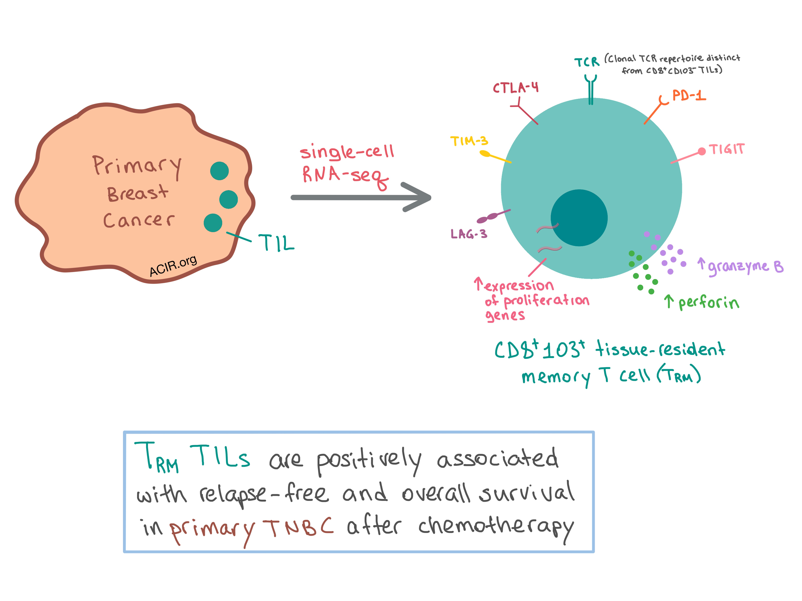
Since TNBC has the highest TIL infiltration among BC subtypes, the researchers chose this subtype to take a closer look at the variation within the T cell infiltrate using a comprehensive single cell mRNA transcriptome analysis. They analyzed 6311 T cells isolated from two human primary TNBC samples and identified ten clusters with unique gene expression profiles, which were similar across the two patients. One cluster that stood out, with 400 differentially expressed genes compared with the other clusters, was the CD8A+CD103+ tissue-resident memory (TRM)-like genotype. These cells had lower expression of tissue egress genes, which is required for retention of the TRM cells within tissue. The TRM cells expressed high levels of immune checkpoints (PD1, TIM3, CTLA4, TIGIT, and LAG3) as well as increased expression of granzyme B and perforin compared with CD8A+CD103- T cells. Another cluster of CD8A+CD103+ T cells was enriched for genes involved in cell proliferation. Overall, the cluster analysis identified a CD8+ TRM-like subset that is cytotoxic and proliferative within the tumor microenvironment.

Expanding their analysis to the entire patient cohort, the team found that a subset of T cells expressed CD103 across all BC subtypes, with significantly more CD8+ T cells than CD4+ T cells expressing this marker. The CD8+CD103+ T cells expressed higher levels of PD-1 and were more frequently double-positive for PD-1 and CTLA-4 than other CD8+ T cell subsets, which was consistent with single-cell analysis. Overall, the data showed that CD8+CD103+ T cells were enriched in BC tumors with a high number of TILs, and bulk RNA-seq of CD8+CD103+ and CD8+CD103- T cells from another three tumor samples confirmed the TRM-like characteristics of the CD8+CD103+ T cells. In vitro co-culturing of TILs with autologous BC cells with or without TCR stimulation revealed that CD8+CD103+ T cells expressed significantly more granzyme B than CD8+CD103- T cells, regardless of TCR stimulation, demonstrating the cytotoxic functionality of this T cell subset.

The team then performed TCRβ sequencing on sorted CD8+CD103+ and CD8+CD103- T cells and revealed distinct TCR repertoires between TRM and TEM subsets in primary tumors, with the former exhibiting higher TCR clonality. These results suggest that the two T cell subsets recognize different antigens. To understand how this came to be, the researchers analyzed normal breast tissue obtained from prophylactic mastectomies and found that the TRM cells make up a significant portion of CD8+ T cells present in the tissue. These observations suggest that the antigen specificity of TRM cells may originate from the microenvironment found in normal, pre-neoplastic, and early neoplastic breast tissue.

Having confirmed the prevalence of the TRM subset in BC TILs, especially primary TNBC, the researchers sought to determine the prognostic value of this subpopulation. Using publicly available gene expression and clinical outcome data from 329 primary TNBC patients, the team found that the TRM gene expression signature was significantly associated with increased relapse-free and overall survival in primary TNBC after chemotherapy treatment.

Overall, Savas and Virassamy et al. demonstrated that a distinct CD8+CD103+ TRM subset of TILs in BC is cytotoxic, proliferative, and clonally enhanced within the tumor microenvironment, and is associated with improved survival in TNBC, pointing to the potential role of the TRM subset as the key player in clinical outcomes that have been observed in BC tumors with a high quantity of TILs.

by Anna Scherer

References:

Savas P., Virassamy B., Ye C., Salim A., Mintoff C.P., Caramia F., Salgado R., Byrne D.J., Teo Z.L., Dushyanthen S., Byrne A., Wein L., Luen S.J., Poliness C., Nightingale S.S., Skandarajah A.S., Gyorki D.E., Thornton C.M., Beavis P.A., Fox S.B.; Kathleen Cuningham Foundation Consortium for Research into Familial Breast Cancer (kConFab), Darcy P.K., Speed T.P., Mackay L.K., Neeson P.J., Loi S. Single-cell profiling of breast cancer T cells reveals a tissue-resident memory subset associated with improved prognosis. Nat Med. 2018 Jul.

In the Spotlight...

Reprogramming human T cell function and specificity with non-viral genome targeting

Roth et al. developed a rapid genome-targeting system that involves co-electroporation of CRISPR-Cas9 ribonucleoprotein complexes and short or long, double- or single-stranded DNA. They successfully demonstrated this strategy in human primary T cells, targeting up to three different genes simultaneously. They also utilized this system to correct IL2RA mutations in cells from patients with an autoimmune disease. Finally, they applied this method to replace the endogenous TCR locus with a new, tumor-targeting TCR, resulting in cells that led to antitumor response in vitro and in vivo.

Antigen-Presenting Cell-Intrinsic PD-1 Neutralizes PD-L1 in cis to Attenuate PD-1 Signaling in T Cells

Using in vitro reconstitution of vesicles, cellular reconstitution, and cell culture assays, Zhao et al. found that in antigen-presenting cells (APCs) – including tumor cells – co-expressing PD-1 and PD-L1, the two bind in cis with high affinity, preventing PD-L1 from engaging in trans with T cells. Selective anti-PD-1 antibody blockade of cis signaling between PD-1 and PD-L1 on APCs freed up PD-L1 to engage with T cells and triggered exhaustion. This research indicates that PD-1 negatively regulates PD-L1 in cis and that the net outcome of the PD-1/PD-L1 pathway depends on PD-1 expression on both APCs and T cells.

MHC proteins confer differential sensitivity to CTLA-4 and PD-1 blockade in untreated metastatic melanoma

In biopsies of previously untreated melanoma, Rodig et al. found that loss of MHC-I expression on more than 50% of malignant cells was common, was associated with transcriptional repression of MHC-related genes, and predicted resistance to anti-CTLA-4 therapy, supporting a role for anti-CTLA-4 in CD8+ T cell priming. The expression of MHC class II on more than 1% of malignant cells or an IFNγ-related immune signature predicted superior response to anti-PD-1 therapy. Patients with the best overall responses to anti-PD-1 had higher baseline signatures for innate NK cells, γδ T cells, and IL-15.

IFN-γ-induced chemokines are required for CXCR3-mediated T cell recruitment and anti-tumor efficacy of anti-HER2/CD3 bispecific antibody

Using mice with HER2-overexpressing tumors, Li et al. demonstrate that an anti-HER2/CD3 bispecific antibody converts the tumor phenotype from poorly infiltrated to inflamed by inducing intratumoral CD8+ T cell proliferation and recruiting CD8+ T cells to the tumor, with the latter being the dominant mechanism and required for antitumor effect. The treatment led to rapid upregulation of IFNγ, which in turn triggered expression of CXCR3 on T cells and production of CXCR3 ligands (CXCL-9, -10, -11) by undetermined cells within the tumor, thereby increasing intratumoral T cell infiltration.

Enhancement of peptide vaccine immunogenicity by increasing lymphatic drainage and boosting serum stability

Building on prior work, Moynihan et al. explored their highly immunogenic “albumin-hitchhiking” amphiphilic peptide vaccine and found that alternative albumin-binding moieties were nearly as effective as the original lipid tail. Various linkage strategies and molecular adjuvants also showed equivalent functionality. Conjugated PEG improved the stability of peptides in serum, and conjugated peptides reached distal lymph nodes and were presented for longer in vivo. Cross-presenting (Batf3+) DCs were critical for enhanced immunogenicity, and human DC presentation of conjugated peptides was comparable to that of free peptides.

Selective FcγR Co-engagement on APCs Modulates the Activity of Therapeutic Antibodies Targeting T Cell Antigens

Adding a new wrinkle to the mechanism(s) of action of T cell-targeted immunomodulatory mAbs, Waight et al. demonstrate that engagement of the Fc portion of antibodies (such as those targeting the inhibitory receptors CTLA-4 and TIGIT on superantigen-stimulated T cells and the CD45RB isoform on Tregs) by FcγR on APCs significantly modulates the resulting T cell response. Human FcγRIIIA (and the comparable FcγRIV in mice) were the key receptors driving superior IL-2 production, and in humans, a CD11chi DC population was involved. The Fc/FcγR-mediated stimulation was shown to be independent of Treg depletion.

Everything New this Week In...

Close Modal

Small change for you. Big change for us!

This Thanksgiving season, show your support for cancer research by donating your change.

In less than a minute, link your credit card with our partner RoundUp App.

Every purchase you make with that card will be rounded up and the change will be donated to ACIR.

All transactions are securely made through Stripe.