T cell exhaustion is accompanied by a distinct and relatively well described epigenetic state, but whether that state is reversible is not quite clear. Investigating this, Yates et al. explored the chromatin landscape of CD8+ T cells in the setting of active and cured chronic infection, and published the results in Nature Immunology.
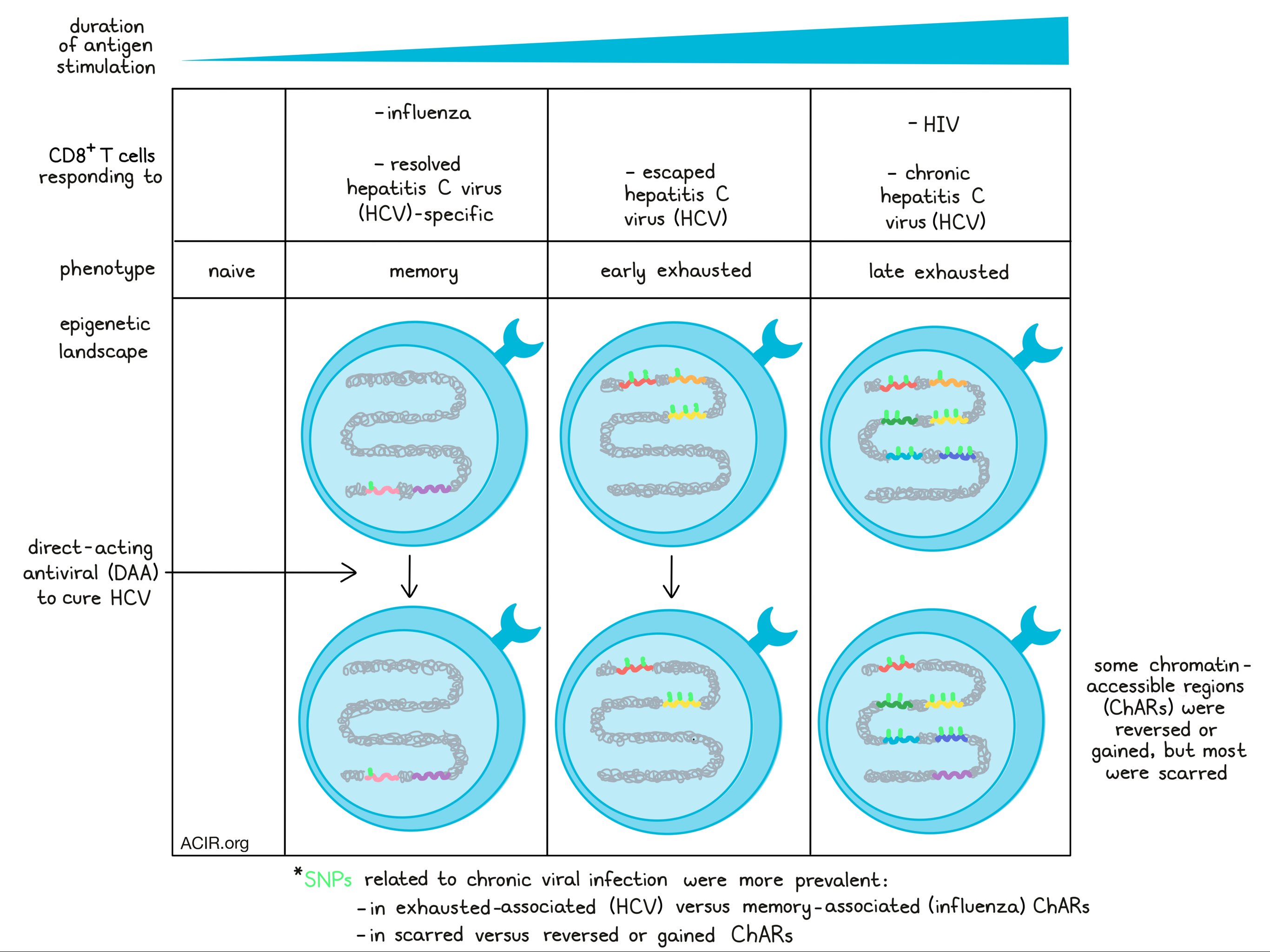
As a model system of T cell exhaustion, Yates et al. isolated antigen-specific CD8+ T cells responding to chronic infection of hepatitis C virus (HCV), which does not clear spontaneously in a majority of patients, but does clear spontaneously in a minority. These were then compared to CD8+ T cells responding to influenza (as a model of memory T cell responses) and to naive CD8+ T cells from the same patient.
ATACseq was performed to evaluate chromatin accessibility, which generally correlated to increased mRNA expression. Analysis of chromatin-accessible regions (ChARs) showed that within patients, T cells responding to chronic HCV were distinct from those responding to influenza. T cells from different patients whose HCV infections had resolved spontaneously were more similar to influenza-specific T cells, sharing more ChARs and showing increased accessibility of genes related to memory, suggesting that T cells from resolved HCV may acquire a more memory-like epigenetic state. In contrast, T cells responding to chronic HCV showed more accessibility of key genes associated with exhaustion, including EOMES and NFATC2.
Next, the researchers analyzed potential relationships between disease-associated single-nucleotide polymorphisms (SNPs) and ChARs, and found that SNPs related to chronic viral infection were significantly overrepresented in the HCV-specific ChARs relative to the influenza-specific ChARs. This suggested that some SNPs may modulate enhancer activity in HCV-specific responses to chronic infection.
To determine whether the epigenetic changes observed in T cells responding to chronic HCV infection were, in fact, related to exhaustion, Yates et al. turned to HIV as a second model. HIV-specific CD8+ T cells showed an epigenetic state similar to what was seen with chronic HCV. Their shared regulatory program included ChARs near exhaustion-specific inhibitory receptor genes, including those related to CD39, NFAT signaling, and PD-1 upregulation. Further, the same chronic infection-related SNPs identified in ChARs in HCV overlapped in HIV. Together these results suggested that epigenetic exhaustion in CD8+ T cells is consistent across multiple chronic viral infections. An investigation of LCMV in mice also showed the same epigenetic pattern, suggesting that it is evolutionarily conserved.
Recently, direct-acting antivirals (DAAs) have been developed to cure chronic HCV infection without the use of interferon, allowing the researchers to investigate whether a non-immune-based removal of the source of chronic antigen stimulation and inflammation could remodel or reverse epigenetic exhaustion. Looking at patients 12 weeks after cessation of DAA therapy (which occurs 20 weeks after viral clearance), the researchers found that while a few ChARs near exhaustion-associated genes (including those near CTLA-4) were reversed, the majority of exhaustion-specific ChARs, (including those near BATF and ENTPD1) were evidently unchanged, or “scarred”. A handful of ChARs were also gained after the clearance of infection, mimicking some changes associated with a memory-like state.
Looking into possible factors that might contribute to whether ChARs are reversed, scarred, or gained, the researchers found that scarred ChARs had higher sequence conservation across mammals compared to reversed regions. Scarred, but not reversed or gained ChARs were also enriched for SNPs related to chronic viral infections compared with all SNPs, and the majority of HCV-associated ChARs that overlapped with chronic viral infection-associated SNPs were scarred rather than reversed after treatment.
Dissecting the relative contribution of persistent inflammation versus chronic antigen stimulation in inducing an exhaustion-associated epigenetic landscape, Yates et al. found that DAA induced almost no changes in naive and influenza-specific T cell populations and that these cell subsets were similar to matched subsets from healthy donors, suggesting that the inflammatory environment imprints minimal genetic changes in bystander populations.
Turning towards the contributions of chronic antigen stimulation, the researchers compared typical cases of chronic HCV infection to cases of HCV infection in which the virus mutated and escaped from the previously dominant T cell epitope, thereby abrogating or diminishing TCR signaling. Prior to DAA therapy, T cells from escaped HCV expressed less PD-1 and weren’t as enriched for signatures of exhaustion, with their epigenetic landscapes more closely resembling influenza-specific memory T cells. DAA therapy and clearance of the viral infection induced minimal changes in these T cells, suggesting that chronic stimulation is likely the primary driver of epigenetic exhaustion programming.
Comparing scarred ChARs between T cells responding to standard versus escaped chronic HCV infection, the researchers found that some scarred ChARs were shared between the two populations, suggesting that these scars were likely acquired early during HCV infection. Most of the scarred ChARs, however, were specific to T cells from standard chronic HCV infection, suggesting that longer exposure to chronic antigen stimulation drove further epigenetic scarring and exhaustion.
Next, Yates et al. developed a novel strategy to accurately identify super-enhancer-associated genes from open chromatin regions as evaluated through ATACseq. Using this strategy to evaluate epigenetic scars, they found that TOX was the top super-enhancer-associated gene within the scarred regions, and was higher in T cells responding to chronic HCV, versus resolved or escaped HCV. HIF-1α, ID2, and NFAT, were also identified, along with other transcription factors, which may represent previously unknown regulators of exhaustion. A large fraction of super-enhancer-associated transcription factors had motifs that were overrepresented in scarred, versus reversed, regions. Interestingly, some of these transcription factors themselves are differentially regulated by epigenetic scars, suggesting a possible positive feedback loop driving T cell exhaustion, and potentially mediating the failure to reverse exhaustion programming after the resolution of infection.
To determine whether epigenetic scarring might reverse over an extended period of time, the researchers evaluated four patients 60-80 weeks after the cessation of DAA therapy. T cells from these patients clustered more closely with cells taken immediately after therapy versus those taken before treatment. While some ChARs did slowly close, the majority, including the region containing the super-enhancer near TOX, remained open and accessible long-term.
Together, these results suggest that exhaustion programming driven by chronic TCR stimulation is mostly irreversible. While these studies focused on chronic infection, the same basic biology is likely applicable to T cells in cancer. This has implications regarding the mechanism of action underlying checkpoint blockades, whether T cell exhaustion programming is reversible at the epigenetic level, and how that can be approached therapeutically.
Write-up and image by Lauren Hitchings
MEET THE RESEARCHER
This week, co-lead author Sen Debattama answered our questions.
What prompted you to tackle this research question?
Our lab’s work has focused on the transcriptional and epigenetic pathways that cause cytotoxic T cells (CD8+ T cells) to lose their ability to control disease in the case of chronic viral infections (like HIV and HCV) or tumors. We call this dysfunctional state “T cell exhaustion”, and it means that cytotoxic cells can no longer fight persistent illnesses. We have previously found that this exhausted state is conserved in both chronic viral infection and tumors, and even between mice and human cells. The key drivers of this state are found in the epigenome, with exhaustion-specific enhancers changing the expression of inhibitory receptors and specific transcription factors.
In this paper, we took advantage of newly developed direct-acting antiviral (DAA) therapies to ask if the exhausted state persists after the viral load is cleared from the body. To do this, we followed a cohort of HCV patients with samples before and after they were cured through DAA therapy and looked at changes in chromatin regulation within the same patient. Therefore, we were able to conclusively demonstrate that the underlying dysfunction of T cells in both chronic viruses and tumors in humans cannot be alleviated just by removing the disease – the cells remain exhausted.
By comparing the T cell response across a range of viruses that are either cleared and produce long-term memory (like Flu) or go chronic and lead to dysfunction (like HCV and HIV), we also produced a definitive map of where exhaustion-specific epigenetic changes occur. This will enable precision editing of the chromatin by allowing us to target specifically the regions relevant to exhausted T cells and minimize the off-target effects in other T cell populations. We hope our work will help develop more targeted therapies that focus on the epigenetic drivers of the exhausted state, and thus reinvigorate tumor and/or virus-specific T cell responses.
What was the most surprising finding of this study for you?
In this study, our major focus was asking whether a cure of chronic infection would be enough to reverse the regulation of T cell dysfunction. Surprisingly, we found that even after clearing the virus, a patient’sT cells maintain many of these exhaustion-specific epigenetic features, essentially leaving a “scar” of exhaustion in the chromatin landscape. In fact, we show that even a year after cure of HCV infection, the T cells remained locked in the exhausted state. In combination with the collaborative papers from Dr. Georg Lauer’s lab and Dr. John Wherry’s lab, our data suggest that these scars might be “locking” the exhausted T cell and preventing return to proper function, even if the chronic infection in the patient is cured through other means. Thus, restoring the function of these cells will likely require direct editing of the epigenome to remove/inactivate these scarred regions and “unlock” the cell’s functionality.
What was the coolest thing you’ve learned (about) recently outside of work?
Given all that’s happening in the world, I’ve been drawn to experiences outside of work that offer moments of peace or self-reflection. One moment that stands out is from a recent visit with my family to the Peabody Essex Museum in Salem, MA. The museum has a great repertoire, including a rare collection of Asian art, and is well worth visiting for those pieces alone. But I was also impressed by the fact that the Peabody is the only museum I know of that hosts a Visiting Neuroscience Researcher. This philosophy of combining art and science was most clearly reflected in a particular exhibition by Anila Quayyum Agha called “All the Flowers Are for Me” (see picture below) that really struck a chord with all of us. It was simple, but incredibly immersive, and spending some time taking in the installation felt like a meditative experience. I’d highly recommend a trip for those in the area!