Weekly Digests
‹ Back to August

Exosomal PD-L1: the unsung villain of immune evasion

August 29, 2018

In a recent paper published in Nature, Chen et al. set out to elucidate the details of how PD-L1 upregulation by the tumor, and the subsequent immune evasion, predicts patient response, and how treatment efficacy could be improved. Building on prior data indicating that extracellular vesicles contain bioactive molecules, including PD-L1, the researchers began by isolating exosomes from human primary and metastatic melanoma cell lines and found that PD-L1 was indeed present in these exosomes, that the extracellular domain was exposed on the surface (recapitulating the topology of PD-L1 on tumor cell surface), that levels were increased following IFNγ treatment of tumor cells, and that the presence and levels of PD-L1 in exosomes were significantly higher in metastatic compared to primary melanomas. Similar findings were observed in the murine B16F10 metastatic melanoma cell line.

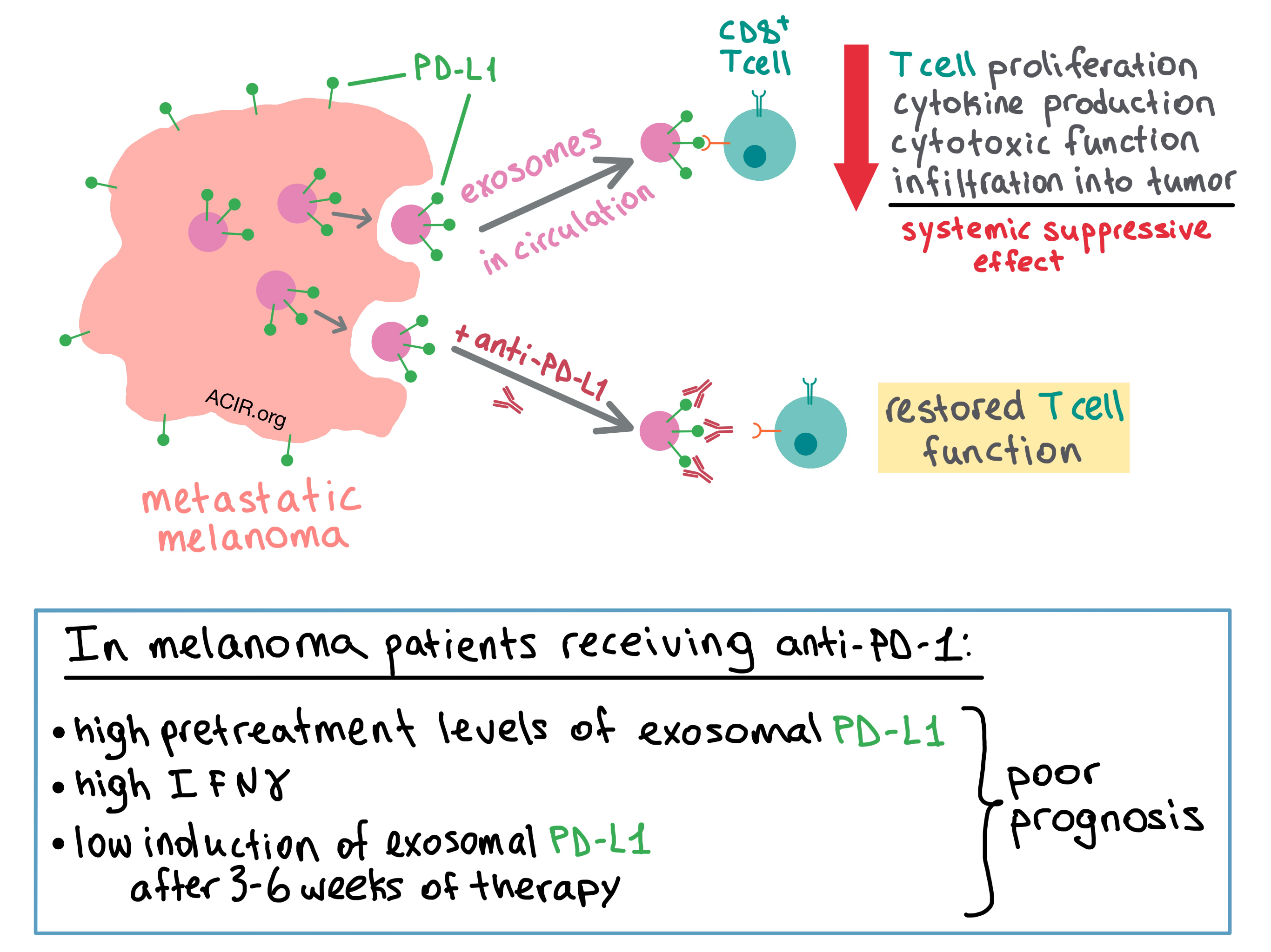
To examine the behavior of the exosomes in vivo, the team utilized a human melanoma xenograft model in nude mice. They isolated exosomes from the blood of the mice and found that the level of circulating exosomal PD-L1 positively correlated with tumor size. Next, the researchers utilized a syngeneic mouse model with a variant of B16F10 in which PD-L1 expression was knocked down; this abrogated the inhibitory effect of tumor-derived PD-L1 on antitumor T cells and slowed tumor growth. Injection of PD-L1-expressing exosomes re-established the inhibitory effect and promoted tumor growth of the B16F10 variant, while pretreating the exosomes with anti-PD-L1 antibodies reduced this effect. The presence of exosomes reduced the infiltration of CD8+ T cells into the tumor, as well as the proportion of proliferating PD-1+CD8+ T cells in the spleen and lymph nodes, suggesting a systemic suppressive effect on antitumor immune response.

The researchers then sought to find out whether exosomal PD-L1 inhibits CD8+ T cells. Chen et al. first used confocal microscopy to demonstrate that the melanoma-derived exosomes and T cells physically interact with each other, and then confirmed with flow cytometry that the interaction was stronger for activated versus non-activated CD8+ T cells. Treating the exosome-producing cells with IFNγ further increased exosomal binding to the T cells. PD-L1-expressing exosomes inhibited proliferation, cytokine production (IFNγ, IL-2, TNF), and cytotoxic function (granzyme B) of CD8+ T cells. These effects were abrogated when the exosomes were pretreated with anti-PD-L1 antibodies.

Convinced of the biological impact of PD-L1-containing exosomes, the team turned their attention to the exosomal PD-L1 levels in melanoma patients. While the number of circulating exosomes was similar between healthy controls and melanoma patients, the level of exosomal PD-L1 was significantly higher in the latter. The researchers then analyzed the PD-L1 expression on circulating exosomes in melanoma patients during anti-PD-1 therapy (pembrolizumab). The pretreatment level of exosomal PD-L1 was significantly higher in non-responders than in responders, and the elevated pretreatment level was associated with lower objective response rate (ORR). Non-responders also had higher pretreatment levels of IFNγ, which may have contributed to upregulated exosomal PD-L1. Overall, the level of circulating exosomal PD-L1 positively correlated with IFNγ levels and tumor burden, both of which indicated poor prognosis.

Although responders had lower baseline levels of exosomal PD-L1, the increase in the expression of PD-L1 during therapy (which was apparent at 3 weeks and peaked at 6 weeks in responders) was more enhanced in responders than in non-responders. In fact, a fold change of 2.43 in exosomal PD-L1 at 3-6 weeks of treatment stratified patient clinical response, with fold change >2.43 being associated with better overall response rate, progression-free survival, and overall survival.

Together, the results of this study suggest that melanoma cells systemically undermine the antitumor response by releasing PD-L1-expressing exosomes into the tumor microenvironment and circulation, where the exosomal PD-L1 binds with PD-1 on CD8+ T cells, suppressing their function. Disrupting the binding between exosomal PD-L1 and CD8+ T cells via anti-PD-1 or anti-PD-L1 antibodies may be one of the previously unknown mechanisms of actions of checkpoint blockade therapy. The authors suggest that circulating exosomal PD-L1 may serve as a blood-based biomarker and predictor of clinical response to anti-PD-1 therapy.

by Anna Scherer

References:

Chen G., Huang A.C., Zhang W., Zhang G., Wu M., Xu W., Yu Z., Yang J., Wang B., Sun H., Xia H., Man Q., Zhong W., Antelo L.F., Wu B., Xiong X., Liu X., Guan L., Li T., Liu S., Yang R., Lu Y., Dong L., McGettigan S., Somasundaram R., Radhakrishnan R., Mills G., Lu Y., Kim J., Chen Y.H., Dong H., Zhao Y., Karakousis G.C., Mitchell T.C., Schuchter L.M., Herlyn M., Wherry E.J., Xu X., Guo W. Exosomal PD-L1 contributes to immunosuppression and is associated with anti-PD-1 response. Nature. 2018 Aug.

In the Spotlight...

Targeted delivery of a PD-1-blocking scFv by CAR-T cells enhances anti-tumor efficacy in vivo

Rafiq et al. modified CAR-T cells to secrete PD-1-blocking single-chain variable fragments (scFvs) and demonstrated improved survival of mice with syngeneic and xenogeneic hematologic and solid tumors compared to standard CAR-T cells. Compared with the combination of standard CAR-T cells and systemic anti-PD-1, the modified CAR-T cells were equally or more effective. The treatment led to functionally relevant bystander binding of scFvs to tumor-specific T cells, and to the formation of memory response. As the secreted PD-1-blocking scFvs remain local to the tumor, systemic toxicities may be reduced.

Sequestration of T cells in bone marrow in the setting of glioblastoma and other intracranial tumors

Chongsathidkiet et al. found that treatment-naive patients and mice with glioblastoma have severely reduced naive T cell counts in the blood. Surprisingly, these cells were not sequestered in the spleen or thymus (both organs were contracted), but instead accumulated in the bone marrow (BM). This was also observed with other cancer types in mice, but only if the tumors were introduced intracranially. Sequestration of T cells in the BM was due to the loss of S1P1 on their surface, and was reversible when S1P1 internalization was blocked. Releasing the T cells from the BM enabled previously ineffective immunotherapies to improve survival in mice.

A fully-virulent retargeted oncolytic HSV armed with IL-12 elicits local immunity and vaccine therapy towards distant tumors

In an immunocompetent mouse model, Leoni and Vannini et al. tested R-115 – an IL-12-armed, HER-2-retargeted, fully virulent oncolytic herpes simplex virus (oHSV) – administered locally in HER2-transgenic/tolerant, immunocompetent mice bearing HER2-Lewis lung carcinoma tumors. The treatment showed a good safety profile and inhibited the growth of primary tumors; surviving mice were protected from tumor rechallenge at distant sites. Treatment led to increased tumor infiltration by CD4+, CD8+, CD335+, CD141+, and Foxp3+ cells, increased activation of effector cells, Th1 polarization, and durable antitumor reactivity in responders.

Blood-based tumor mutational burden as a predictor of clinical benefit in non-small-cell lung cancer patients treated with atezolizumab

Addressing the need for a non-invasive test for response to atezolizumab (anti-PD-L1 antibody), Gandara et al. evaluated a targeted sequencing capture assay that measures tumor mutation burden (TMB) from circulating free DNA in a baseline blood sample (bTMB). Correlation was high between TMB in tissue and blood samples. Analysis of bTMB from the randomized NSCLC POPLAR study selected a bTMB cut-point of >16 as associated with atezolizumab efficacy and meaningful clinical outcomes. Analysis of the OAK study validated bTMB as a predictive biomarker of improved progression-free survival, independent of the biomarker of high PD-L1.

The chimeric TAC receptor co-opts the T cell receptor yielding robust anti-tumor activity without toxicity

Helsen et al. constructed an MHC-independent T cell antigen coupler (TAC), a chimeric receptor that consists of an antigen-binding domain, a TCR recruitment domain, and a CD4+ or CD8+ co-receptor, and signals through the endogenous TCR. TAC-engineered T cells were effective against solid and liquid tumor targets in murine xenograft models with no observed autoactivation. TAC-engineered T cells accumulated faster and more exclusively within tumor tissues than first and second generation CAR T cells, were proliferative only within the tumor, and induced more efficient antitumor responses with less toxicity.

Human iPSC-Derived Natural Killer Cells Engineered with Chimeric Antigen Receptors Enhance Anti-tumor Activity

Li et al. utilized human-derived induced pluripotent stem cells (iPSCs) to generate NK cells engineered with chimeric antigen receptors (CARs) specifically designed for NK cells with the NKG2D transmembrane domain, the 2B4 costimulatory domain, and the CD3ζ signaling domain to support antigen-specific NK cell activation and cytotoxicity. In an murine xenograft model of human ovarian cancer, the CAR-expressing iPSC-derived NK cells outperformed other NK cell variants in antitumor activity. Persistence and tumor control were comparable to that of CAR T cells, however, the modified NK cells were substantially less toxic and prolonged survival.

Everything New this Week In...

Close Modal

Small change for you. Big change for us!

This Thanksgiving season, show your support for cancer research by donating your change.

In less than a minute, link your credit card with our partner RoundUp App.

Every purchase you make with that card will be rounded up and the change will be donated to ACIR.

All transactions are securely made through Stripe.