Weekly Digests
‹ Back to August

Anti-PD-L1 unleashes the power of the macrophage

August 15, 2018

In a paper recently published in Cancer Immunology Research, Hartley et al. set out to explore how PD-L1 signaling affects the function of tumor-associated macrophages (TAMs), and in the process they discovered that PD-L1 blockade and PD-1 blockade confer distinct immunological effects.

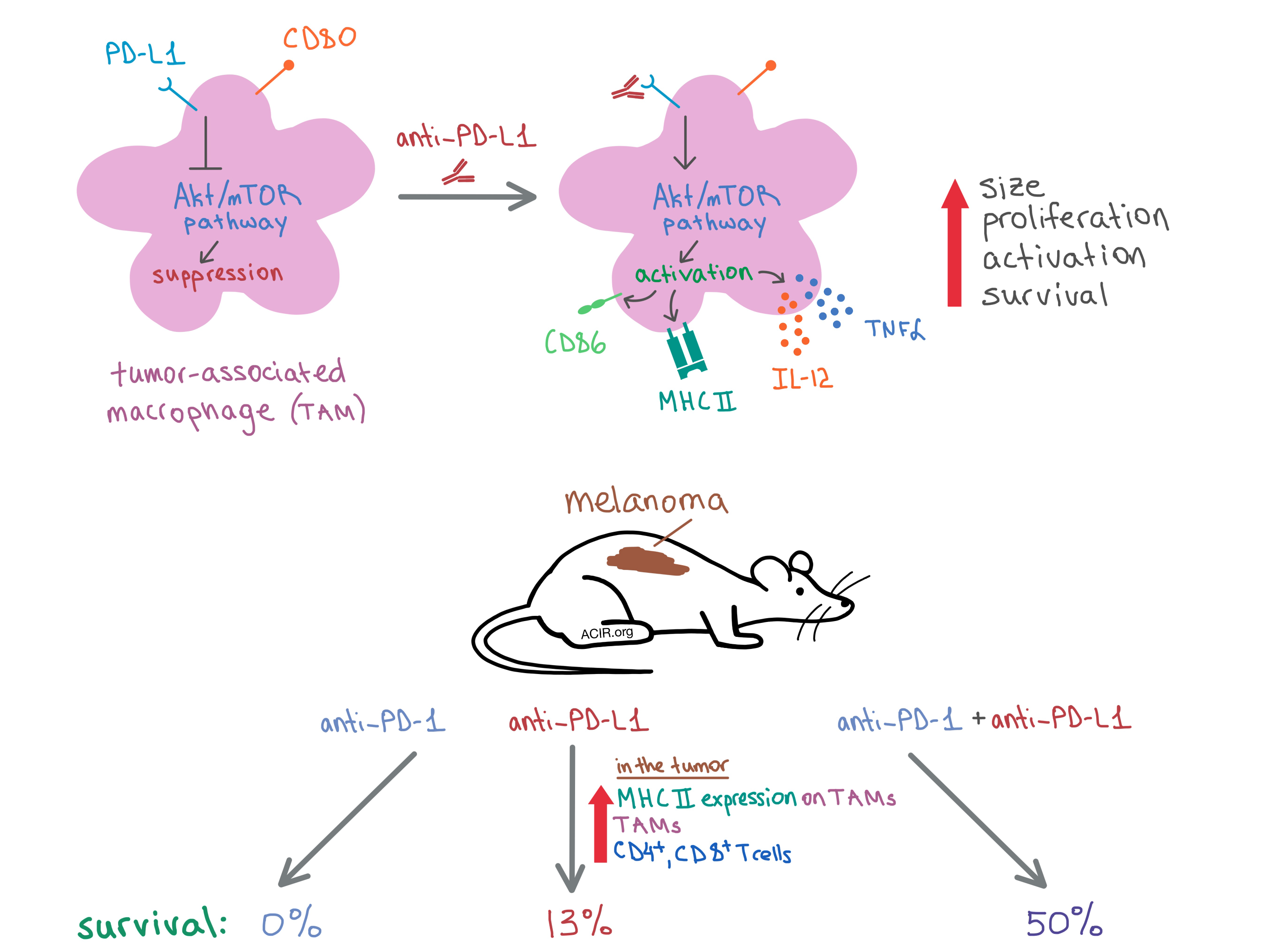
To begin, the researchers examined bone marrow-derived mouse macrophages treated with an anti-PD-L1 antibody and found that the macrophages expressed high levels of PD-L1 and CD80, but not any detectable levels of PD-1 compared to an isotype control antibody. Culturing the macrophages with anti-PD-L1 increased their size, proliferation, and survival. The team also demonstrated in vitro that anti-PD-L1 induced activation of murine macrophages, as indicated by the upregulation of MHC II and the costimulatory molecule CD86. In addition, PD-L1 blockade led to a proinflammatory macrophage phenotype, as treated macrophages produced more TNFα and IL-12. Similar (though slightly less dramatic) effects were observed in human monocyte-derived macrophages.

To see if other PD-L1 ligands could reproduce the effect of the PD-L1 antibody, Hartley et al. treated the mouse macrophages with soluble PD-1 (sPD-1) or soluble CD80 (sCD80). As with anti-PD-L1, both treatments increased the size of the macrophages. Treatment with sPD-1 increased the expression of CD86 only, while sCD80 upregulated CD86, MHC II, and TNFα. Meanwhile, treatment with neither FcRII/III antibody nor CD11b antibody had any effect on the macrophages’ size, proliferation, activation, or survival; pre-treatment with the FcRII/III antibody did not abrogate the effect of anti-PD-L1 antibody treatment.

Digging deeper to discover the mechanism behind the anti-PD-L1 effect, the researchers examined macrophages from PD-L1-/- mice and found that these cells had increased proliferation and MHC II expression compared to macrophages from wild-type mice, indicating constitutive, negative PD-L1 signaling in wild-type macrophages. These results suggest that the anti-PD-L1 antibody interrupts PD-L1 signaling, leading to macrophage activation and other phenotypic changes.

Since PD-L1 signaling is involved in the mTOR pathway in mouse tumors, the researchers treated mouse macrophages with mTOR inhibitors rapamycin or torin2, which inhibited anti-PD-L1-induced proliferation, MHC II expression, and TNFα production. Meanwhile, anti-PD-L1 increased the levels of phosphorylated Akt and mTOR in macrophages. Together, these results indicate that constitutive PD-L1 signaling in macrophages inhibits the mTOR pathway, which regulates cellular metabolism, and that blocking PD-L1 removes the brakes from mTOR signaling, leading to macrophage activation.

The researchers then examined the anti-PD-L1-induced changes on a genetic level via RNAseq and transcriptome analysis. The data indicated that macrophages treated with anti-PD-L1 had upregulated inflammatory, survival, and proliferation pathways and downregulated anti-inflammatory and apoptosis genes, as well as pathways related to cellular processes.

Next, the team performed a series of experiments on mice with tumors to observe the effects of PD-L1 in vivo. In mice with B16 melanoma, anti-PD-L1 increased the infiltration of macrophages into the tumor, but not in spleen or lymph nodes. TAMs from treated mice had an increased expression of MHC II. More CD4+ and CD8+ T cells infiltrated the tumor post-treatment as well. In RAG-/- mice (lacking functional T cells) with PyMT breast carcinoma, the number of TAMs did not change with anti-PD-1 treatment, but CD86 and MHC II expression increased and tumor growth was delayed, indicating that anti-PD-L1 directly activates macrophages independent of T cells.

Since the results of the in vivo experiments demonstrated a T cell-independent antitumor response to PD-L1 blockade, Hartley et al. hypothesized that PD-L1 and PD-1 antibodies may have non-redundant antitumor effects. To that end, they treated mice with established B16 melanoma with either PD-L1, PD-1, or a combination of the two antibodies. They found that while monotherapies induced complete response in only 6% of mice, the combination treatment resulted in complete tumor rejection in 50% of animals. None of the anti-PD-1-treated mice survived to 68 days post treatment, while 13% of anti-PD-L1-treated mice and 50% of mice treated with the combination treatment were alive at that time.

Overall, Hartley et al. demonstrate that anti-PD-L1 treatment acts directly on TAMs to induce a proinflammatory phenotype and increase activation, proliferation, and survival via the activation of the mTOR signaling pathway. In addition, they show that PD-1 and PD-L1 antibody treatments result in distinct antitumor effects, and the combination of the two antibodies synergizes to produce a greater antitumor response than either therapy alone, suggesting another therapeutic opportunity.

by Anna Scherer

References:

Hartley G.P., Chow L., Ammons D.T., Wheat W.H., Dow S.W. Programmed Cell Death Ligand 1 (PD-L1) Signaling Regulates Macrophage Proliferation and Activation. Cancer Immunol Res. 2018 Jul 16.

In the Spotlight...

CD4+ T cell help in cancer immunology and immunotherapy

Borst et al. comprehensively review how CD4+ T cells enhance the cytotoxic T lymphocyte (CTL) antitumor response. Proper CD4+ T cell help leads to clonal expansion and differentiation of CTLs into effector and memory T cells, and increases the cytotoxicity of CTLs, resulting in enhanced antitumor response. Topics reviewed include types and spatiotemporal stages of dendritic cell involvement and the intricate interplay with T cells, co-presentation of CD4+ and CD8+ epitopes, T cell costimulatory ligands (CD40L) and receptors (CD28, CD27), and cytokine (IL-12, IL-15) and chemokine (XCL1) production.

Recombinant Listeria promotes tumor rejection by CD8+ T cell-dependent remodeling of the tumor microenvironment

Deng et al. demonstrate that live-attenuated Listeria monocytogenes lacking two virulence genes and expressing an endogenous retroviral antigen induced KLRG1+PD1loCD62- effector CD8+ T cells, which localized to the spleen, were functional and not exhausted, produced IFNγ, infiltrated the tumor, and converted the tumor microenvironment from immunosuppressive to inflamed by repolarizing the tumor-associated macrophages from M2 to M1, decreasing Tregs, and increasing proinflammatory cytokines. The treatment led to durable tumor rejection and formation of immunological memory in mice.

CD11c+ MHCII(lo) GM-CSF-bone marrow-derived dendritic cells act as antigen donor cells and as antigen presenting cells in neoepitope-elicited tumor immunity against a mouse fibrosarcoma

Investigating the use of antigen-presenting cells (APCs) as adjuvants, Ebrahimi-Nik et al. found that neoantigen-pulsed bone marrow-derived dendritic cells (BMDCs) differentiated with either GM-CSF or FLT3L, and monocyte-derived dendritic cells (but not macrophages) acted as strong in vivo adjuvants. Within GM-CSF-BMDCs, the CD11c+MHCIIlo subset had superior adjuvanticity and antigen uptake capacity compared to the more mature CD11c+MHCIIhi subset. The adjuvanticity of DCs could be attributed mainly to their roles as “antigen reservoirs” (i.e. - serving as antigen donors to other APCs), though they also contributed directly as APCs.

Stress-testing the relationship between T cell receptor/peptide-MHC affinity and cross-reactivity using peptide velcro

Gee et al. used a yeast display to identify a peptide (“velcro”) that, when conjugated to a very low-affinity, TCR-specific peptide, enhanced the affinity of the binding peptide, converting it from a non-agonist to an agonist without altering the TCR/peptide:MHC interaction. Surprisingly, the velcro did not increase the peptide repertoire that bound a specific TCR, despite the increased affinity, indicating a conserved cross-reactivity profile of even weak affinity TCRs. This tool could be utilized to determine specificities of engineered TCRs, to discover new T cell agonists, and to increase the functional avidity of low-affinity peptides.

Reversal of indoleamine 2,3-dioxygenase-mediated cancer immune suppression by systemic kynurenine depletion with a therapeutic enzyme

In the TME, tryptophan (Trp) is catabolized by IDO1 and TDO into kynurenine (Kyn) at an increased rate, leading to immune suppression. To determine whether Kyn accumulation was responsible for this effect, Triplett et al. used pegylated bacterial KYNases to break down Kyn into an inert metabolite. In vitro, the PEG-KYNase reversed Kyn-induced cell death of activated T cells. In various murine tumor models, the enzyme led to enhanced CD8+ T cell infiltration, proliferative capacity, polyfunctionality, and cytotoxicity, and improved survival time and complete response rates, especially in combination with anti-PD-1, anti-CTLA-4, or a cancer vaccine.

Co-expression of CD39 and CD103 identifies tumor-reactive CD8 T cells in human solid tumors

Duhen et al. show that various human primary and metastatic tumors (but not peripheral blood) contain CD8+ T cells that express both CD103 and CD39 as a result of prolonged TCR stimulation in the presence of TGFβ. These TILs exhibit an exhausted, tissue-resident memory phenotype, possess a highly clonal TCR repertoire, and express relatively low levels of IFNγ and TNFα, however they proliferate within the tumor and produce granzyme B. Following in vitro expansion, CD103+CD39+CD8+ TILs were tumor-reactive and cytotoxic, and their frequency correlated with increased survival in patients with head and neck squamous cell carcinoma.

Autoantigen-Harboring Apoptotic Cells Hijack the Coinhibitory Pathway of T Cell Activation

Apoptotic cells (ACs) cleared by phagocytosis are routinely able to evade stimulating an anti-self immune response despite harboring numerous self-antigens. After demonstrating that ACs did not affect VEGF-A, Arg2, costimulatory CD28, or coinhibitory PD-L1 or PD-L2, Yakoub et al. uncovered that apoptotic cells induce the upregulation of coinhibitory ligand CD80 on macrophages mildly upon cell-cell recognition, and strongly upon phagocytosis of the ACs, thus suppressing T cell activation. This effect was specific to ACs (not observed with necrotic cells), and was independent of the concentration and cell type of the apoptotic cells.

TCR signal strength controls the differentiation of CD4+ effector and memory T cells

Investigating how early TCR activation events affect the differentiation of CD4+ T cells, Snook et al. utilized a set of in vivo-derived TCRs specific for a viral epitope and found that strong TCR signaling corresponded with early high CD25 expression and these cells gave rise almost exclusively to terminally differentiated effector T helper (TH1) cells. Weaker TCR signaling corresponded with lower CD25 expression and differentiation towards T follicular helper cell and memory phenotypes. Memory T cells derived from CD25hi and CD25lo T cells were equally functional upon restimulation. SHP-1 knockdown favored terminal differentiation.

Everything New this Week In...

Close Modal

Small change for you. Big change for us!

This Thanksgiving season, show your support for cancer research by donating your change.

In less than a minute, link your credit card with our partner RoundUp App.

Every purchase you make with that card will be rounded up and the change will be donated to ACIR.

All transactions are securely made through Stripe.