Weekly Digests
‹ Back to September

PDL1 Signals through Conserved Sequence Motifs to Overcome Interferon-Mediated Cytotoxicity

September 6, 2017

PD-L1 is doubling down on defense. It is well-known that cancer cells often upregulate PD-L1 as a mode of defense against the immune system, however, this new research by Gato-Cañas et al. suggests that PD-L1 may also play a direct defensive role within the tumor cell by conferring resistance to IFN-mediated cytotoxicity and cell death.

Trying to develop a melanoma cell line that secreted IFNβ while PD-L1 was silenced with a microRNA, Gato-Cañas et al. found the cells could be selected but could not survive. To explore the cause of the lethality, they surprisingly found that cells could only survive the secretion of IFNβ when PD-L1 expression was not silenced. They further noted that overexpression of PD-L1 abrogated the lethality of the IFNβ secretion/PD-L1-silencing microRNA combo. The researchers determined that cancer cells must utilize PD-L1 expression to negatively regulate IFNβ signal transduction. Intrigued by their findings, they delved deeper.

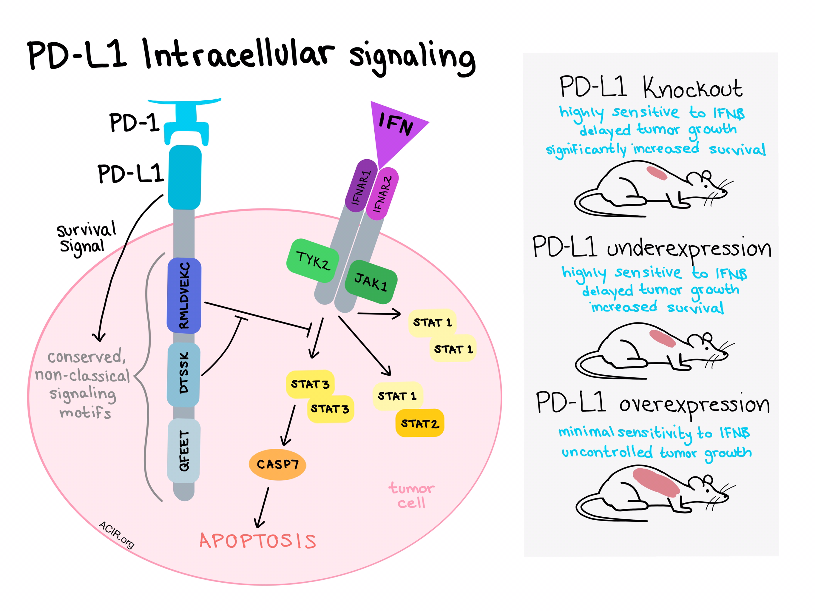
Further experiments showed that when the extracellular domain of PD-L1 engages with PD-1, it can activate an intracellular survival signal. The PD-L1 intracytoplasmic domain then transduces the signal and interferes with IFNβ cytotoxicity. When Gato-Cañas et al. analyzed the intracellular domain in search of signaling domains, however, they found none, suggesting that PD-L1 uses non-conventional signaling motifs.

In an effort to identify the mysterious signal transduction functional domains, the team compared ten mammalian PD-L1 molecules and found three phylogenetically conserved regions - “RMLDVEKC”, “DTSSK”, and “QFEET”. Experiments in which each of these motifs was deleted showed that RMLDVEKC was essential for PD-L1 to function against IFNβ cytotoxicity in the tumor cell, while DTSSK was found to be a negative regulatory motif of this activity. At least two mutations in the “DTSSK” regulatory motif were hyperactive in blocking the negative regulator, enhancing resistance to IFN-mediated cytotoxicity. Collectively, this suggested that PD-L1 intrinsic signaling within cancer cells would confer resistance to IFNs.

With the signaling domains of PD-L1 established, the next step was to determine how PD-L1 signaling crosstalks with the IFN pathway. IFNs kill cells by first engaging with their receptor, IFNAR1/IFNAR2. This recruits JAK1 and TYK2, which phosphorylate STAT1, STAT2, and STAT3, which then associate into homo- and heterodimers. IFNs also cause caspase-dependent apoptosis. By silencing various components of the known IFN pathway, Gato-Cañas et al. showed that PD-L1 interferes with the IFN pathway by inhibiting STAT3 phosphorylation and preventing STAT3 upregulation of CASP-7, stopping the signal cascade. When CASP-7 was silenced, tumor cells evaded IFNβ-mediated apoptosis. Direct blockade of PD-L1 with an anti-PD-L1 antibody sensitized murine and human cancer cells to IFNβ in vitro, indicating that cancer cells can utilize PD-1/PD-L1 engagement and subsequent signal transduction to counteract IFN toxicity.

The final step in this study was to test the IFN-resistance function of PD-L1 in vivo, independently of its role in the inhibition of T cell function. B16 tumor cells were engineered to over-or under-express PD-L1 or a PD-L1 variant lacking the cytoplasmic domain. Established tumors were then dosed directly with lentivectors expressing IFNβ. In the construct with reduced PD-L1 expression, tumor growth was delayed, and mice with the PD-L1 expression knocked out entirely showed significantly increased survival. In models that overexpressed PD-L1, tumors were protected against IFN-mediated cell death compared to controls.

Collectively, these results suggest that PD-L1 signal transduction contributes significantly to the protection of cancer cells from type I IFNs and that upregulation of PD-L1 serves as an intrinsic tumor defense mechanism against an immune attack, independent of its role in suppression of the T cell response. Blocking the IFN pathway or upregulating PD-L1 helps tumors by favoring their escape from immune attack.

by Lauren Hitchings

References:

Gato-Cañas M., Zuazo M., Arasanz H., Ibañez-Vea M., Lorenzo L., Fernandez-Hinojal G., Vera R., Smerdou C., Martisova E., Arozarena I., Wellbrock C., Llopiz D., Ruiz M., Sarobe P., Breckpot K., Kochan G., Escors D. PDL1 Signals through Conserved Sequence Motifs to Overcome Interferon-Mediated Cytotoxicity. Cell Rep. 2017 Aug 22.

In the Spotlight...

Bispecific T cell engager (BiTE(R)) antibody constructs can mediate bystander tumor cell killing

An EGFR-CD3ε bispecific antibody can mediate bystander killing of EGFR-negative tumor cells (and potentially normal stromal cells) proximal to EGFR-expressing tumor cells via secreted cytokine induction of ICAM-1 and FAS on target cells, allowing activated T cells, expressing LFA-1 and FAS-ligand, to engage and target EGFR-negative cells in vitro and in a reconstructed xenograft model.

Multidimensional pooled shRNA screens in human THP-1 cells identify candidate modulators of macrophage polarization

Surdziel et al. used the human monocytic THP-1 cell line for large-scale shRNA lentiviral library screening of the genes affecting human macrophage M1/M2 polarization, and identified known and novel regulators, including O-linked N-acetylglucosamine transferase, a mediator of M2 polarization and a suppressor of the M1 macrophage phenotype. These results were further validated by CRISPR knockout, small molecule inhibition, and transcriptional comparison of polarized human macrophage marker genes.

The immune system prevents recurrence of transplanted but not autochthonous antigenic tumors after oncogene inactivation therapy

Oncogene inactivation therapy works with a low rate of relapse in tumor transplant models, however, autochthonous tumors often relapse after therapy is discontinued. In a slowly developing autochthonous tumor model, Anders et al. showed that dormant tumor cells are tolerated by the co-evolved host immune system during long-term regression, and that adoptive transfer of effector T cells can eradicate them, preventing relapse.

cGAS-STING and Cancer: Dichotomous Roles in Tumor Immunity and Development

Activation of the cGAS-STING pathway, involving cytosolic DNA sensing and induction of a type I IFN response, promotes potent antitumor effects and is part of the mechanism of action of various anticancer therapeutics. But, cGAS-STING induced inflammation can also mediate tumor growth and metastasis in a tumor type- and tumor stage-specific manner. Ng et al. review the dichotomous role of this pathway in cancer and how it can be adapted for therapeutic use.

Heterogeneous Tumor-Immune Microenvironments among Differentially Growing Metastases in an Ovarian Cancer Patient

This case study examined the differential progression of primary tumor and several metastases during an off-treatment period in a heavily pretreated patient with high-grade serous ovarian cancer. Tumor progression/regression status correlated with T cell infiltration or exclusion, and T cell clonal expansion, but not with mutation burden or neoepitope number, indicating distinct tumor-immune microenvironments coexisting at different metastatic sites within the same patient. (Link to partially patient-produced VIDEO abstract.)

IL-6/STAT3 signaling as a promising target to improve the efficacy of cancer immunotherapy

The proinflammatory cytokine IL-6 activates STAT3 signaling in dendritic cells, reducing antigen presentation via downregulation of MHC class II/HLA-DR (by degradation within the lysosome), and attenuates Th1 CD4+ responses via reduced IL-12 production. The data, thoroughly reviewed by Kitamura et al., suggest that blockade of IL-6 signaling is a promising target for cancer immunotherapy.

Everything New this Week In...

Close Modal

Small change for you. Big change for us!

This Thanksgiving season, show your support for cancer research by donating your change.

In less than a minute, link your credit card with our partner RoundUp App.

Every purchase you make with that card will be rounded up and the change will be donated to ACIR.

All transactions are securely made through Stripe.