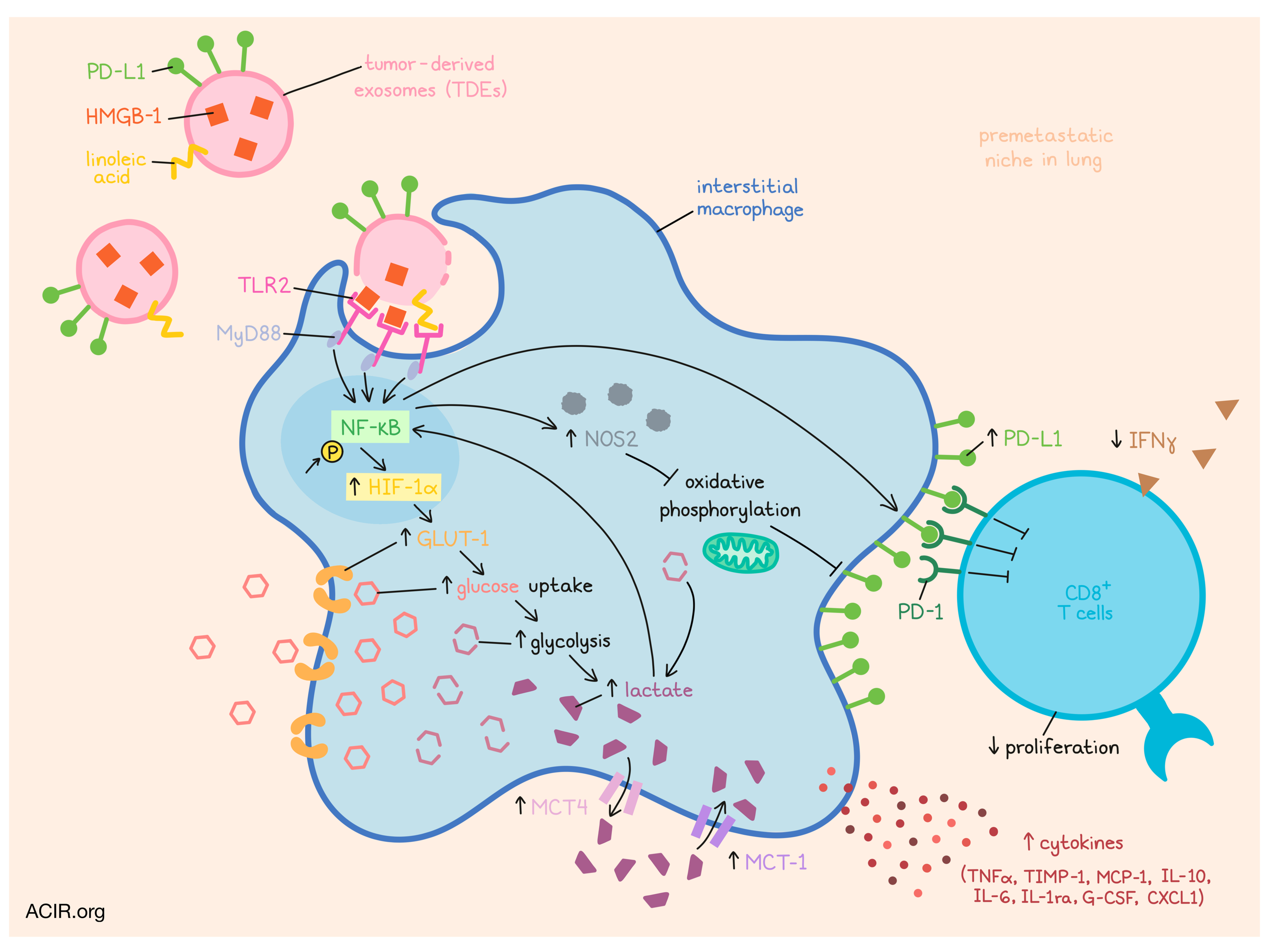
Before cancer makes any metastatic moves, it drives the development of pre-metastatic niches in certain tissues, priming a local environment to support a future tumor. These pre-metastatic niches often contain polarized immunosuppressive macrophages; however, exactly how these macrophages acquire that phenotype is not well understood. In a recent study, Morrissey et al. explored this mechanism and found that tumor-derived exosomes drive metabolic changes in macrophages that polarize them towards an immunosuppressive phenotype characterized by increased PD-L1. Their results were recently published in Cell Metabolism.
To begin, the researchers isolated tumor-derived exosomes (TDEs) from murine Lewis lung carcinoma (LLC) cells and injected them into mice that had previously been injected with s.c. LLC tumors. Mice treated with the LLC exosomes showed increased micro-metastases and increased PD-L1 expression on myeloid cells in the lungs compared to mice treated with control (non-tumor-derived) exosomes. PD-L1 was particularly upregulated in a dose-dependent manner in interstitial macrophages (IMs), which also had the highest uptake of LLC exosomes among lung macrophages.
Looking more into the functionality of macrophages impacted by TDEs, Morrissey et al. found that TDE-stimulated macrophages increased secretion of a variety of cytokines, including TNFα TIMP-1, MCP-1, IL-10, IL-6, IL-1ra, G-CSF, and CXCL1. Arg-1, VEGF, and iNOS expression were also notably increased. When these macrophages were cocultured with CD8+ T cells, the T cells showed reduced proliferation and reduced IFNγ production, which could be partially attributed to PD-1/PD-L1 signaling. Together, these results suggested that TDE-stimulated macrophages were polarized towards an immunosuppressive phenotype.
To better understand how TDEs induce immunosuppressive polarization, the researchers turned their investigation towards toll-like receptors (TLRs) and found that TLR2 was required for the TDE-induced increase in TNFα, IL-6, and PD-L1. HMGB-1 on exosomes was found to be the primary ligand and main agonist responsible for the TDE-induced TLR2-dependent increase in PD-L1, while linoleic acid contributed to a lesser extent.
Given that PD-L1 and TLR ligation are associated with activation of the NF-κB pathway, the researchers investigated this pathway and found that TDEs, but not control exosomes, increased phosphorylation of NFkBp65, and that inhibition of NF-κB abrogated the TDE-induced increase in PD-L1. TDEs also induced an increase in glucose uptake, which further contributed to increased PD-L1. TDE-polarized macrophages also showed increased levels of GLUT-1, HIF-1α, LDHA, and PDK1 validating a role for glycolysis in increased PD-L1 expression, despite normoxic conditions. Looking more closely at this metabolic state, the researchers found that oxygen was being used in the generation of NOS2, which inhibits oxidative metabolism. As oxidative metabolism inhibits PD-L1, the generation of NOS2 would thus support increased PD-L1.
NOS2 production was dependent on MyD88 signaling, but not HIF-1α signaling, suggesting HIF-1α may contribute to PD-L1 upregulation in its own way. The researchers showed that HIF-1α is downstream of NF-κB and contributes to TDE-induced upregulation of GLUT-1 and increased PD-L1. This suggested that while NOS2 inhibits mitochondrial oxidative phosphorylation, HIF-1α helps to drive glucose uptake, both of which support a glycolytic metabolic profile.
Increased glycolysis in TDE-treated macrophages also increased the production of lactate from pyruvate. Both NOS2 and HIF-1α contributed to increased lactate production, which increased PD-L1 and also fed back to increase NF-κB. Increased expression of the lactate exporter MCT4 and the lactate importer MCT-1, and a decrease in PD-L1 when MCT-1 was blocked, suggested that both intra- and extracellular lactate may contribute to this phenotype.
Next, Morrissey et al. investigated whether these in vitro observations held up in vivo. In mice treated with TDEs, they confirmed an increase in glucose uptake, PD-L1, HIF-1α, and MCT-4 in lung macrophages. In mice bearing primary tumors, IM populations were dramatically expanded and showed upregulated PD-L1 within pre-metastatic environments. Within these tissues, the team also saw decreased monocytes and increased MDSCs. CRISPR/Cas9 knockdown of RAB27a, a protein critical for exosome secretion, reduced these effects. Instead MDSCs decreased, percentages of CD4+ and CD8+ T cells increased, PD-L1 and PD-1 decreased, and an overall more active immune profile was induced.
Next, the researchers investigated whether their observations held true for TDEs derived from human tumor cell lines, and found that exosomes derived from several tumor types increased PD-L1 on CD14+ monocytes, and enhanced their capacity to suppress CD4+ and CD8+ T cell responses. As expected, HMGB-1 increased PD-L1, while blocking TLR2, inhibiting NF-κB, or inhibiting glycolysis each reduced PD-L1. Lactate secretion was again dependent on TLR2 stimulation and contributed to PD-L1 expression via the NF-κB pathway.
To further interrogate clinical relevance of this mechanism, the researchers obtained dLN samples from patients that were negative for tumor cell infiltration as a model of a pre-metastatic niche, and LNs from lung transplant donors as healthy controls. The dLNs from patients with cancer showed markers of an activated M2 phenotype, and co-expression of CD206 and PD-L1 was roughly doubled on CD68+ macrophages compared to controls. Coexpression of CD206 and PD-L1 also positively correlated with PD-1 expression on CD8+ T cells in dLNs, and CD206hiPD-L1hi macrophages had increased GLUT-1 expression compared to CD206loPD-L1lo macrophages.
Finally, turning to TCGA data, the researchers found that two exosome release genes, YKT6 and TSG101, had higher expression in primary tumors of lung adenocarcinoma patients with positive compared to negative nodal metastasis; a similar correlation was observed for YKT6 in colon adenocarcinoma. Further, the top quartile of lung adenocarcinoma patients expressing YKT6 had worse survival outcomes than the bottom quartile. To interrogate this observation experimentally, the researchers transfected A549 lung adenocarcinoma cells with a GFP-YKT6 fusion plasmid and showed that the treated cells secreted 5-fold more exosomes than GFP-transfected cells. In their own NSCLC patient cohort, the team showed that tumor cells expressed increased YKT6 and TSG101 compared to immune cells, and that each showed a trending positive correlation to PD-L1 expression in dLNs.
Overall, Morrissey et al. showed that exosomes derived from primary tumors travel to potential metastatic sites, where they engage macrophages via TLR2, activating NF-κB and leading to increased glucose uptake and increased NOS2 production, both of which contribute to increased glycolysis and lactate production and drive upregulation of PD-L1. This unique metabolic reprogramming polarizes macrophages towards an immunosuppressive phenotype, which contributes to the formation of pre-metastatic niches. Targeting this mechanism at various points could potentially serve to inhibit tumor metastasis and improve outcomes in patients, and warrants further investigation.
By Lauren Hitchings
Meet the researcher
This week, first author Samantha Morrissey and senior author Jun Yan answered our questions.
What prompted you to tackle this research question?
SM: This project started back in 2014 when PD-L1 and immunotherapy were just beginning to take off. At the time, exosome biology was also developing as an emerging field. There had been a few papers discussing a role for tumor-derived exosomes in macrophage activation, but none linking the fields of immune checkpoint blockade and exosome science together, particularly within the context of pre-metastatic niche formation. Once we discovered that tumor-derived exosomes increase PD-L1 expression on macrophages, we sought to learn by what mechanism. One of the major mechanisms that governs macrophage phenotype and function is metabolism. Therefore, given the short timeframe in which exosomes influence PD-L1 expression, we hypothesized that there could be a metabolic link to the phenotype.
JY: My laboratory focuses on the understanding of immune cell infiltrates in the tumor microenvironment, particularly innate immune cells. Certainly it was intriguing to know how these innate cells also promote tumor metastasis. I discussed this project with Samantha to see whether tumor-derived exosomes play a critical role in this process.
What was the most surprising finding of this study for you?
SM: To be completely honest, I originally thought that tumor-derived exosomes were going to polarize macrophages to a classic M2 phenotype. We had started off the project with the immunosuppression assays and cytokine profiling. Given the suppressive phenotype coupled with the increased IL-10 production, I was confident that moving into the metabolic assays, we would see a predominant reliance on mitochondrial oxidative phosphorylation for energy production. The glucose consumption data was the first result that suggested our hypothesis might be incorrect. I was even further surprised by the oxygen consumption Seahorse data. We repeated those experiments multiple times to ensure the data were valid, because it was such a striking and unexpected result. I think the Seahorse experiments were a real turning point in the paper – one that highlighted just how important of a role metabolism was going to play in our story.
JY: When Samantha showed me that macrophages stimulated with tumor-derived exosomes displayed a glycolytic dominant metabolic profile, I was surprised, since this data was opposite to most of reports in the literature. We termed these macrophages as immunosuppressive “non-classical M1” macrophages. Another surprising finding was to see the phenotypic changes in myeloid cells from draining lymph nodes of non-small cell lung cancer patients. These patients did not have tumor metastasis in the lymph nodes by pathology report. These data imply that early detection of tumor metastasis cannot fully rely on the pathology. It may be necessary to have comprehensive immunophenotyping on these tissues.
What was the coolest thing you’ve learned (about) recently outside of work?
SM: I recently visited The Ernest Hemingway Home and Museum in Key West, Florida. Aside from its iconic literary owner, the home is famously known for the polydactyl (six-toed) cats who live there. Back in the day, six-toed cats were considered auspicious on ships, and Hemingway, being a very superstitious fellow, convinced a sea captain to give him a polydactyl white cat, appropriately named Snow White, for luck. The property is now home to over 60 polydactyl cats, some of whom are the living descendants of the original Snow White. This historic site provides a nice and beautiful blend of genetics, literature, and American history.
JY: I love fishing and cooking. I enjoy outdoor activities as a way to relax myself. Any movements in my fishing rod make me excited. I also enjoy reading biography books such as Code Breaker, written by Walter Isaacson.


