As one of the the main components of tumor stroma, cancer-associated fibroblasts (CAFs) play essential roles in the tumor microenvironment (TME), including in remodeling of the extracellular matrix, metastasis, and inflammation, making them interesting therapeutic targets. However, much remains unknown about their phenotypes, functions, and relationships with the immune infiltrate. In work recently published in The EMBO Journal, Wu et al. explored the phenotypes of stromal cells, using single-cell RNAseq (scRNAseq), immunohistochemistry (IHC), signaling network analysis, and functional studies to reveal key insights into the role of these stromal cell in the setting of triple-negative breast cancer (TNBC).
Wu et al. began by using scRNAseq to assess non-tumor cells in primary tumors from five patients with TNBC. The researchers compared their scRNAseq-obtained stromal and immune clusters with published cell type signatures from the XCell database using an area-under-the-curve (AUC) approach to annotate and remove immune cells. This left endothelial cells and two clusters enriched for fibroblast-like cells that shared stromal markers such as PDGFRB, S100A4, ITGB1, and THY1. Reclustering of these two stromal clusters resulted in two main fibroblast and perivascular-like (PVL) lineages, each containing two major cell subsets.
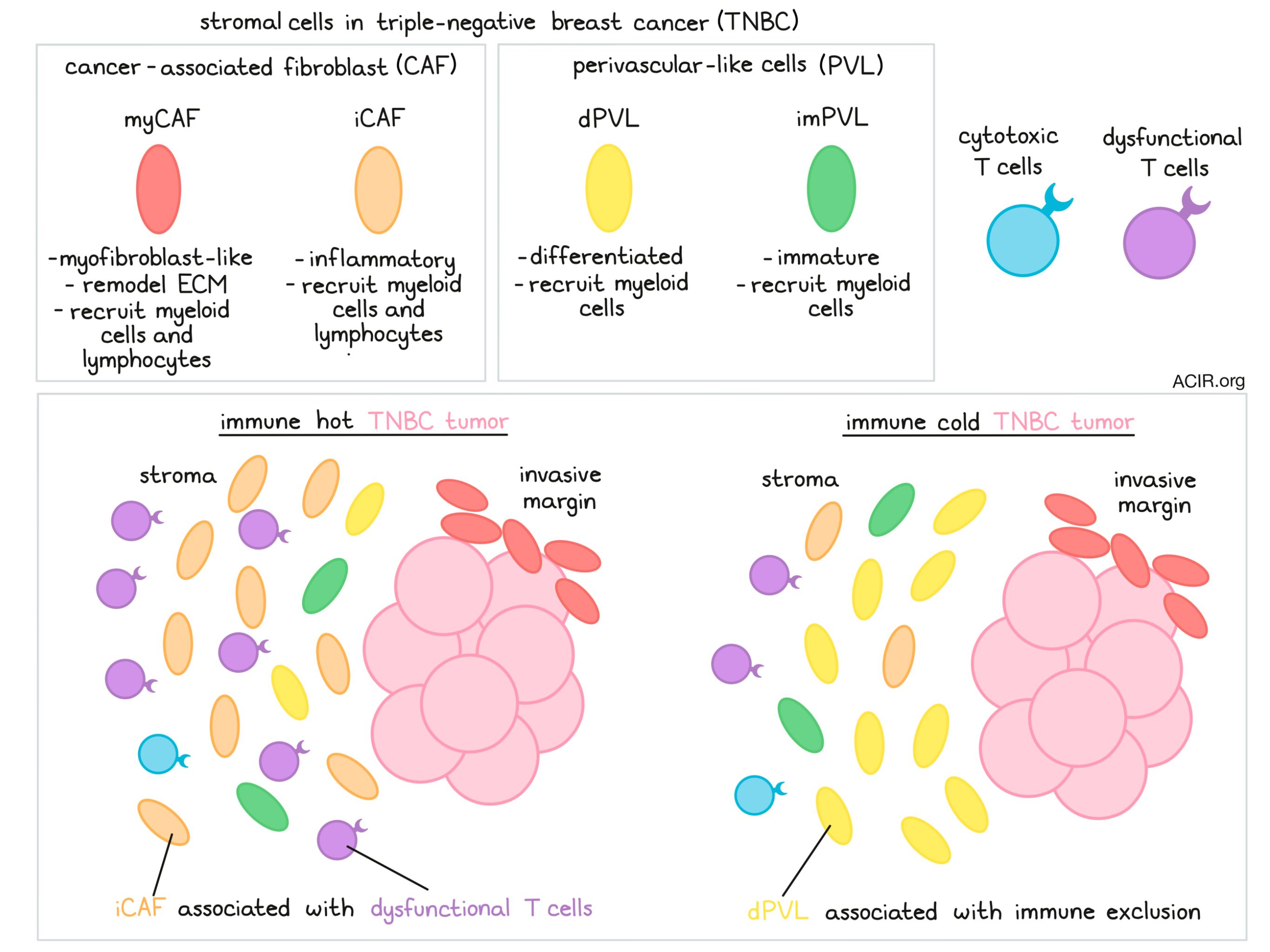
The first fibroblast lineage subset consisted of CAFs with high expression of markers associated with activated fibroblasts (ACTA2, FAP, PDPN) and collagen-related genes (COL1A1 and COL1A2), which were classified as myofibroblast-like CAFs (myCAFs). The second subset resembled inflammatory CAFs (iCAFs) and was enriched for CXCL12 and CXCL13. Similar myCAF and iCAF phenotypes were found within previously published gene signatures of pancreatic ductal adenocarcinoma (PDAC). Interestingly, there was not a perfect correspondence of marker enrichment, and a few of the PDAC CAF markers were detected in the opposing annotated phenotype in TNBC. No signature of antigen-presenting CAFs, as found in PDAC, were observed in TNBC, which could be due to the rarity of this subset, or these cells could be unique to PDAC.
The second stromal cluster was enriched for perivascular markers, such as MCAM, a marker used to differentiate perivascular cells from fibroblasts. These PVL cells also formed two main clusters; one that could be annotated as differentiated-PVL (dPVL), enriched for myogenic differentiation genes, and a second cluster that was enriched for genes associated with an immature phenotype and was classified as immature-PVL (imPVL). Both PVL clusters were also enriched for the PDAC myCAF signature, suggesting common features in these cells’ genetic profiles. The four stromal clusters were found in all five patients but in varying proportions.
To understand the functional differences between the subsets, Wu et al. performed differentially enriched genes (DEG) and gene ontology (GO) analysis. Collagen biosynthesis and extracellular matrix (ECM) regulatory pathways were enriched in myCAFs, and iCAFs were enriched for developmental signaling pathways and chemotactic regulation, stem cell markers, and EGFR. In contrast, dPVL cells were enriched for muscle system and contractility pathways, and imPVL cells for focal and substrate adhesion pathways. Since many of the identified genes and pathways related to cell activation and contractility, the authors hypothesized that stromal subclusters resemble cell differentiation stages rather than distinct subpopulations. To confirm this, they performed cell trajectory analysis across pseudotime using Monocle and found that relevant marker genes transitioned throughout CAF or PVL differentiation, and transcription factor expression analysis identified unique drivers for each subpopulation.
To assess the functionality of the subsets, the researchers generated cell-derived matrices to compare the ability of each subset to produce collagen using second harmonic generation microscopy. The myCAFs had increased production of collagen and released more uniformly aligned collagen fibers than iCAFs. PVL cells produced the least collagen, suggesting myCAFs were the main regulators of ECM remodeling.
Next, the spatial localization of the four subsets was determined by IHC on formalin-fixed paraffin-embedded (FFPE) blocks from the tumor and adjacent normal tissues of the patients. The myCAFs localized close to the invasive tumor border, while iCAFs were found more distal, in an area that also contained immune cells. PVL cells were detected throughout the stromal regions, with no apparent colocalization to the invasive border. Samples from normal, healthy patient tissue identified the same subpopulations, but in different abundances and/or locations.
Using a published set of curated human ligand-receptor pairs, the researchers investigated how the spatially distinct stromal subclasses interact with other cells in the TME. By constructing a cell-to-cell communication network, they found that myCAFs and iCAFs had the highest predicted ligand activity. To assess whether these stromal subsets might have immunoregulatory properties, Wu et al. predicted ligand–receptor interactions of stromal and immune cells, focusing on cytokines and checkpoint molecules. They found an enriched interaction between iCAFs and myeloid cells via complement activation. Additionally, myCAFs and iCAFs had predicted enriched TGFB–TGFBR interactions with myeloid cells, and even though PVL cells had lower ligand expression profiles, multiple interactions with myeloid cells were predicted. These data suggested that stromal cells might be involved in the recruitment of myeloid cells – potentially immunosuppressive myeloid cells – into the TME.
Assessing lymphoid cell interactions, Wu et al. found that chemoattractant pathways involving CXCL12–CXCR4 and CXCL13–CXCR5 were strongly upregulated in iCAFs. In myCAFs, enrichment for secreted immunoregulatory and checkpoint molecules were also found, including CXCL9–CXCR3, CXCL11–CXCR3, and PD-1–PD-L1. Therefore, these CAF populations might be involved in the recruitment of T and B cells into the TME, and their functionality.
To further explore this, Wu et al. assessed the association between stromal gene signatures and immune content in three large, independent, published TNBC cohorts with associated bulk gene expression data. They used the computational model TIDE (tumor immune dysfunction and exclusion), which evaluates cytotoxic T lymphocyte (CTL) levels in each sample based on gene expression, to stratify patients into high and low CTL groups. These data were used to study two main mechanisms of immune invasion: CTL dysfunction and exclusion.
Only iCAFs were enriched for prognostic genes associated with CTL dysfunction. In patients with low levels of the iCAF signature, high CTL infiltration had a survival benefit. In contrast, in patients with high iCAF signatures, CTL levels were not prognostic, consistent with dysfunction of these cells. The researchers confirmed the heterogeneity of dysfunctional CD8+ T cells within their scRNAseq dataset using the AUC approach to score a published T cell exhaustion signature, including PDCD1, LAG3, TIGIT, and CTLA4.
Second, the researchers focused on CTL exclusion, often referred to as a cold “immune-desert” phenotype. The collective bulk stromal signature correlated negatively with CTL levels in four TNBC cohorts, but only dPVL independently correlated with lower CTL levels in three of the four cohorts. To further confirm these findings, the researchers analyzed the T cell infiltrate by IHC in the five patients for which scRNAseq data was available. Similar to the larger datasets, they found that the two patients with exceptionally low CTL infiltration had the highest proportions of dPVL profiles, while the patient with the highest CTL content had the lowest proportion of dPVL.
In conclusion, by scRNAseq Wu et al. confirmed that the stroma of TNBC tumors contained four subsets of stromal cells that could be split into two fibroblast (myofibroblast-like and inflammatory) and two perivascular-like phenotypes (immature and differentiated). The specialized stromal cell phenotypes had variable functionality and localization and were associated with signatures predictive of tumor immune evasion, with a low inflammatory CAF signature in patients with high CTL infiltration correlating with improved survival. These data, once further confirmed in larger studies, might aid in designing combinatory treatment strategies targeting stromal cells.
Write-up by Maartje Wouters, image by Lauren Hitchings
Meet the researcher
This week, first author Sunny Wu answered our question.

What prompted you to tackle this research question?
We have known for quite some time now that solid tumors are actually a complex ‘ecosystem’ of diverse cell types, including recruited host stromal cells, which support many aspects of cancer progression. Although these stromal cells, such as fibroblasts, are considered to be strong candidates for targeted therapy, they have yet to make significant advances in the clinic for patient care. These developments have been impeded by a poor understanding of the functional heterogeneity of stromal cells, where our knowledge has been largely derived from more traditional low-resolution methods and model in vitro and in vivo experimental systems. Through close clinical collaborations, we sought to apply the latest single-cell sequencing methods to powerfully study the transcriptome of individual stromal cells directly in patient tumors. We hypothesised that diverse subclasses of stromal cells are found in the tumor microenvironment of breast cancers, each with distinct roles in promoting tumorigenesis and helping aid the evasion of the immune system.
What was the most surprising finding of this study for you?
Although studies profiling the stroma of breast cancers often focus on fibroblasts, we were surprised to find that it was also comprised of functionally distinct perivascular cells, including pericytes and smooth muscle cells. Interestingly, these perivascular cells were not necessary localised to blood vessel regions, where we might expect them to be from their classical roles in supporting the function of the vasculature. Rather, they could be found dispersed or ‘lost’ in the tumor stroma alongside fibroblasts. As they also share many common markers with fibroblasts, it suggests the strong possibility that they have been previously misclassified in the literature. Furthermore, we found an association between these perivascular cells and tumors that have a low T cell infiltration. We hypothesise that this may be related to dysfunctional blood vessel structures with low perivascular coverage and integrity, which may prevent the extravasation (movement) of T cells into tumor tissues. We are now investigating this in more detail using experimental models!
What was the coolest thing you’ve learned (about) recently outside of work?
I’ve learnt that I absolutely love the simpler things in life. A juicy podcast during a walk in the sun, a glass of whiskey with a book by the fire, a bush walk through nature, gazing up at the stars on a clear night, and switching my phone off to enjoy some quiet. I’ve also found my love for cooking again, and have learnt a few new recipes that I have cooked for my partner, family, and mates. My faves include making wontons from scratch, a nice fish fillet & shrimp smoked under the fire, and juicy little vego Japanese curry


