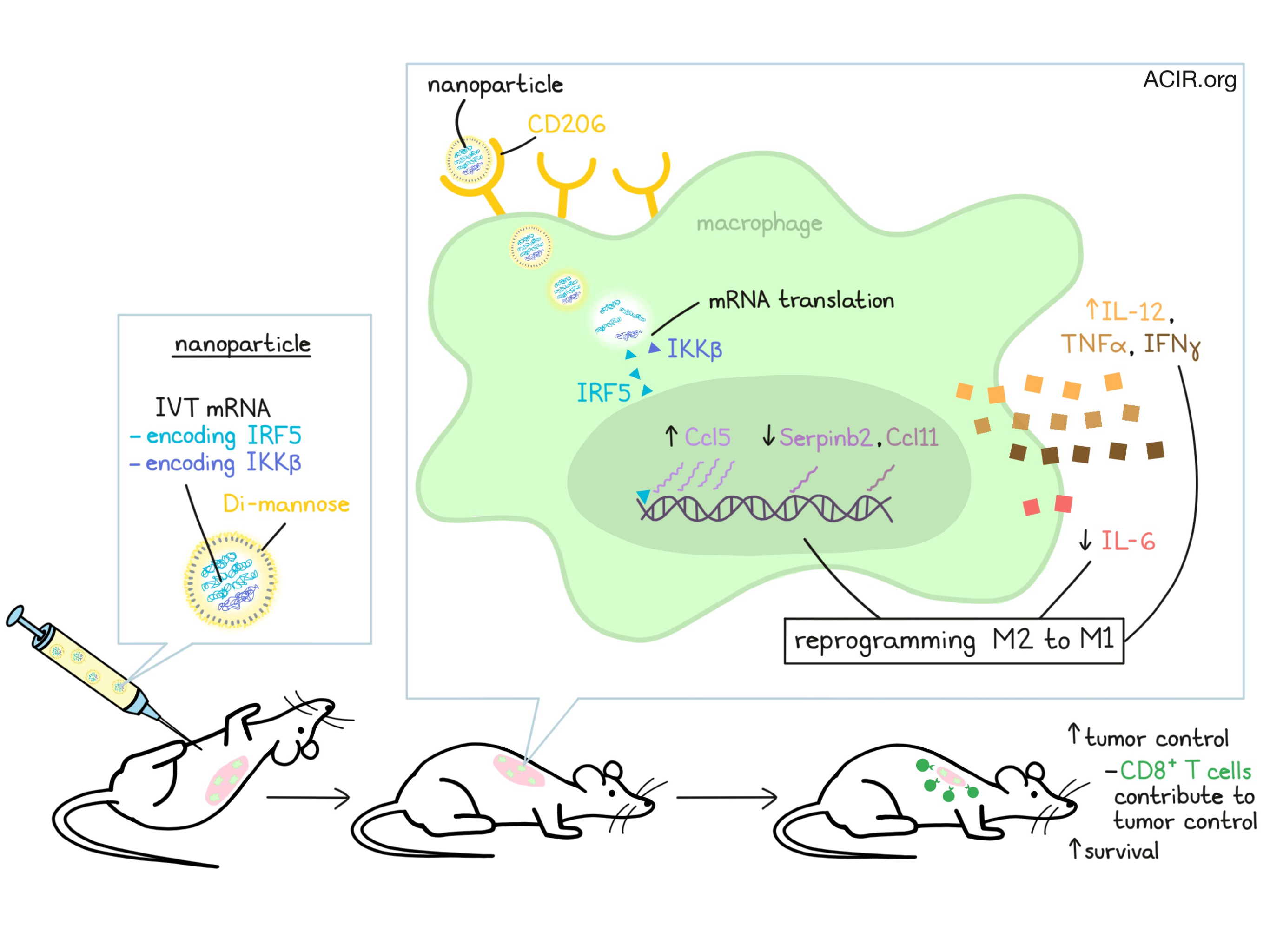
An immunosuppressive tumor microenvironment is one of the biggest hurdles to successful immunotherapy. In many tumors, the presence of M2-polarized macrophages is to blame for immunosuppression. For Zhang et al., the possibility of reprogramming macrophages from the pro-tumor M2 phenotype to a pro-inflammatory M1 phenotype without inducing systemic inflammation was the goal in their recent study, published in Nature Communications.
In order to reprogram macrophages from an M2 to an M1 phenotype, Zhang et al. developed targeted nanocarriers that deliver in vitro-transcribed (IVT) mRNA encoding M1-polarizing transcription factors to phagocytic cells expressing CD206, a mannose-binding lectin upregulated on tumor-associated macrophages. The researchers chose to encode the IRF regulatory factor 5 (IRF5), which can drive macrophage polarization towards M1. They also encoded IKKβ, an activating kinase for IRF5. Packaging these mRNAs at a 3:1 ratio in nanoparticles decorated with Di-mannose and delivering them to macrophages in vitro induced expression of the intended proteins. As expected, this expression was transient, though IRF5 levels did remain upregulated for up to 5 days before returning to baseline.
To determine whether induced expression of IRF5 and IKKβ effectively reprogrammed macrophages from M2 to M1, researchers generated M2 macrophages by culturing bone marrow-derived macrophages (BMDMs) with IL-4. When M2 macrophages were transfected with the IVT mRNA-loaded nanoparticles, gene expression profiles revealed a shift from M2 to a phenotype that more closely resembled the phenotype of inflammatory macrophages (generated separately by culturing BMDMs with TLR4). Signature M2 genes like Serpinb2 and Ccl11 were downregulated following nanoparticle treatment, while signature M1 differentiation genes, like Ccl5, were upregulated.
To test whether macrophage reprogramming was effective in vivo, Zhang et al. tested their IVT mRNA-loaded delivery system in a mouse model of late-stage, unresectable ovarian cancer. Compared to PBS control and a nanoparticle control, weekly intraperitoneal delivery of nanoparticles loaded with IRF5/IKKβ IVT mRNA induced disease regression in all mice, with eventual tumor clearance and long-term survival in 40% of treated animals. The overall median survival in treated mice was 142 days, compared to 60 days in controls.
Zhang et al. next went on to investigate the mechanism by which IRF5/IKKβ mRNA nanoparticles mediated tumor control. Biodistribution testing following intraperitoneal delivery showed that nanoparticles typically concentrated in organs within the peritoneum, though some entered systemic circulation. As expected, they found that the nanoparticles preferentially transfected cells expressing CD206, with macrophages and monocytes being the primary targets of the nanoparticles, and with some transfection of DCs and neutrophils.
Phenotypic and functional analysis of macrophages and monocytes from the peritoneum of mice following multiple doses of mRNA NPs showed that the population of immunosuppressive macrophages reduced from an average of 43% in controls to 2.5% in treated mice. Meanwhile, M1-like macrophage populations were found to increase from an average of 0.5% in control mice to 10.2% in mice treated with IRF5/IKKβ mRNA nanoparticles. Treated mice also showed an increase in inflammatory monocytes from 4.5% to 73.4%. Analysis of cytokine secretion identified an increase in the release of proinflammatory/antitumor cytokines including IL-12, IFNγ, and TNFα, and a decrease in the release of IL-6, which is associated with differentiation towards M2.
In addition to the changes observed in macrophage and monocyte populations, the researchers also identified dense clusters of lymphocytes within or surrounding the tumor mass. Depletion of CD8+ T cells reduced, but did not completely abrogate the antitumor activity observed with IRF5/IKKβ mRNA nanoparticle treatment, indicating that CD8+ T cells as well as other factors contribute to antitumor immunity. The researchers did not observe evidence of inflammation, necrosis, or systemic toxicity, suggesting that nanoparticle delivery is safe for repeated dosing.
Next, Zhang et al. explored whether their nanoparticles could be delivered systemically to control disseminated disease. Using a mouse model of pulmonary melanoma metastases, researchers confirmed that tumor engraftment coordinated with phagocyte accumulation in tumors. Intravenous infusion of IRF5/IKKβ mRNA nanoparticles showed preferential biodistribution to organs with a high number of resident macrophages/phagocytes. Treatment reduced tumor burden in the lungs, reduced the number of individual metastases, and improved overall survival. Flow cytometry revealed a shift from immunosuppressive macrophages towards activated phagocytes. No obvious signs of toxicity or systemic inflammation were observed.
Taking on a more challenging tumor model, Zhang et al. established gliomas in mice, a model with a very prominent M2-like macrophage component. As a monotherapy, IRF5/IKKβ mRNA nanoparticles only moderately slowed tumor progression. In combination with standard-of-care radiotherapy, however, tumor growth was substantially reduced. The addition of nanoparticles increased survival compared to radiotherapy alone, and more than doubled survival compared to untreated mice.
To determine whether their nanoparticle delivery system could be used to reprogram human macrophages, the researchers generated M2 macrophages from the human monocytic THP-1 cell line and treated them with IVT mRNA loaded nanoparticles. The nanoparticles effectively transfected human macrophages, leading to increased secretion of IL-1β and increased surface expression of the M1 macrophage marker CD80.
Overall, Zhang et al. conclude that nanoparticle delivery of IVT mRNA encoding IRF5 and IKKβ could be used to reprogram M2 macrophages to an M1 phenotype in support of antitumor immune responses. Based on in vitro evidence and evidence in models of ovarian cancer, melanoma, and glioma, the researchers are planning to launch a phase I clinical trial in which nanoparticles will be delivered intraperitoneally to patients with chemotherapy-resistant ovarian carcinoma.
by Lauren Hitchings
Meet the researcher
This week, first author Fan Zhang answered our 3 questions.

What prompted you to tackle this research question?
Since my graduate school, I've been fascinated with the intrinsic property of nanoscale materials, such as polymeric nanoparticles, to interact with the immune system. After joining my postdoc position, I always think about how to use this property of nanomaterials to deliver therapeutic reagents to the immune cells and prime the immune system against cancer. In this project, we designed a nanocarrier that could successfully deliver therapeutic mRNAs to the tumor-associated macrophages in a targeted manner. These mRNAs, once released into the cytoplasm of macrophages, could translate into transcription factors that reprogram macrophages into tumor-clearing cells.
What was the most surprising finding of this study for you?
It was amazing to see that 40% of the ovarian tumor mice had tumor-free survival without significant systemic toxicity after receiving multiple doses of these macrophage-reprogramming nanoparticles. Similar approaches that are either in the clinics or under clinical trials suppress tumor-associated macrophages in the whole body, leading to the disruption of homeostasis in the immune system.
What was the coolest thing you’ve learned (about) recently outside of work?
I've had some time recently to really think about the future direction of my research and how to build a research program that focuses on using nanotechnology as an approach to modulate the immune activity against diseases. This has let me appreciate that sometimes it is important to find opportunities to take a break from your work and reorganize yourself.


