Weekly Digests
‹ Back to October

What’s happening under the hood: The cellular kinetics of a CAR T cell therapy

October 25, 2017

Therapies in which T cells are engineered to express a chimeric antigen receptor (CAR) have proven to be incredibly effective against certain hematological malignancies. In recent months, two CAR T cell therapies have earned FDA approval, and it is likely this groundbreaking strategy will continue to expand. In an effort to understand the patterns of behavior of CAR T cell therapy within patients, Mueller et al. analyzed data from patients who had received the genetically-modified autologous T cell therapy tisagenlecleucel (CTL019) as a treatment for CD19+ relapsed or refractory chronic lymphocytic leukemia (CLL) or acute lymphoblastic leukemia (ALL). CTL019 is a cellular therapy in which a patient’s own T cells are engineered ex vivo with a transgene for a CD19-targeting CAR and re-infused into the patient.

Differing from a pharmacokinetic analysis, which tracks the behavior of traditional molecular drugs within a system, Mueller et al.’s “cellular kinetic analysis”, published in Blood, accounts for the in vivo proliferation of CTL019. The researchers analyzed data from a cohort of 103 pediatric and adult patients with ALL and CLL. They tracked CTL019 levels over time using two methods: qPCR tracked integration of the transgene into the genome of CD3+ cells, while flow cytometry measured surface expression of the CAR construct. Data from the two methods generally agreed, though in some patients there was evidence of integration of the transgene but not of functional expression of the CAR, suggesting some hindrance to either transcription or translation of the transgene.

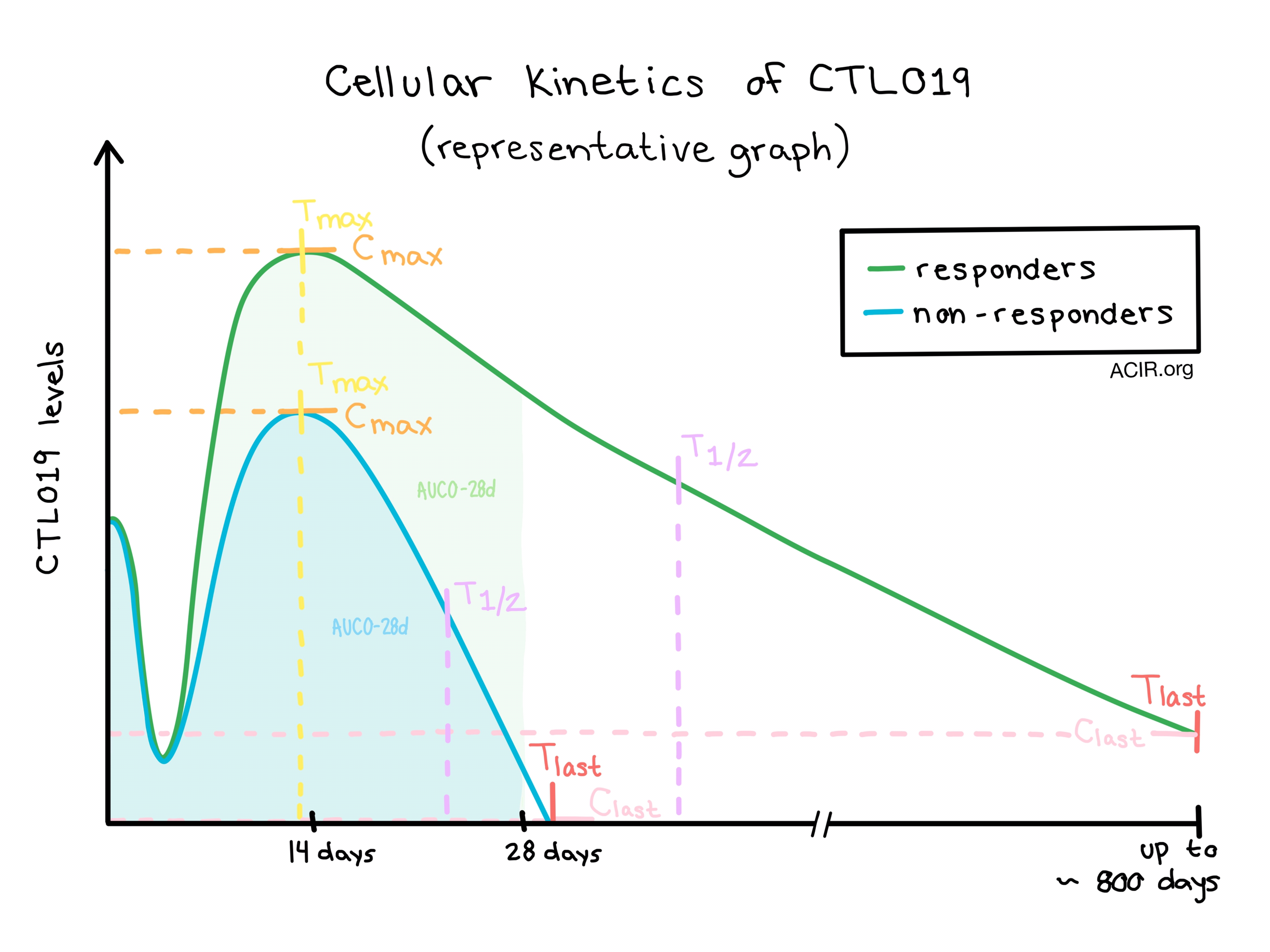
The team tracked parameters of expansion, including maximum concentration (Cmax) and time of maximum concentration (Tmax), and parameters of persistence, including the terminal half life (T1/2), last measured concentration (Clast), and time of last measured concentration (Tlast). They also calculated the area under the curve for days 0-28 (AUC0-28d) as a measure of cumulative exposure to the therapy in the first 28 days following infusion.

Though there was variation between individual patients and subgroups, the cellular kinetics of CTL019 generally followed the same basic trend: immediately following intravenous infusion, CTL019 levels were high, then swiftly but transiently declined, possibly as cells distributed throughout the peripheral blood, bone marrow, and other tissues. CTL019 cells then expanded, reaching Cmax within two weeks of the initial infusion and declined slowly over weeks, months, or years.

Significant differences were observed between responders (patients with complete response, or complete response with incomplete blood count recovery) and non-responders; on average, responders had higher Cmax values indicating greater expansion, and higher T1/2 and Tlast values, indicating longer persistence. Patients who experienced a complete response generally had high Cmax and AUC0-28d values, and patients with longer persistence tended to maintain longer event-free survival.

While expansion of and exposure to CTL019 cells generally correlated with positive clinical outcomes, it is also worth noting that patients with more exposure to therapy were also more likely to experience cytokine release syndrome (CRS) and suffer from neurological symptoms. Measurements of serum cytokine levels showed that cytokines including IFN-γ and IL-6 increased following infusion and remained high in patients who experienced CRS. Interestingly, higher pre-infusion tumor burden emerged as a significant predictor of the expansion of CTL019 as well as of CRS severity in patients with ALL, suggesting that greater exposure of infused CTL019 cells to target cells led to enhanced proliferation and activation. Tocilizumab, which was used to manage CRS in some patients, did not appear to hinder the expansion of CTL019.

As a measure of CTL019 trafficking, Mueller et al. tested for transgene levels in the bone marrow and cerebrospinal fluid and found that CTL019 cells did traffic to these tissues. Similar to results from peripheral blood, higher levels and longer persistence of CTL019 cells in the bone marrow correlated with improved patient outcomes. The observation that CTL019 cells trafficked to the cerebrospinal fluid could prove to be an important finding, as none of the observed patients with pediatric B-ALL patients had central nervous system relapses.

While this study was based on data from a fairly large cohort, differences in age, cancer type, pre-infusion tumor burden, exposure to prior therapies, and missing long-term data from non-responders still leave unanswered questions. The general kinetic trends and the differences in trends observed between responders and non-responders with respect to safety and efficacy, however, were clear and very consistent across diseases, and reflected the proposed mechanism of action of CAR T cells.

by Lauren Hitchings

References:

Mueller K.T., Maude S.L., Porter D.L., Frey N., Wood P., Han X., Waldron E., Chakraborty A., Awasthi R., Levine B.L., Melenhorst J.J., Grupp S.A., June C.H., Lacey S.F. Cellular kinetics of CTL019 in relapsed/refractory B-cell acute lymphoblastic leukemia and chronic lymphocytic leukemia. Blood. 2017 Sep 21.

In the Spotlight...

Tumor and Microenvironment Evolution during Immunotherapy with Nivolumab

Riaz et al. examined the effects of 4 weeks of nivolumab therapy in ipilimumab (IPI)-naïve or -progressor patients stratified according to good or poor response to nivolumab. In responding patients, mutation and neoantigen load contracted, presumably due to therapy-induced immunoediting, and the intratumoral TCR repertoire expanded in number (IPI-progressors) or became more unequally distributed (IPI-naïve), suggesting antigen-driven changes in the T cell population.

Interleukin-10-regulated tumour tolerance in non-small cell lung cancer

In patients with NSCLC, increased expression of IL-10 in the region surrounding the tumor positively correlated with tumor diameter. In the subset of patients with adenocarcinoma (ADC), tumor-infiltrating Foxp3+ Treg and tumor cells expressed IL-10R, which inversely correlated with PD-L1/PD-1 expression; in an ADC cell line, IL-10 downregulated PD-1, PD-L1, and FAS/FASL, indicating that IL-10/IL-10R expression may be linked to PD-1/PD-L1 blockade resistance. Meanwhile, in patients with squamous cell carcinoma, IL-10R directly correlated with PD-L1 expression.

Hypothesis: stimulation of trained immunity as adjunctive immunotherapy in cancer

This review by Netea et al. serves as both a general history of cancer immunotherapy and a call to acknowledge and study trained immunity (TI) – the paradigm-shifting idea that the innate immune system (primarily macrophages and monocytes) is capable of immunological memory, enabled by epigenetic and metabolic reprogramming that induces enhanced long-term function against infection. They propose a future research agenda to assess whether TI could enhance cancer immunotherapy.

IL-10 release upon PD-1 blockade sustains immunosuppression in ovarian cancer

Blockade of PD-1 on murine ovarian tumor-infiltrating dendritic cells (TIDC) led to an increased release of IL-10, which in turn increased PD-1 expression on TIDCs, creating an immune escape feedback loop. While either monotherapy was ineffective, combination of anti-PD-1 and anti-IL-10/IL-10R prolonged survival by enhancing the infiltration of activated tumor-specific T and B cells and by reducing the recruitment of myeloid-derived suppressor cells into the ascites of tumor-bearing mice.

Tumor-associated myeloid cells: new understandings on their metabolic regulation and their influence in cancer immunotherapy

Hijacking of key metabolic and tissue repair activities of the abundant tumor-infiltrating myeloid cells is another example of adaptive immune suppression. Porta et al. reveal the cooperative metabolic programming between tumor and myeloid cells to reduce inflammatory responses, including T cell anti-tumor activity, and strengthen the pro-tumor environment, affecting energy substrates and metabolites and leading to epigenetic effects on gene expression.

An anti-glypican 3/CD3 bispecific T cell-redirecting antibody for treatment of solid tumors

Ishiguro et al. developed a humanized IgG T cell-redirecting antibody bispecific for CD3 and glypican 3, a highly tumor-specific antigen expressed on a variety of solid tumors. They showed that the antibody was effective even against tumors with low immunogenicity, which are unresponsive to checkpoint therapy. Aside from a manageable and reversible transient cytokine increase, the safety profile was favorable in monkey studies. Phase I clinical trials are ongoing.

The 2017 complete overhaul of adjuvant therapies for high-risk melanoma and its consequences for staging and management of melanoma patients

Recent “game-changing” melanoma clinical trials demonstrated that adjuvant nivolumab was superior to adjuvant ipilimumab in resected stage IIIB/C-IV patients (regardless of BRAF status) with respect to efficacy and safety, dabrafenib+trametinib was superior compared with placebo in stage III patients with BRAF mutations, and IFN-α therapy was beneficial only in patients with ulcerated melanoma. Collectively, these results are predicted to change clinical practice, shifting away from the use of ipilimumab and IFN-α in the adjuvant setting.

A drug development perspective on targeting tumor-associated myeloid cells

Tumor-associated myeloid cells (M2 macrophages and myeloid-derived suppressor cells) represent the most abundant yet heterogeneous and plastic immune populations within tumors, and their complexity is only beginning to be unraveled. Majety et al. review and comment on the clinical strategies (depletion, recruitment, reprogramming) that are being pursued to reverse the potent immunosuppressive effects of this cell population.

Agonist OX40 immunotherapy improves survival in glioma-bearing mice and is complementary with vaccination with irradiated GM-CSF-expressing tumor cells

Using a challenging intracranial glioma model, Jahan et al. demonstrate that combination of a vaccine (irradiated tumor cells and GM-CSF-expressing K562 cells) with an agonist-OX40 antibody improved survival, increased the number of Th1 polarized splenocytes (particularly granzyme B-expressing CD4+ cells), shifted the brain infiltrating CD4+ T cells to a Th1 phenotype, increased the CD8+/Foxp3+ ratio, and prevented exhaustion as monitored by co-expression of both PD-1 and either TIM-3 or LAG-3 on intratumoral T cells.

Expression of LAG-3 defines exhaustion of intratumoral PD-1+ T cells and correlates with poor outcome in follicular lymphoma

Yang et al. found that intratumoral PD-1+ T cells in follicular lymphoma are heterogeneous, with the LAG-3- subset being activated and functional, and the LAG-3+ subset being functionally exhausted and correlated with poor outcome. IL-12, which is elevated in lymphoma patients, upregulated and maintained LAG-3 expression on T cells. Dual blockade of PD-1 and LAG-3 improved the functionality of tumor-infiltrating CD8+ T cells ex vivo better than either blockade alone.

Everything New this Week In...

Close Modal

Small change for you. Big change for us!

This Thanksgiving season, show your support for cancer research by donating your change.

In less than a minute, link your credit card with our partner RoundUp App.

Every purchase you make with that card will be rounded up and the change will be donated to ACIR.

All transactions are securely made through Stripe.