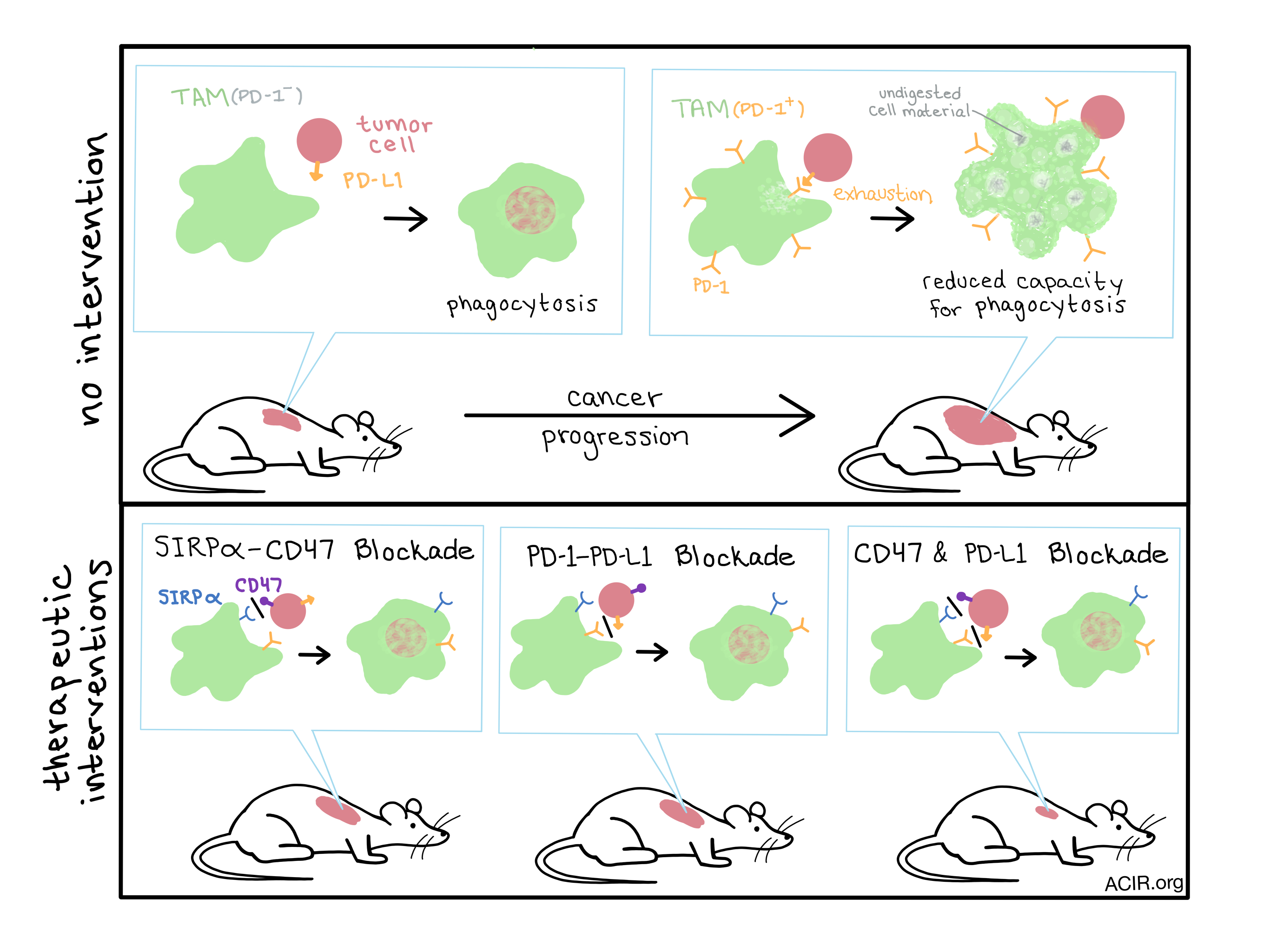
The presence of tumor-associated macrophages (TAMs) in a tumor microenvironment (TME) is usually associated with worse outcomes for patients. However, recent research and ongoing clinical trials have demonstrated that therapies blocking the interaction between SIRPα and CD47 can induce TAMs to phagocytose tumor cells, providing therapeutic benefit.
Gordon et al. set out to explore other mechanisms that might regulate the function of macrophages in the TME. Knowing that macrophages have been shown to express PD-1 in some instances of infection, the team wondered whether the same could be true of macrophages in tumors. Using a murine colon cancer model, they found about 50% of the tumor-infiltrating macrophages expressed surface PD-1, confirming their hypothesis.
TAMs are typically distinguished as either inflammatory “M1” or pro-tumor “M2” macrophages. Using flow cytometry, the researchers found that while most PD-1- TAMs resembled M1 cells, almost all PD-1+ TAMs expressed an M2 phenotype, indicating that PD-1 expression is likely associated with pro-tumor traits. The researchers also noted that the PD-1+ TAM population only began to emerge two weeks after tumor engraftment and then accumulated over time during tumor progression. Similarly, in human colorectal cancer samples, the frequency of PD-1+ macrophages within the tumor increased with disease stage and was associated with an M2 phenotype. The time-dependent increase in PD-1+ TAMs was due to circulating bone marrow-derived macrophages homing in on the inflammatory TME rather than from the expansion of resident immune cells, as shown with bone marrow transplantation experiments.
Compared to their PD-1- counterparts, PD-1+ TAMs showed altered expression patterns and morphology. They expressed more M2-associated scavenger receptor CD206, less MHC class II, and more CD111c. Stained PD-1+ TAMs appeared large and foamy, indicating that they were filled with vacuoles and/or lipids, and electron microscopy confirmed that this foamy appearance was due, at least in part, to an accumulation of uncleared phagocytic material and lysosomes in the cytoplasm.
A series of ex vivo and in vivo experiments revealed that, much like its role in T cells, PD-1 expression on TAMs induced ‘exhaustion’ and limited the effector functions of activated TAMs by decreasing their ability to phagocytose tumor cells. Experiments in mice lacking an adaptive immune system confirmed that this decrease in phagocytosis by PD-1+ TAMs could be overturned by a knockout of PD-L1 in the tumor. Mice whose tumors did not express PD-L1 also showed reduced tumor burden after three weeks compared to mice with tumors that overexpressed PD-L1.
The researchers next treated mice with either a PD-1 or PD-L1 blockade and observed a significant reduction in tumor growth. Depletion of TAMs repealed this effect, indicating the direct role of TAMs in the antitumor efficacy. Further, the researchers tested these blockades in combination with anti-CD47 immunotherapy, which is already being tested in early clinical trials, to explore how the two therapy types would interact. Individually, the PD-L1 and CD47 blocking therapies equivalently reduced tumor size, while the combination of the two therapies showed a trend toward greater reduction of tumor burden and greater survival benefit.
Although blockade of the PD-1 axis is typically considered a T cell-directed therapy, the PD-1 expression observed on B cells, dendritic cells, natural killer cells, and now on tumor-associated macrophages, indicates that this axis is important to both the innate and adaptive arms of the immune system. The breadth of cell types expressing PD-1 may explain the effects of PD-1/PD-L1 inhibition in situations where T cell activity might be compromised.
by Lauren Hitchings


