Weekly Digests
‹ Back to May

Dendritic cells go viral to achieve immortality

May 16, 2018

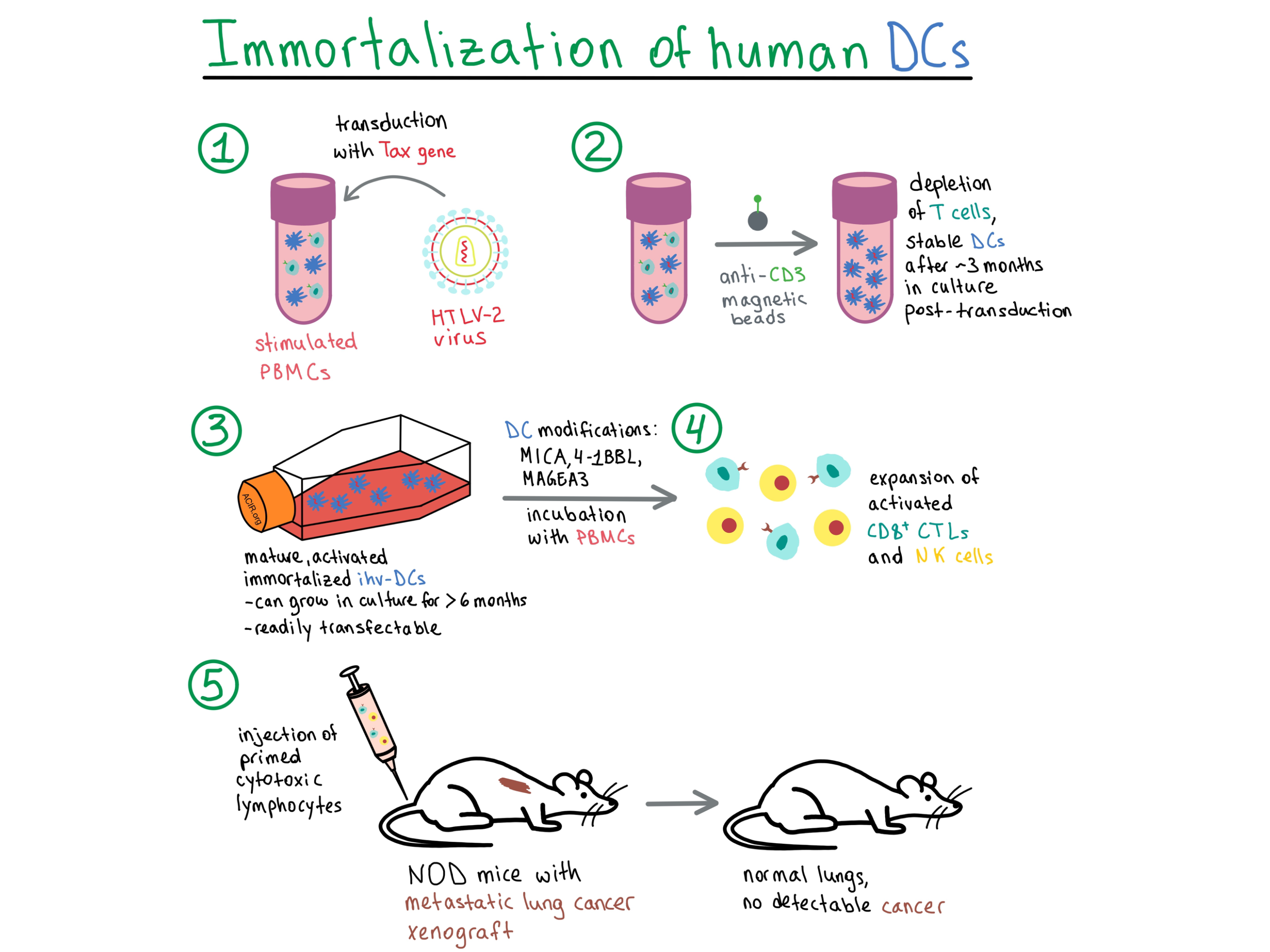
Current methods for preparing dendritic cells (DCs) for cancer vaccine immunotherapy, utilizing antigen-loaded monocyte-derived DCs (MoDCs) that are activated via a complex process, result in DCs that are difficult to maintain in culture, have low growth potential, and require repeated preparations for several rounds of vaccine delivery. In a paper recently published in PNAS, Wu et al. developed a method for establishing immortalized and constitutively activated human primary blood dendritic cell lines (ihv-DCs) that utilizes the viral protein Tax, a transcriptional regulator from human T cell leukemia virus type 2 (HTLV-2), which is not known to be pathogenic in humans.

To begin, Wu et al. transduced stimulated peripheral blood mononuclear cells (PBMCs) with Tax and, two to three weeks later, negatively selected for the CD3 marker to deplete T cells. Two cell lines, named ihv-DC1 and ihv-DC2, grew continuously in culture for over 6 months, and displayed DC-related markers (CD11c, CD141, CD205) and DC maturation and activation markers (CD83, CD80, CD86, CD70, CCR7, HLA-DR), with an immortalization rate of about 20%. To separately validate the ability of Tax to promote DC growth, the researchers in parallel generated MoDCs by stimulating adherent monocytes from PBMCs with GM-CSF/IL-4. The MoDCs that presented dendrites were transduced with Tax, detached during culture, and maintained proliferation for up to 3 months, but were not immortalized. Since the MoDC-Tax cells had a mature and activated phenotype, this experiment validated the ability of Tax to promote the growth of DCs in standard culture conditions.

Although various TLR receptors were upregulated in Tax-transduced DCs, they were no longer required for inducing maturation and activation in these constitutively activated DC lines that already persistently expressed high levels of costimulatory molecules. Both ihv-DC lines and the Tax-transduced MoDCs produced the inflammatory cytokines IL-1A and TNFα, as well as IL-15, which is involved in the development of cytotoxic CD8+ T lymphocytes (CTLs). Thus, the cytokine and TLR expression in transduced DC lines was similar to that in DCs generated by conventional methods. In addition, the transduced DCs upregulated the pro-survival protein Bcl-xL, as well as other growth-promoting, signaling, and cell cycle progression pathways, pointing to their immortalization.

Aiming to enhance antigen presentation of a desirable target epitope, the team transduced ihv-DC2 cells with a fusion construct encoding pTERT, which consisted of a fragment from human telomerase reverse transcriptase (hTERT) linked to a proteasomal target sequence of IκBα. hTERT contained six HLA-A2-restricted CTL epitopes and was degraded within DCs to facilitate epitope presentation. Mixing ihv-DC2-pTERT cells with autologous PBMCs led to priming of autologous CD8+ T cells, which produced large amounts of IFNγ and TNFα. However, the lymphocyte expansion was modest. To enhance the immune response to hTERT, the researchers introduced two HLA-DRB1 molecules into the ihv-DC2-pTERT cells. Such modified alloreactive DCs activated CD4+ T cells, which in turn enhanced the proliferation of CD8+ CTLs, enabling lysis of TERT+ bone osteosarcoma, melanoma, and lung cancer cells in an HLA-A2-restricted manner.

Because cancer cells are often heterogeneous in their expression of HLA molecules, it is desirable to induce both an HLA-specific CTL response and an HLA-independent natural killer (NK) cell response. To this end, Wu et al. engineered ihv-DC1 cells to express high levels of the NK cell activators MICA and 4-1BBL, in addition to the endogenously expressed CD58, in order to efficiently activate NK cells. These modified DCs were also transduced with the HLA-A2.1 molecule and made to overexpress the testis antigen MAGEA3, which is present in many cancer types. The altered DCs (ihv-DC1.2-MAGEA3) were incubated with HLA-A2+ matched naive PBMCs from various donors for at least 2 weeks, leading to stimulation of both CTLs and NK cells. The primed CTLs were able to lyse various types of MAGEA3+ cancer cells in an HLA-A2-restricted manner, while the activated NK cells were highly cytotoxic to HLA-A2- cancer cells.

Moving on to in vivo experiments, the researchers infused NSG mice with A549-Luc/A2.1 cells to establish lung metastases, and then treated the mice with ihv-DC1.2-MAGEA3-primed cytotoxic lymphocytes. The mice in the treatment group had lungs that were normal in appearance and weight compared with the enlarged lungs found in the control group. Cancer cells were undetectable in the lungs of treated mice. Both CTLs and NK cells were found in the lung tissues of treated mice, and the CTLs were also detected in the spleens and lymph nodes, and weakly in the livers.

In summary, Wu et al. developed a method to establish immortalized and constitutively activated human primary blood DC lines, and explored various modifications to the DCs to enhance their immunotherapeutic potential.

by Anna Scherer

References:

Wu L., Zhang H., Jiang Y., Gallo R.C., Cheng H. Induction of antitumor cytotoxic lymphocytes using engineered human primary blood dendritic cells. Proc Natl Acad Sci U S A. 2018 May 8.

In the Spotlight...

The function and dysfunction of memory CD8(+) T cells in tumor immunity

In a set of highly informative articles focused on immunologic memory published in Immunological Reviews, Reading et al. tackle the formation and loss of effective memory CD8+ cells and the unique impact of an evolving tumor microenvironment. Conversion of helpful, but transient, responses to checkpoint or adoptive T cell therapy into durable benefit depends critically on understanding and controlling the multiplicity of factors affecting CD8+ memory cell formation. Other notable topics reviewed include the role of cytokines and stroma, other immune cells, and metabolism.

Role of MDA5 and interferon-I in dendritic cells for T cell expansion by anti-tumor peptide vaccines in mice

Exploring the in vivo mechanism of cytotoxic T cell generation following a prime and boost with a peptide vaccine and poly-ICLC adjuvant, Sultan et al. found that dendritic cells are essential not only for peptide processing and cross-presentation, but also for producing IFN-I, which induces IL-15 in an unidentified bone-marrow-derived cell type to promote CTL expansion. Although both poly-IC and poly-ICLC were sufficient for stimulating TLR3 in the endosomal compartment for production of IL-12, poly-ICLC could also stimulate MDA5 in the cytoplasm, inducing higher levels of IFN-I production.

Sustained cross-presentation capacity of murine splenic dendritic cell subsets in vivo

Ho et al. tested the capability of CD8α+ DCs, CD8α- DCs, plasmacytoid dendritic cells (pDCs), and macrophages to store and present antigen delivered in vivo to C57BL/6 mice as OVA antigen/IgG antibody complexes. CD8α+ DCs were most efficient at MHC-I cross-presentation and induction of cytotoxic CD8+ T cells and demonstrated prolonged (≥ 1 week) antigen storage. Only CD8α- DCs were capable of prolonged MHC-II presentation to CD4+ T cells. Neither pDCs or macrophages were capable of protein antigen presentation.

Elimination of established tumors with nanodisc-based combination chemoimmunotherapy

Kuai et al. developed synthetic high-density lipoprotein (sHDL)-like nanodiscs loaded with chemotherapeutic doxorubicin (DOX) as a targeted way to induce immunogenic cell death in cancer cells (via upregulation of “eat me” and “danger” signals). sHDL-DOX triggered a strong antitumor CD8+ T cell response, including recognition of neoantigens, and exerted antitumor efficacy as monotherapy in mice. Combined with PD-1 blockade, sHDL-DOX led to complete regression in 88% and 80% of mice with CT26 and MC38 tumors, respectively, and protection from re-challenge.

Genomic Features of Response to Combination Immunotherapy in Patients with Advanced Non-Small-Cell Lung Cancer

Using whole exome sequencing, Hellmann et al. demonstrated that high tumor mutation burden (TMB), comprising non-synonymous mutations and indels, was the strongest predictor of response to combination PD-1/CTLA-4 blockade in non-small-cell lung cancer patients. High TMB was associated with greater objective response rate, durable clinical benefit, and progression-free survival. Predicted neoantigen burden was not more predictive than TMB. High TMB and PD-L1+ tumors predicted significantly better outcome than the presence of only one or neither of these variables.

Macrophages: The Road Less Traveled, Changing Anticancer Therapy

Reviewing the complex roles of macrophages in immune oncology, Jennifer Guerriero delves into a variety of factors including ontogeny, tissue residency, phenotype, transcriptional activity, epigenetic diversity, and the highly dynamic adaptation within the tumor microenvironment. She addresses the inadequacy of the current toolbox and the ambiguity of markers, and criticizes oversimplification, particularly in regards to the M1-M2 paradigm. Despite the very limited clinical efficacy thus far, she encourages further exploration of strategies that minimize pro-tumor functions and maximize the antitumor potential of macrophages.

Cancer-Germline Antigen Expression Discriminates Clinical Outcome to CTLA-4 Blockade

Shukla et al. retrospectively analyzed RNA-seq data from melanoma biopsies and found that increased expression of a subcluster of MAGE-A cancer-germline genes predicted resistance to CTLA-4 (but not PD-1) blockade and correlated with lower OS and PFS. Further analysis suggested that MAGE-A was negatively associated with expression of LC3B and mutually exclusive with presence of HMGB1, indicating that MAGE-A suppressed autophagy and hence reduced T cell priming, contributing to clinical resistance to CTLA-4 blockade.

The Transcription Factor Runx3 Establishes Chromatin Accessibility of cis-Regulatory Landscapes that Drive Memory Cytotoxic T Lymphocyte Formation

By mapping regions of hyper-accessible chromatin following TCR stimulation and identifying transcription factor motifs in the remodeled regions, Wang et al. found that in TCR-stimulated naive CD8+ T cells, chromatin accessibility reprogramming is mediated by the transcription factor Runx3. In a series of gain- and loss-of-function experiments, they showed that Runx3 drives differentiation of cytotoxic T lymphocytes, but represses terminal differentiation, and was essential in the development of memory precursor CTLs, in part by preventing high expression of T-bet and by slowing proliferation.

Everything New this Week In...

Close Modal

Small change for you. Big change for us!

This Thanksgiving season, show your support for cancer research by donating your change.

In less than a minute, link your credit card with our partner RoundUp App.

Every purchase you make with that card will be rounded up and the change will be donated to ACIR.

All transactions are securely made through Stripe.