Positive and negative costimulatory molecules on T cells are known to play a role in T cell activation, however, whether they play a role in T cell differentiation (along with TCR signal strength and cytokine signaling) is not as well defined. Following observations that genetic loss or blockade of CTLA-4 can affect CD4+ T cell phenotypes, Wei and Sharma et al. investigated the role of negative regulators CTLA-4 and PD-1 in T cell differentiation and determination of phenotype; their findings were recently published in Immunity.
To better understand the role of CTLA-4 in T cell differentiation, Wei and Sharma et al. profiled lymphocytes from a Ctla4-/- mouse model via mass cytometry, and conducted unsupervised clustering to identify phenotypes. Compared to wild type Ctla4+/+ and heterozygous Ctla4+/- controls, the distribution of phenotypes across lymphocytes from Ctla4-/- was distinct. Within both the CD8+ and CD4+ T cell compartments, multiple phenotypes appeared to be enriched in Ctla4-/- mice, including terminally differentiated CD8+ T cells and an ICOS+ Treg population that was very minor in control mice. More interestingly though, some phenotypes within the CD4+ compartment were entirely unique to the Ctla4-/- mice, including populations of unskewed ICOS+ cells, ICOS+ Th1-skewed Tregs, ICOS+ Th1-like effectors, and Th17-like cells. The emergence of these unique, non-canonical CD4+ T cell subsets in the absence of CTLA-4 expression suggests that CTLA-4 signaling limits the boundaries of phenotypic expression within the CD4+, but not the CD8+ T cell compartment, in peripheral lymph node tissues. Archetypal analysis (a computational approach) confirmed these findings and defined these distinct new cell types as new archetypes with a significantly different set of phenotypes not observed in control mice.
In an effort to understand how these non-canonical phenotypes arise in the absence of CTLA-4, Wei and Sharma et al. investigated a number of factors. First, the researchers investigated whether unrestricted activation and proliferation may have contributed to the novel phenotype populations. To this end, CTLA4-/- and control were vaccinated with ovalbumin to induce a strong T cell response against a number of antigens, and while some T cell phenotypes did expand due to the enhanced T cell activation, the novel phenotypes did not, indicating that their emergence was not an indirect effect of CTLA-4 loss by enhanced T cell activation. Next, the researchers examined the high ICOS expression that was shared across the novel phenotypic subsets and found that while ICOS expression was essential to the functionality of these cells, it did not play a role in driving their development. Finally, the researchers analyzed the TCRβ repertoire in thymic and lymph node tissue to determine whether changes in the central or peripheral repertoire might underlie differentiation towards novel CD4+ T cell phenotypes. While no major differences in the thymic TCR repertoire were observed, T cell clonality was increased in lymph node tissue in Ctla4-/- mice. Interestingly, specific self-reactive T cell clones were found to be recurrently expanded or in a few cases, contracted, across Ctla4-/- animals, and this at least partially influenced differentiation towards atypical CD4+ T cell phenotypes.
In order to identify specific molecular events that are restricted by the expression of CTLA-4 in T cells, the researchers computationally constructed T cell differentiation pathways based on quantitative mass spectrometry data. Changes in key marker expression could be observed tracking differentiation from a naive-like state and resulting in the discrete archetypes observed only in CTLA-4-deficient mice. The researchers found that while CTLA-4 does not keep any pathways fully dormant, it does impose maximum limits on the expression levels of active pathways. Genetic absence of CTLA-4 therefore allowed cells to extend beyond the phenotypic boundaries of their destined cell fate. Ratios of key transcription factors (e.g. FoxP3, RORγT, TBET, GATA3, BCL6) changed when a normal differentiation boundary was approached. Analyzing cytokine production by T cells from Ctla4-/- mice in comparison to controls, the researchers identified an increased frequency of cytokine-producing cells and increased expression of IFNγ, TNFα, and IL-17α, particularly by ICOS+ cells. This indicated that loss of CTLA-4 increased the frequency of functionally active CD4+ effector cells. Genetic loss of Ctla4 also led to an increased frequency of cytokine-producing Tregs, however, it did not enhance their cytokine production capacity, and in fact decreased their suppressive functionality.
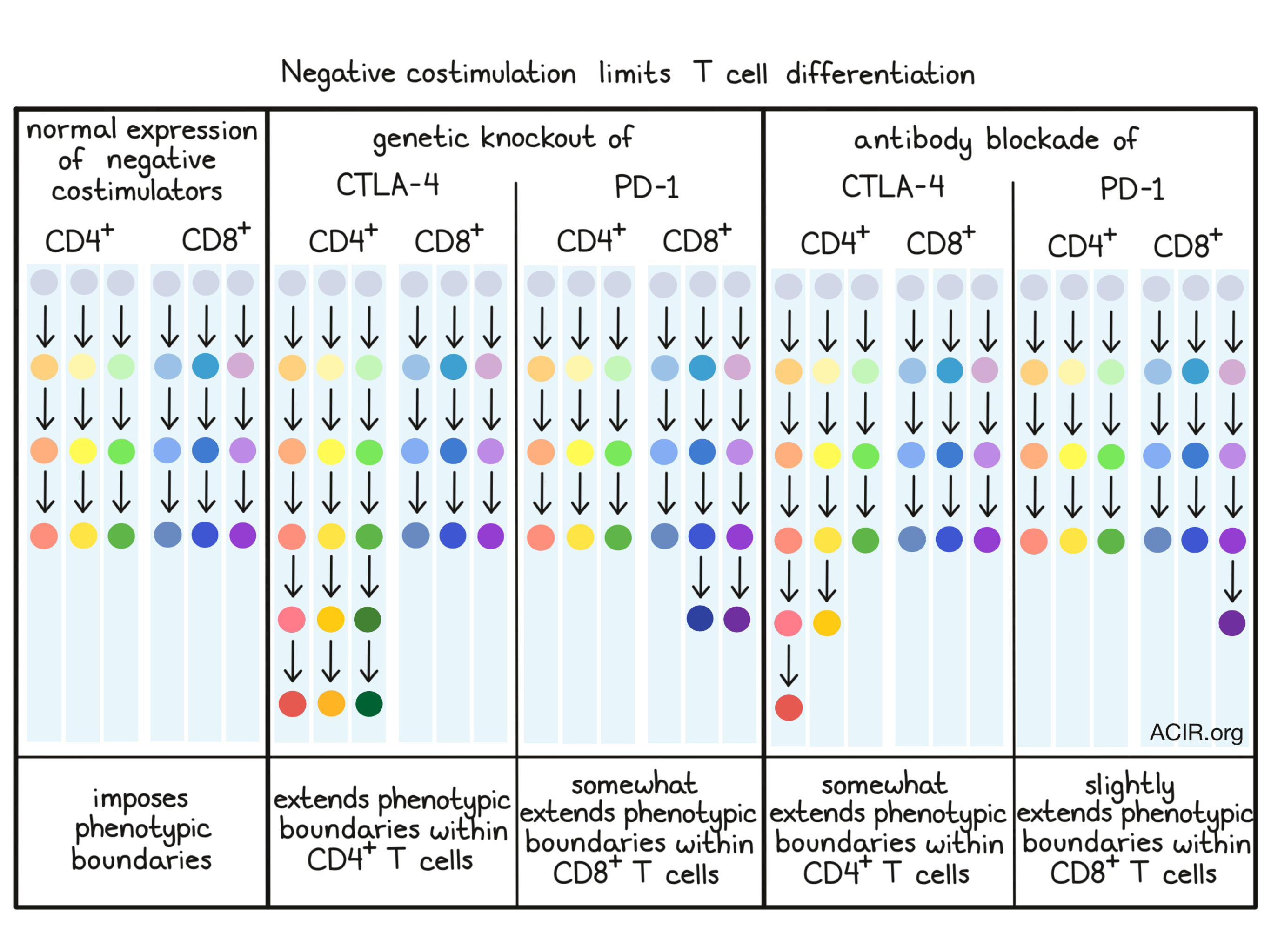
To determine whether the capacity to restrict phenotypic boundaries within T cells applied to negative costimulatory molecules beyond CTLA-4, the researchers turned to PD-1. In contrast to observations in Ctla4-/- mice, the phenotypic boundaries of CD4+ T cells were not expanded in Pdcd1-/- mice compared to controls, whereas the phenotypic boundaries of CD8+ T cells were somewhat expanded (though this effect was only seen in older mice). These results were consistent with a model in which CTLA-4 regulation is induced immediately after T cell priming, while PD-1-mediated regulation is engaged only after the initiation of an active immune response. Based on this, it is likely that both CTLA-4 and PD-1 impose phenotypic boundaries in T cells, but they do so through different mechanisms and in different T cell subsets, and act to a different extent.
Next, the researchers naturally wondered whether antibody blockade of CTLA-4 or PD-1 had similar effects to genetic deletion of these negative costimulators. They found that CTLA-4 blockade enriched T cells for a T-BET+TCF1+CD4+ T cell archetype, and that it did not enrich any CD8+ T cell archetype. Meanwhile, PD-1 blockade enriched a PD-1+PD-L1intCD8+ T cell archetype. Unlike in the genetic studies, however, none of the enriched archetypes were unique compared to controls. This less dramatic phenotypic expansion is likely attributed to incomplete loss of signaling from antibody blockade, compared to the complete loss of signaling afforded by genetic engineering.
Overall, the evidence uncovered by Wei and Sharma et al. shows that CTLA-4 and PD-1, in addition to their effects on activation, impose boundaries on T cell phenotypes during peripheral differentiation. T cells engineered to lack these molecules are therefore able to differentiate further down active pathways, leading to the acquisition of phenotypes not observed under natural circumstances. Similarly, antibody blockade of these molecules may similarly reduce some of the limitations on T cell differentiation.
by Lauren Hitchings
Meet the Researcher
This week, Spencer C. Wei and Roshan Sharma, first co-authors on this study, answered our questions.

What prompted you to tackle this research question?
There is so much that we still do not know about how T cell activity is regulated and insights into basic biological processes are absolutely essential for improving immunotherapy approaches. Here we wanted to understand how T cell phenotypes are defined and whether T cell costimulation plays a role in this process.
What was the most surprising finding of this study for you?
How profound of a role CTLA-4 plays in regulating CD4+ T cell phenotypic limits. This function complements, but is distinct from its well-known role in regulating T cell activation. This changes our perspective of how we think about how CTLA-4 regulates and shapes T cell biology.

What prompted you to tackle this research question?
The primary question we tackle in this project can be stated as "Do negative costimulatory molecules play a role in T cell differentiation?". We arrived at this question with inspiration and motivation from an earlier collaboration between the Pe'er and Allison lab, which utilized mass cytometry data and machine learning tools to dissect the cellular mechanisms of anti-CTLA-4 and anti-PD-1 treated tumor-infiltrating T cells. The study revealed that anti-CTLA-4 and anti-PD-1 checkpoint blockade act through distinct cellular mechanisms. In particular, it was found that anti-PD-1 treatment resulted in an expansion of exhausted CD8+ T cells subsets while anti-CTLA-4 treatment resulted in a predominant expansion of Th1-like CD4+ T cell subsets. In addition, highlighting the cellular underpinnings of checkpoint blockade, the study hinted at the fact that molecules such as CTLA-4 and PD-1, also known as negative costimulatory molecules, might play a role in T cell differentiation along with their well-known and essential role in T cell activation. Therefore, we wanted to take a more basic approach to target this question directly.
What was the most surprising finding of this study for you?
The most interesting conclusion of the study was that negative costimulatory molecules indeed play a role in T cell differentiation in addition to their role in T cell activation. What was surprising was the way they seem to influence differentiation, which is by putting restriction on the differentiation limits of the T cells. Essentially, our work highlights that CTLA-4 and PD-1 act as "gate-keepers", which restrict the expansion of phenotypic states of differentiating T cells. Once they are deleted, the cells can expand along a continuum to occupy phenotypic states not attainable under normal circumstances. Further analysis led us to observe that protein expression behavior is largely maintained intact despite the loss of CTLA-4, that is, our study argued against the possibility of activation of otherwise dormant signaling pathways under normal circumstances. In summary, loss of negative costimulatory molecules allow normal signaling behavior but at an exaggerated or expanded level.
What was the coolest thing you’ve learned (about) recently outside of the lab?
I recently learned about a paper titled "A network approach to topic models" from a colleague. The paper essentially presents a marriage of network science with topic models, typically thought of as a distinct branch of computer science.


