IL-12 is of great interest for immunotherapy for its broad immune-activating effects. However, systemic treatment induces severe toxicities, limiting its application. Approaches to safely deliver cytokines to the tumor microenvironment are therefore warranted. Jones and Nardozzi et al. investigated a method to tether cytokines via antibodies to tumor-specific T cells used in adoptive therapy. Their results were recently published in Science Advances.
The researchers started by assessing surface receptors on T cells, including cytokine receptors, integrins and related surface adhesion molecules, and inhibitory receptors (e.g., PD-1) that could be used for tethering cytokines. Profiling T cells revealed that cytokine receptors and inhibitory receptors had low antibody binding capacity, even after activation, while integrin molecules showed a very high capacity, which further increased after stimulation. Several of these receptors may serve as ‘handles’ for cell surface cytokine tethering.
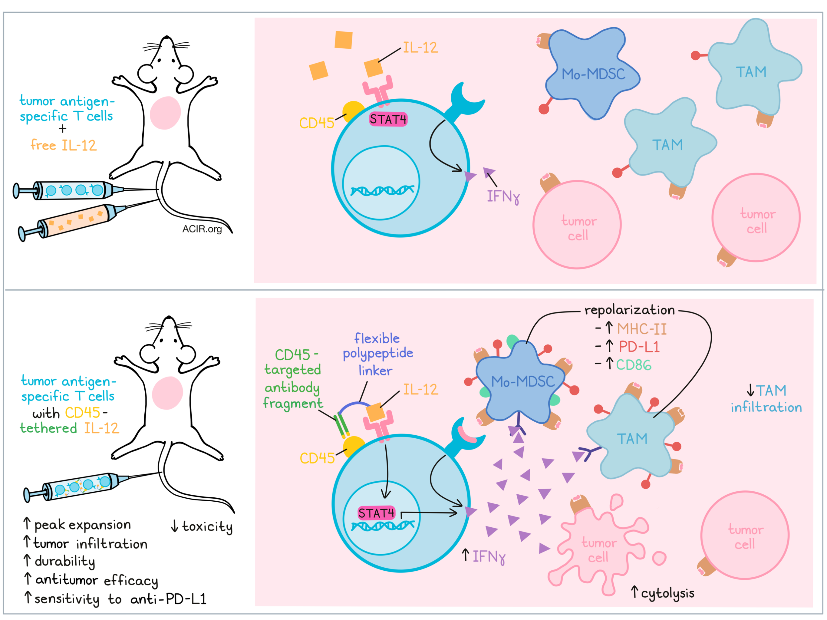
To assess this, cytokine “tethered fusion proteins” were constructed by linking a cytokine to a receptor-targeted antibody. IL-12 and IL-15 and antibodies directed against CD11a, CD18, or CD45 were created by fusing the cytokine to the C terminus of the antibody light chain in Fab fragments using a flexible polypeptide linker. Each cell surface receptor facilitated uniform dose-dependent loading of the cytokines onto the T cell surface. The highest tethering was achieved with CD45.
To assess whether the tethering resulted in sustained cytokine stimulus to the T cells, IL-15 tethered fusions were assessed for T cell expansion in vitro. Each fusion product resulted in dose-dependent T cell expansion, while blocking the target of the tethered fusion abolished proliferation. Higher affinity antibodies had stronger proliferation potency. Pulse incubation (1 r exposure followed by washing) with CD45-tethered IL-15, but not free IL-15, activated STAT5, and similar effects were found with IL-12 and STAT4. Conditioned supernatant from cells tethered with IL-12 showed that these cytokines could dissociate from the loaded T cell and activate other T cells.
IL-12 tethered to CD45 was assessed further to determine whether T cell activation could be enhanced without inducing off-target effects. Human CD8+ T cells specific for the HLA-A*02:01-restricted MART-1 peptide were generated by ex vivo stimulation of T cells from a healthy donor with autologous monocytic dendritic cells (mDCs) presenting the MART-1 peptide. After these cells were CD45-tethered with IL-12, they were co-cultured with a MART-1-expressing melanoma cell line (SK-MEL-5). This revealed a dose-dependent increase in cytolysis and IFNγ production, which did not occur when cells were cocultured with tumor cells lacking MART-1 expression.
To determine whether the cell-tethered IL-12 could improve antitumor activity in vivo, CD8+ T cells obtained from PMEL-1 TCR-transgenic mice were activated and expanded ex vivo and subsequently tethered with IL-12 before adoptive cell transfer (ACT) into mice bearing B16F10 tumors. These transferred cells were more efficacious than PMEL-1 T cells alone, and the delivery route did not affect this (intravenous or intratumoral). Jones and Nardozzi et al. then assessed the effects of tethered fusions with different CD45 binding affinities. Similar effects were found on tumor growth inhibition, suggesting the Fab antibody fragment used had sufficient affinity for antitumor effects.
Antitumor efficacy did not improve when untethered PMEL-1 T cells were coadministered with a four-fold higher dose of intravenous IL-12. Therefore, tethered IL-12 was more effective than coadministered systemic IL-12, and higher IL-12 loading resulted in more durable tumor regression. The tethered IL-12 also increased peak expansion of the transferred T cells and had increased long-term engraftment. The effects were dose-dependent on surface-tethering levels, as saturating and sub-saturating IL-12 loading had similar effects on engraftment.
These effects were observed in the absence of body weight loss, and systemic IFNγ production was moderate and transient. A higher cell dose resulted in transient peak body weight loss that reversed to baseline by day 7. Additionally, none of the doses resulted in liver or renal function blood marker increases, except for a temporary, moderate increase in alanine aminotransferase. Therefore, it was safe to repeat the treatment, which increased the durability of the antitumor response.
Next, the authors evaluated the antitumor effects of tethering T cells with endogenously stimulated T cells of variable reactivity obtained from tumor-draining lymph nodes of BALB/c mice bearing CT26 tumors. Isolated T cells were stimulated ex vivo to obtain a polyclonal population of tumor antigen-reactive T cells. CD45-tethering with IL-12 improved antitumor efficacy over ACT with untethered T cells. Smaller tumors were eradicated in 6/8 mice, while only 3/8 mice in the untethered group (systemic co-administration of IL-12) achieved complete remissions. Surviving mice also rejected a rechallenge, suggesting durable immunity was induced.
IL-12-tethered PMEL T cells demonstrated increased infiltration and tumoral IFNγ compared to T cells with untethered systemic IL-12. To assess how this affected the tumor immune microenvironment, myeloid lineage cells were profiled. Monocytic myeloid-derived suppressor cells (Mo-MDSCs) and tumor-associated macrophages (TAMs) were the most abundant myeloid populations in B16F10 tumors. Infiltration of T cells CD45-tethered with IL-12 reduced the infiltration of TAMs and recovered their expression of MHC-II, an activation marker associated with the M1-like phenotype. The tethered T cells did not affect Mo-MDSCs infiltration, but increased the expression of activation markers involved in antigen presentation and T cell costimulation (MHC-II and CD86) on these cells. Depleting Mo-MDSCs with anti-Ly6 antibody reduced the antitumor efficacy of the tethered T cells, suggesting the repolarization of Mo-MDSCs might be required for the efficacy of this therapy.
ACT with IL-12-tethered T cells resulted in the upregulation of PD-L1 on various immune cell populations, including Mo-MDSCs and TAMs. This effect was most pronounced in the repolarized MHC-II+CD86+ population of the Mo-MDSCs. When a neutralizing IFNγ antibody was used, there was less repolarization of Mo-MDSC and TAMs, and less PD-L1 expression induction.
The B16F10 model does not respond to PD-1/PD-L1 checkpoint blockade, even when combined with PMEL T cell ACT. However, combining ACT of IL-12-tethered T cells with anti-PD-L1 checkpoint blockade improved antitumor efficacy, resulting in more mice with complete responses.
This study shows the applicability of tethering IL-12 and IL-15 to adoptive T cell transfer (CARs, TILs, engineered TCRs) while overcoming toxicities associated with these cytokines, in particular, generating a pathway to capitalize on the immune-stimulating effect of IL-12. This provides a rationale for testing this therapy in combination with other immunotherapeutic strategies for various cancer types.
Write-up by Maartje Wouters, image by Lauren Hitchings.
Meet the researcher
This week, first author Douglas Jones 2nd answered our questions.

What prompted you to tackle this research question?
IL-12 is a powerful cytokine that is ideally suited for combining with cell therapies. The challenge has been how to deliver it safely. We asked ourselves the question: could you control IL-12 dosing and focus its activity by directly tethering the cytokine to the surface of the T cell therapy?
What was the most surprising finding of this study for you?
We were impressed by how directly tethering IL-12 to the surface of the T cells prior to adoptive transfer drove such pronounced activity, specifically in the tumor. The activity was both more potent and more focused than what you get with conventional systemic IL-12 dosing. For example, it drove high levels of IFNγ (itself a potent immune-activating cytokine) specifically in the tumor, and repolarized mMDSCs and TAMs – two key suppressive tumor immune cell types – from suppressive to inflammatory phenotypes. Repertoire Immune Medicines, a clinical-stage cell therapy company, is currently investigating the use of cell-tethered IL-12 in an ongoing Phase 1 study of human papillomavirus-specific T cells.
What was the coolest thing you’ve learned (about) recently outside of work?
We recently took a road trip from Utah to Seattle and along the way we happened across a historic site of the Oregon Trail, which was a heavily used migration route in the mid-1800s. What was fascinating was that so many wagons had traveled this route that you could still see the ruts in the ground from this mass migration ~150 years ago. As someone who knew about the Oregon Trail mostly from the popular 1980s-era computer game, it was an intriguing piece of history brought to life.


