Motivated to extend the benefit of PD-1 blockade to more patients, Chow et al. explored the role that chemokines play in resistance to anti-PD-1 and found that the chemokine receptor CXCR3 and its ligand CXCL9 were crucial for the response to anti-PD-1 therapy in mouse tumor models. The results were recently published in Immunity.
The researchers began by showing that anti-PD-1 significantly reduced the growth of MC38 colon adenocarcinoma or D4M.3A.3 UV3 melanoma and improved survival in wild-type (WT) mice compared with Cxcr3-/- mice, demonstrating that CXCR3 plays a role in PD-1 blockade. Given that CXCR3 plays a role in the recruitment of T cells into the tumor in adoptive cell transfer models, the researchers hypothesized that the subpar efficacy of anti-PD-1 in Cxcr3-/- mice may be due to reduced intratumoral CD8+ T cell infiltration. However, pre-treatment MC38 tumor analysis showed no difference in the percentage, absolute number, cytokine production, or phenotype of intratumoral CD8+ T cells between WT and Cxcr3-/- mice. Thus, in this model, CXCR3 was not required for the early recruitment and function of CD8+ T cells.
A different scenario emerged post-treatment. Anti-PD-1 significantly increased the number of intratumoral CD8+ T cells, as well as the percentage of IFNγ+TNFα+ and Ki-67+granzymeB+ CD8+ T cells in WT, but not in Cxcr3-/- mice. To understand the mechanism behind these different outcomes, Chow et al. blocked the egress of T cells from lymphoid organs with FTY720 before anti-PD-1 treatment and showed that although peripheral T cell numbers were dramatically reduced, neither the intratumoral CD8+ T cell expansion nor the antitumor effect in WT mice were affected. Monitoring of DNA replication demonstrated that anti-PD-1 functioned primarily by expanding the pre-existing intratumoral CD8+ T cells, and not by recruiting more CD8+ T cells into the tumor and that this intratumoral T cell expansion was dependent on CXCR3. The researchers confirmed the requirement for CXCR3 signaling in PD-1 blockade response using chimeric mouse models, in which anti-PD-1 preferentially expanded and increased the effector function of WT neoantigen-specific intratumoral CD8+ T cells compared with their Cxcr3-/- counterparts.
The team further analyzed the role of CXCR3 using CXCR3-GFP reporter mice bearing MC38 tumors. They found that CXCR3-PD-1hiCD8+ T cells were more exhausted and produced lower levels of IFNγ and TNFα than their CXCR3-expressing counterparts. In addition, the percentage of CXCR3+CD8+ T cells decreased as the tumor grew over time, suggesting that the expression of CXCR3 on CD8+ T cells is inversely correlated with tumor growth.
The researchers then analyzed the expression of CXCR3 ligands – CXCL9 and CXCL10 – in WT mice. Anti-PD-1 increased the levels of both ligands in MC38 tumors, and the anti-PD-1 antitumor effect was abrogated when both ligands were blocked with antibodies. To tease out the specific role of each ligand, the team utilized separate Cxcl9- and Cxcl10-knockout mice, and found that CXCL9, but not CXCL10, was crucial for the anti-PD-1-mediated CD8+ T cell antitumor response.
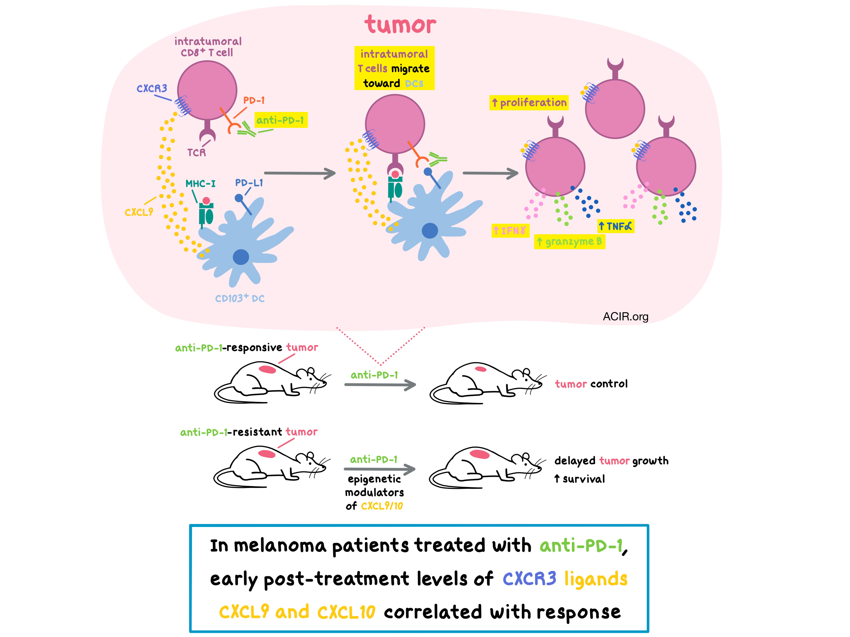
Utilizing CXCL9-CXCL10 dual reporter mice, Chow et al. observed that myeloid cells were the major source of the two ligands within the MC38 tumors, with CD103+ DCs producing CXCL9 and CD11b+ DCs producing CXCL10. PD-1 blockade increased the production of CXCL9 by CD103+ DCs. Diphtheria-toxin-induced depletion of classical, but not monocyte-derived or plasmacytoid DCs, demonstrated that CD103+ DC-derived CXCL9 was functionally important for anti-PD-1-mediated antitumor response and improved survival. Based on these results, the authors hypothesized that PD-1 blockade-mediated increase in CXCL9 expression allowed CD103+ DCs to attract CXCR3+CD8+ T cells that were already in the tumor, thus facilitating DC-T cell interactions and enhancing T cell antitumor responses.
The researchers then screened solid murine tumors and found that the anti-PD-1-responsive tumors had a much higher baseline expression of CXCL9 and CXCL10 in CD11c+MHCII+ DCs than tumors resistant to PD-1 blockade, suggesting that baseline intratumoral expression of CXCR3 ligands could predict response to PD-1 blockade. Combination of anti-PD-1 and epigenetic modulators (DZNeP + 5-AZA-dC), which upregulated DC expression of CXCL9 and CXCL10, delayed tumor growth and extended survival in mice bearing anti-PD-1-resistant AT-3 breast tumors. These results suggested that tumors resistant to PD-1 blockade could become more responsive to anti-PD-1 when it is combined with epigenetic modulation of CXCR3 ligand expression; clinical trials examining the efficacy of this combination are ongoing.
Turning to human data, Chow et al. analyzed plasma samples from melanoma patients treated with anti-PD-1 alone or with anti-CTLA-4, and found that levels of CXCR3 ligands, measured a few months post-treatment, positively correlated with response. This data suggest that CXCR3 ligands could serve as early biomarkers of immunotherapy response.
by Anna Scherer
Meet the researcher
This week, Melvyn T. Chow, first author on this paper, took the time to answer our questions.
1. What prompted you to tackle this research question?
A successful and efficacious immune checkpoint blockade therapy requires the productive interaction of different immune cells to generate an optimal antitumor response. Chemokines likely play key roles in the recruitment of immune cells into tumors as well as their cell-cell interactions within tumors. Therefore, we were interested in understanding the role of the CXCR3 chemokine system in the recruitment and cellular interactions of T cells within the tumor microenvironment, ultimately with the goal of exploring the potential for specific enhancement of effector T cell infiltration and function.
2. What was the most surprising finding of this study for you?
The most surprising finding of this study was the important role the CXCR3 chemokine system plays for the activation of CD8+ T cells within tumors and for the success of PD-1 immune checkpoint blockade therapy.
3. What was the coolest thing you’ve learned (about) recently outside of the lab?
I was traveling in Iceland early March with my wife and one of the coolest things I learned on this trip is that in Iceland, hot water is pumped through tubes under many of the sidewalks to keep the streets free of ice and snow without plowing or shoveling. Furthermore, almost 100 percent of the electricity consumed in Iceland comes from renewable energy, which I find remarkable.


