Weekly Digests
‹ Back to May

M2-like TAMs tamed by anti-CSF1R and anti-PD-1 combination therapy

May 9, 2018

To uncover why anti-PD-1 therapy fails in the majority of melanomas, Neubert et al. investigated the role of colony-stimulating factor 1 (CSF1) in creating a protumorigenic, macrophage-rich microenvironment. CSF1 is an important regulator of both monocytes and macrophages and controls proliferation and survival of macrophages from their precursors. The results of their experiments, published in Science Translational Medicine, indicate the impact of CSF1 expression and its role in limiting the effectiveness of immune checkpoint blockade therapy via enhancing function and accumulation of M2-like tumor-associated macrophages (TAMs).

Neubert et al. began by observing that the levels of CSF1 were higher in peripheral blood of melanoma patients than healthy volunteers and further elevated in Stage IV than Stage IIIB patients. To uncover a direct relationship between CSF1 and melanoma cells, four low passage melanoma cell lines were tested for CSF1 production in vitro. Media conditioned by any of the four melanoma cell lines showed no or low CSF1 expression levels, however after co-culturing the cell lines with MelanA-specific cytotoxic CD8+ T lymphocyte (CTL) clones, CSF1 could be detected in the conditioned media from each line. Additionally, intracellular CSF1 expression increased in the melanoma cells upon encountering melanoma-specific CTLs, while non-melanoma-specific CTLs didn’t cause secretion of CSF1 from the cell lines. Staining studies of eight human melanoma specimens demonstrated that approximately 80% of melanoma cells expressed CSF1, which correlated with regions of high CD8+ T cell density.

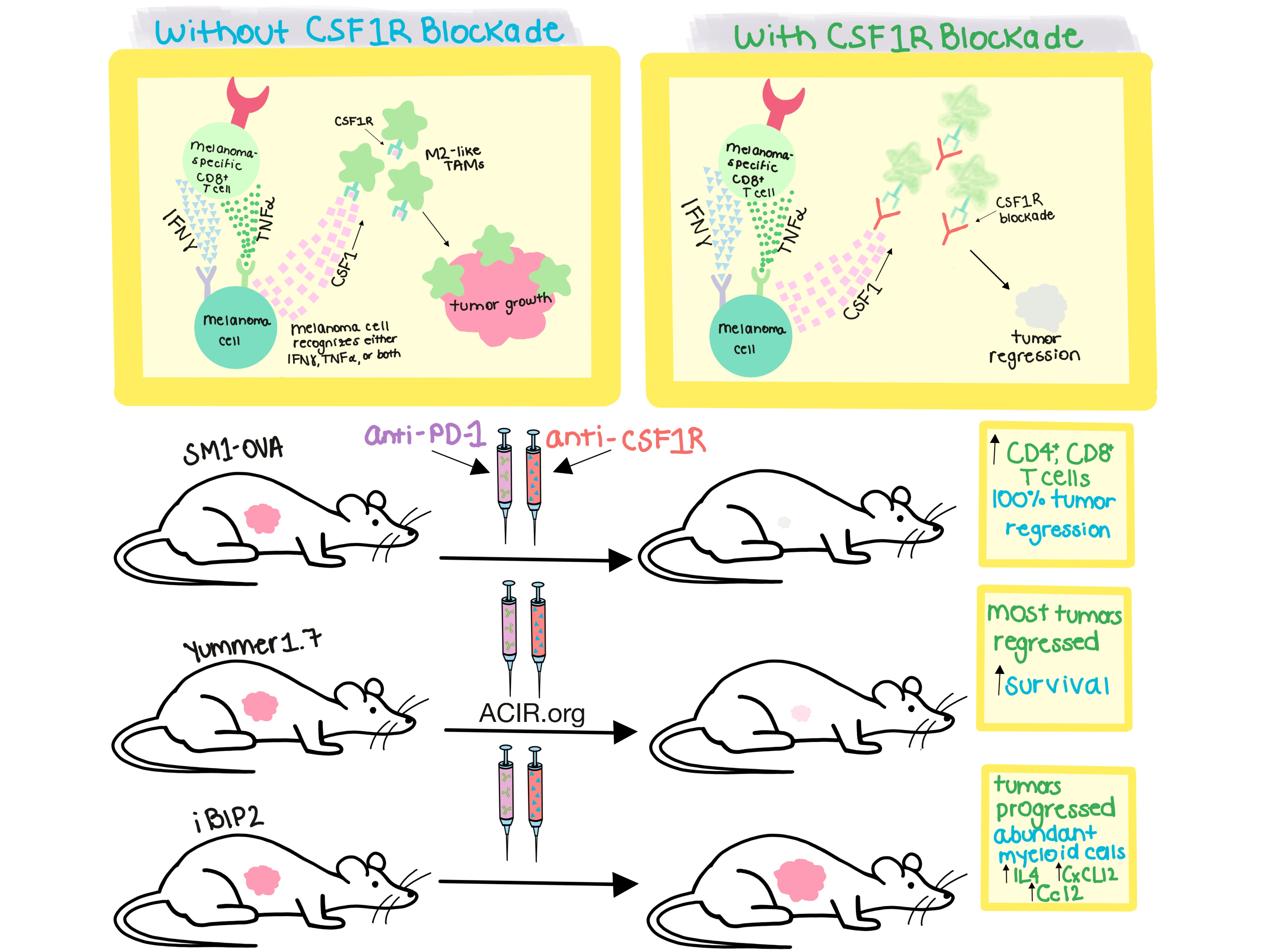
Stimulation was independent of direct cell contact between CTLs and melanoma cells and further experiments determined that the soluble factors derived from CTLs able to induce CSF1 expression in melanoma cells were TNFα and IFNγ. Each cytokine was able to induce CSF1 expression alone, and their combination strongly increased CSF1 expression in all fifteen cell lines tested, with peak concentration of T cell-released IFNγ correlating with the onset of CSF1 expression in cell supernatant. Based on these in vitro studies, TNFα and IFNγ were concluded to be CTL-derived factors able to induce CSF1 expression in melanoma cell lines. The team then utilized a large data set of human metastatic melanoma patient information from The Cancer Genome Atlas (TCGA) to demonstrate that expression levels correlated between CSF1, IFNγ, and TNFα.

TCGA data was also used to examine the relationship between CD8+ T cell and TAM levels and responsiveness to anti-PD-1 blockade. Analysis of the expression of pretreatment tumor biopsies labeled either as responders or nonresponders demonstrated that nonresponders had a strong association between high amounts of CD8+ T cells and expression of CSF1+, CSF1R+, and CD163. These results led the team to conclude that CD8+ T cell presence in human melanoma initiated infiltration of TAMs driven by CSF1, which was produced by melanoma cells following exposure to CTL cytokines, suggesting that cascade of events limited antitumor response. A high ratio of CD8A/CSF1R, indicative of TAM infiltration being less extensive, was found to be indicative of improved overall survival.

The importance of CSF1 was next demonstrated in murine models to “reverse translate” the human observations. The BRAFV600E mutant ovalbumin-expressing SM1 cells (SM1-OVA), which secrete high levels of CSF1 and produce highly macrophage-infiltrated tumors, were monitored for tumor growth inhibition following both single agent anti-PD-1 or anti-CSF1R treatments or combination therapy. The combination therapy proved most effective, with total regression of all tumors after 17 days of treatment and increased CD4+ and CD8+ T cells in the spleens of the mice. A second model, Yummer1.7, which better approximated the high mutational load of human melanoma, demonstrated that combination of anti-CSF1R and anti-PD-1 therapy eradicated the majority of their tumors and significantly extended mouse survival after therapy. In a third transgenic model, iBIP2, combination therapy was unable to inhibit melanoma progression due to an inability to eliminate/repolarize TAMs with the anti-CSF1R antibody. Interestingly, infiltration of multiple myeloid cells was significantly higher in iBIP2 than SM1-OVA tumors; expression analysis indicated high expression of the chemokines Ccl2 and Cxcl12, and the cytokine IL-4 genes, potentially abrogating the impact of the single or combined therapies.

The work presented by Neubert et al. demonstrates the importance of CSF1 in metastatic melanoma, in vitro and in vivo. Anti-CSF1R combination therapy with anti-PD-1 significantly improved tumor regression and overall survival in two of three mouse models, indicating its potential for use in clinical trials. The authors indicate their intention to use CSF1R inhibition with anti-PD-1/PD-L1 in melanomas stratified for an abundance of M2-like TAMs in the tumor microenvironment.

by Brynn Vessey

References:

Neubert N.J., Schmittnaegel M., Bordry N., Nassiri S., Wald N., Martignier C., Tillé L., Homicsko K., Damsky W., Maby-El Hajjami H., Klaman I., Danenberg E., Ioannidou K., Kandalaft L., Coukos G., Hoves S., Ries C.H., Fuertes Marraco S.A., Foukas P.G., De Palma M., Speiser D.E. T cell-induced CSF1 promotes melanoma resistance to PD1 blockade. Sci Transl Med. 2018 Apr 11.

In the Spotlight...

Nivolumab plus Ipilimumab in Lung Cancer with a High Tumor Mutational Burden

In one of 4 publications in the NEJM describing the results of clinical trials evaluating immune checkpoint blockade (ICB), released in concert with the 2018 AACR Annual Meeting, Hellman et al. demonstrated that high tumor mutational burden (> 10 mutations per megabase as determined by the FoundationOne CDx assay) was effective in predicting improved progression-free survival for first line non-small cell lung cancer patients treated with a combination of nivolumab and ipilimumab versus platinum doublet chemotherapy. Please click here for our summary of the other ICB clinical trial results (Eggermont, Gandhi, and Pardoll).

Potent antitumor efficacy of anti-GD2 CAR T cells in H3-K27M+ diffuse midline gliomas

Mount et al. utilized patient-derived xenograft murine models of three subtypes (pons, thalamus, and spinal cord) of diffuse midline gliomas with H3-K27M+ mutations to demonstrate the potent antitumor effects of GD2 CAR T cell therapy. The K27M mutation in histone H3 increased expression of ganglioside synthesis enzymes, providing high levels of target antigen expression, desirable for CAR therapy. Lethal “on target, on tumor” neuroinflammation in this precarious anatomical site dictate a cautious approach in entry to human trials.

Monomeric TCRs drive T cell antigen recognition

Using four different non-invasive imaging techniques with single-molecule sensitivity on living T cells interacting with ligands embedded in a planar lipid membrane, Brameshuber et al. demonstrate that it is TCR-CD3 monomers, not dimers or oligomers, that are primarily responsible for the recognition of antigenic peptide:MHC complexes, contrary to the data previously obtained via invasive imaging. Thus, the signaling mechanism relevant for antigen sensitivity and signal generation is distinct from receptor tyrosine kinase-determined dimerization mechanisms.

Role of PD-1 during effector CD8 T cell differentiation

Ahn et al. found that PD-1 is rapidly expressed alongside activation markers following antigen stimulation (either by viral infection or peptide injection) of naive CD8+ T cells in vivo, even before division begins. This rapid expression was found to be primarily mediated by antigenic stimulation of the TCR and was dependent on NFAT. Early blockade of the PD-1 axis increased the effector function of virus-specific CD8+ T cells, resulting in more rapid viral clearance, followed by an increase in CD127hi KLRG1low memory precursor cells, indicating that PD-1 plays an early role in T cell function and differentiation.

The systemic response to surgery triggers the outgrowth of distant immune-controlled tumors in mouse models of dormancy

By combining a novel tumor dormancy mouse model with a wound-healing model, Krall et al. showed that surgical wounding and healing triggers local and distant outgrowth of breast cancer tumors, which, in the absence of a wound, would be suppressed by the adaptive immune system. Surgery systemically mobilizes inflammatory monocytes, which differentiate into tumor-associated macrophages (TAMs) and promote metastatic outgrowth via PD-L1 expression. Administration of an NSAID peri- and postoperatively significantly reduced tumor growth by shifting TAMs away from the protumor M2 phenotype.

Somatic POLE exonuclease domain mutations are early events in sporadic endometrial and colorectal carcinogenesis, determining driver mutational landscape, clonal neoantigen burden and immune response

Using targeted and whole-genome sequencing on precursors and advanced endometrial and colorectal cancers, Temko et al. show that somatic mutations in the POLE exonuclease domain occur early in tumorigenesis (detectable even at the precancerous stage), create pathogenic mutations in known oncogenic driver genes, and are possibly the initiating oncogenic events. Such mutations drive significant CD8+ T cell infiltration into tumors and precancerous lesions, probably due to an increased mutation burden and consequent high neoantigen frequency.

Co-inhibitory Molecule B7 Superfamily Member 1 Expressed by Tumor-Infiltrating Myeloid Cells Induces Dysfunction of Anti-tumor CD8+ T Cells

Li et al. show that the molecule B7S1, expressed on APCs and some tumor cells, is an inhibitory ligand which acts to suppress activated CD8+ T cells via Eomes overexpression and other pathways that largely overlap with PD-1, possibly explaining the low response rate in patients treated with PD-1 blockade. Blockade of both B7S1 and PD-1 synergistically improved the antitumor response in mice. Coexpression of PD-1 and the putative B7S1 receptor on CD8+ TILs indicated an activated, transitional state, while coexpression of PD-1 and TIM-3 was associated with exhaustion.

Long Terminal Repeat CRISPR-CAR-Coupled "Universal" T Cells Mediate Potent Anti-leukemic Effects

Georgiadis et al. developed a lentivirus construct containing a CD19 CAR-encoding region and a TRAC-targeted sgRNA to achieve simultaneous transgene expression with TCR knockout. Following transduction and electroporation of Cas9 mRNA, a highly homogenous and effective CD19 CAR+TCR- T cell population was produced. When adoptively transferred into a mouse leukemia model, CAR+TCR- T cells rapidly cleared tumors, expanded more readily, and expressed less PD-1 compared to CAR+TCR+ T cells. This strategy could be incorporated into an automated manufacturing platform.

Everything New this Week In...

Close Modal

Small change for you. Big change for us!

This Thanksgiving season, show your support for cancer research by donating your change.

In less than a minute, link your credit card with our partner RoundUp App.

Every purchase you make with that card will be rounded up and the change will be donated to ACIR.

All transactions are securely made through Stripe.