Weekly Digests
‹ Back to February

Local chemo and CTLA-4 blockade: Turning a limb into a cancer vaccine

February 7, 2018

Immune checkpoint blockade is a powerful weapon in the fight against cancer, but for most patients, checkpoint blockade alone is not enough, and standards of care like chemotherapy are still the norm. In a recent paper published in Cancer Immunology Research, Ariyan et al. sought to enhance the efficacy of CTLA-4 checkpoint blockade in poorly immunogenic tumors by combining it with melphalan, a common chemotherapeutic agent that could act as an in vivo vaccine by inducing cell death and inflammation at the tumor site.

Melphalan is known to induce tumor cell apoptosis and the release of inflammatory cytokines, and in preclinical experiments, Ariyan et al. explored the possibility that it could also have an immune effect. The researchers applied melphalan to B16 melanoma cells in vitro and found that it altered two key immune parameters: it increased tumor cell expression of MHC class I molecules by 2.5 fold and increased PD-L1 expression by 7 fold, indicating that melphalan could increase the antitumor response to immunotherapy.

Next, in mice with B16 melanoma, the researchers intratumorally administered melphalan and intraperitoneally administered anti-CTLA-4. While neither agent was effective as a monotherapy, the combination treatment improved survival and protected mice long term against cancer rechallenge. Immune cell phenotyping revealed that the combination therapy enriched the tumor microenvironment for effector T cells and reduced the number of T regulatory (Treg) cells, increasing the CD8/Treg ratio. They also showed that long-term anti-tumor immunity was dependent on host expression of IFNγ. Similar results were seen combining CTLA-4 blockade with gemcitabine chemotherapy in the TRAMP2 murine model of prostate cancer.

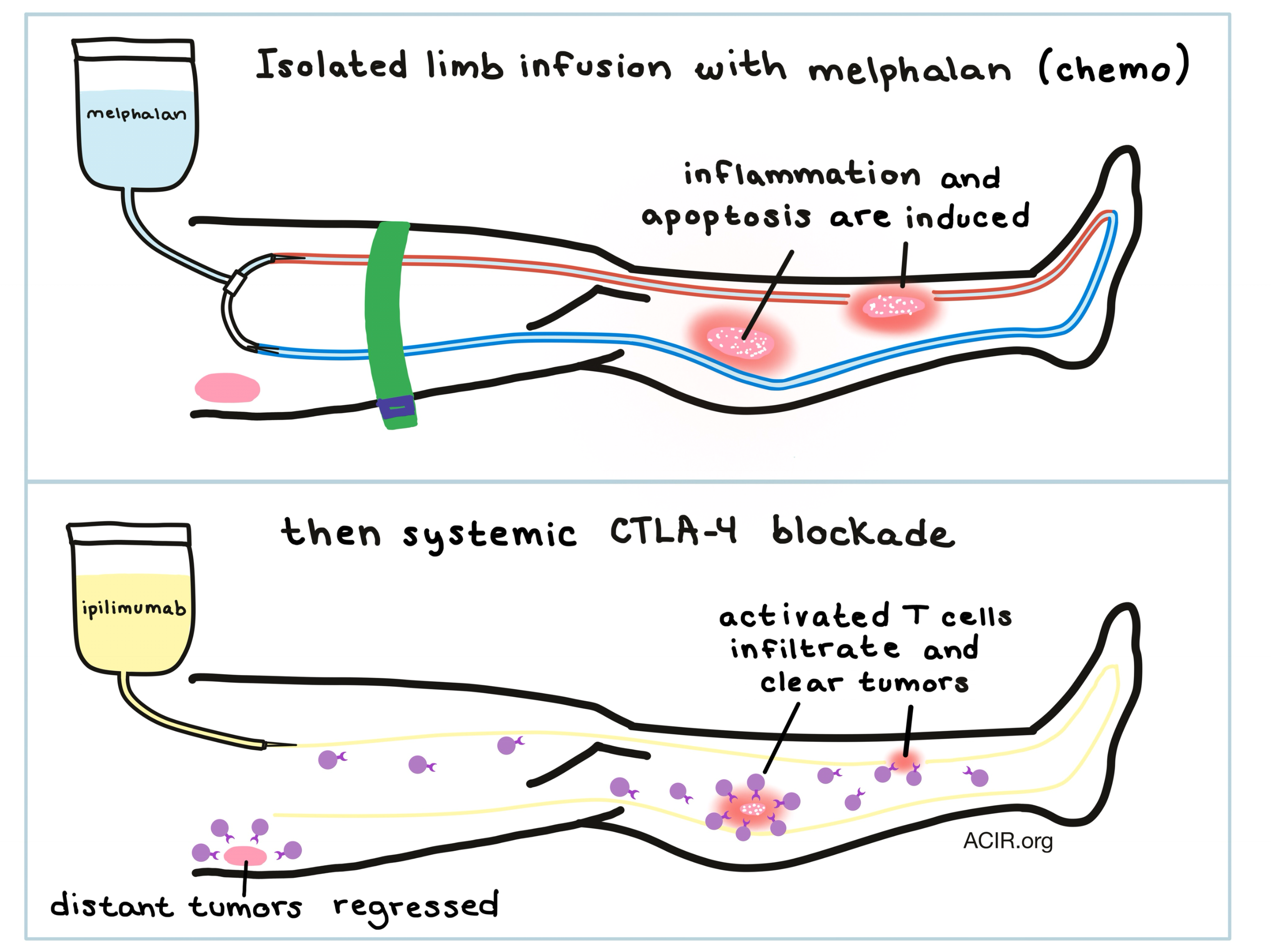
The preclinical efficacy of combining melphalan with anti-CTLA-4 prompted the initiation of a Phase II clinical trial in 26 patients with advanced, recurrent melanoma. Patients were treated locally with melphalan via isolated limb infusion (ILI), a technique that involves creating a closed circuit in an extremity in order to locally deliver chemotherapy and avoid systemic toxicity. Patients were then treated with their first dose of ipilimumab to block CTLA-4; patients went on to receive, on average, 3 out of 4 planned doses of ipilimumab.

Of the 26 patients,16 (62%) had complete responses and 6 (23%) had partial responses in the limb, for an impressive response rate of 85%. At one year, 58% of patients remained progression-free. With a median follow up of 3 years, median survival and median progression-free survival (PFS) were not reached. Patients achieved local and distal responses to the combination therapy, and the two patients who entered the study with stage IV disease showed complete responses lasting at least two years. Importantly, the researchers observed no increase in limb toxicity with the addition of ipilimumab over ILI with melaphan alone, however, most patients did have immune-related adverse events including diarrhea, colitis, pneumonia, and long-term adrenal insufficiency.

To better understand the immune reactions in patients, Ariyan et al. analyzed gene expression of tumors before and after each therapy. After ILI, researchers noted statistically significant upregulation of genes related to innate and adaptive immune responses, chemotaxis, costimulatory ligands and receptors, and MHC class I and class II molecules. The addition of ipilimumab induced upregulation of ICOS and genes related to cytotoxic T cell-effector molecules, including granzymes, IFNγ, and perforins. The researchers also measured circulating cytokines and found that the inflammatory cytokines IL8 and IL6 increased after ILI with melphalan, while IFNγ, IL17, and TNF increased following additional treatment with ipilimumab. Immune phenotyping showed an increase in proliferating CD4+ and CD8+ T cells as well as an increase in ICOS+CD4+ T cells in the blood after combination therapy. Tumor biopsies showed increased infiltration of CD4+ and CD8+ T cells and an increase in PD-L1 in the tumor microenvironment following therapy. Importantly, T cell frequencies were low in all patients before therapy and increased most in patients who achieved PFS for a year or more.

Overall, the results of preclinical and clinical research by Ariyan et aI. support the idea that local treatment with melphalan induces an inflammatory tumor microenvironment that supports an enhanced response to CTLA-4 blockade, which mobilizes cytotoxic effectors. This combination treatment offers a dramatic clinical improvement over CTLA-4 blockade or ILI alone, and the high PFS rate encourages further studies combining CTLA-4 blockade with strategies that prime adaptive immunity.

by Lauren Hitchings

References:

Ariyan C.E., Brady M.S., Siegelbaum R.H., Hu J., Bello D.M., Rand J., Fisher C., Lefkowitz R.A., Panageas K.S., Pulitzer M., Vignali M., Emerson R., Tipton C., Robins H., Merghoub T., Yuan J., Jungbluth A., Blando J., Sharma P., Rudensky A.Y., Wolchok J.D., Allison J.P. Robust Antitumor Responses Result from Local Chemotherapy and CTLA-4 Blockade. Cancer Immunol Res. 2018 Jan 16.

In the Spotlight...

Wide expression and significance of alternative immune checkpoint molecules, B7x and HHLA2, in PD-L1 negative human lung cancers

Cheng et al. analyzed nearly 400 non-small cell lung cancer samples and found that PD-L1 was expressed on less than one third of the tumors and was associated with advanced stage, lymph node involvement, and high TIL score. However, two recently discovered immune checkpoints, B7x and HHLA2, were expressed on two thirds of tumors, usually not overlapping with PD-L1. While antibodies to all three checkpoints suppressed cytokine production, antibodies to B7x and HHLA2 inhibited T cell proliferation to a greater extent than antibodies to PD-L1.

A kinetic investigation of interacting, stimulated T cells identifies conditions for rapid functional enhancement, minimal phenotype differentiation, and improved adoptive cell transfer tumor eradication

Zhou et al. developed a two-step protocol to investigate the short-term kinetics of T cell activation. By analyzing surface markers, gene transcripts, and the single-cell secreted protein profile, they demonstrated a feedback loop in which T cell–T cell interactions following antigen stimulation led to highly polyfunctional, but not significantly differentiated T cells. Such optimally primed T cells efficiently eradicated tumors in mice, suggesting that T cell functionality and phenotype evolution can be temporally decoupled for optimization of adoptive cell therapy.

Downregulation of membrane trafficking proteins and lactate conditioning determine loss of dendritic cell function in lung cancer

Caronni et al. demonstrated the suppression of dendritic cell function by the lung cancer tumor microenvironment (TME), potentially mediated by lactic acid accumulation. Alterations to antigen processing and presentation machinery and inhibition of innate signals producing IFN type-I and IL-12 combine to abrogate T cell priming and antitumor response.

Nitric Oxide Production by Myeloid Derived Suppressor Cells Plays a Role in Impairing Fc Receptor-Mediated Natural Killer Cell Function

Stiff et al. found that nitric oxide production by MDSCs impairs the FcR-mediated signal transduction and downstream effector functions (cellular cytotoxicity, antibody-dependent cell-mediated cytotoxicity, and cytokine production) in NK cells of mice and humans, thus antagonizing NK cell-dependent monoclonal antibody (mAb) therapies. Depletion of MDSCs or inhibition of iNOS (a nitric oxide-producing enzyme) in vivo restored NK cell functions and improved the efficacy of mAb therapies in mouse models.

Combination gemcitabine and WT1 peptide vaccination improves progression-free survival in advanced pancreatic ductal adenocarcinoma: A phase II randomized study

In this phase II randomized clinical study, Nishida et al. tested the combination of gemcitabine and a Wilms’ tumor gene 1 (WT1) peptide vaccine in patients with pancreatic ductal adenocarcinoma. The combination was well-tolerated. Although effects on overall survival were minimal, resulting in a failed primary endpoint, progression-free survival was significantly improved in patients with metastatic disease and in patients demonstrating a response to the vaccine (as measured by delayed type hypersensitivity positivity and increased WT1-specific cytotoxic T cells).

Chemotherapy weakly contributes to predicted neoantigen expression in ovarian cancer

Using computational tools, mutational signature information, as well as whole genome and RNA sequencing of bulk, high grade ovarian carcinoma pre- and post-chemotherapy tumor samples, O’Donnell et al. demonstrated that relapsed tumors have an increase in neoantigen load compared with primary tumors, but most of the increase is due to pre-existing mutational processes and not chemotherapy-related mutagenesis. Pre-chemotherapy neoantigen load and CD8+ T cell infiltration independently predicted survival outcomes.

TIM-3 Regulates CD103(+) Dendritic Cell Function and Response to Chemotherapy in Breast Cancer

De Mingo Pulido et al. found that TIM-3 is highly expressed on tumor-infiltrating CD103+ dendritic cells (cDC1s) and that anti-TIM-3 antibody mediates antitumor activity through the CXCL9/CXCR3 axis, potentially by upregulating CXCL9 expression by cDC1s, which enhances Granzyme B expression by CD8+ T cells. In mouse models of breast cancer, anti-TIM-3 antibody increases cell death within tumors and improves response to paclitaxel chemotherapy without increasing toxicity, indicating a possible clinical application.

CD19 CAR T Cells Expressing IL-12 Eradicate Lymphoma in Fully Lymphoreplete Mice through Induction of Host Immunity

To overcome the toxicity of prior lymphodepletion in CAR T cell therapy, Kueberuwa et al. created a lymphoreplete mouse model of CD19+ B cell lymphoma and demonstrated that murine CD19 CAR T cells expressing IL-12 were able to eradicate the tumor and confer long-term survival in approximately 25% of mice. The antitumor response was due to both direct killing by the CAR T cells and recruitment of host immune cells (a.k.a. epitope spreading), a beneficial effect potentially subverted by the current practice of lymphodepletion.

Everything New this Week In...

Close Modal

Small change for you. Big change for us!

This Thanksgiving season, show your support for cancer research by donating your change.

In less than a minute, link your credit card with our partner RoundUp App.

Every purchase you make with that card will be rounded up and the change will be donated to ACIR.

All transactions are securely made through Stripe.