Even though TILs predict survival and response to immune checkpoint inhibition in non-small cell lung cancer (NSCLC), the origins of these cells remain largely unknown. Gueguen and Metoikidou et al. performed single-cell RNAseq (scRNAseq) and TCRseq (scTCRseq) on tumors, healthy adjacent tissue (juxtatumor), and peripheral blood to investigate CD8+ TIL differentiation and origin. They recently published their results in Science Immunology.
The researchers performed clustering analysis on CD3+ T cell scRNAseq data from all available tumor, juxtatumor, and peripheral blood samples from 11 untreated patients with early-stage disease, resulting in 21 main clusters. The most abundant clusters were CD4+ Tregs and memory T cells, while the most abundant CD8+ T cells were circulating precursor/transitional cells. CD4-CCR8 and CD8-LAYN clusters were the main clusters expressing immune checkpoint and stimulatory receptors.
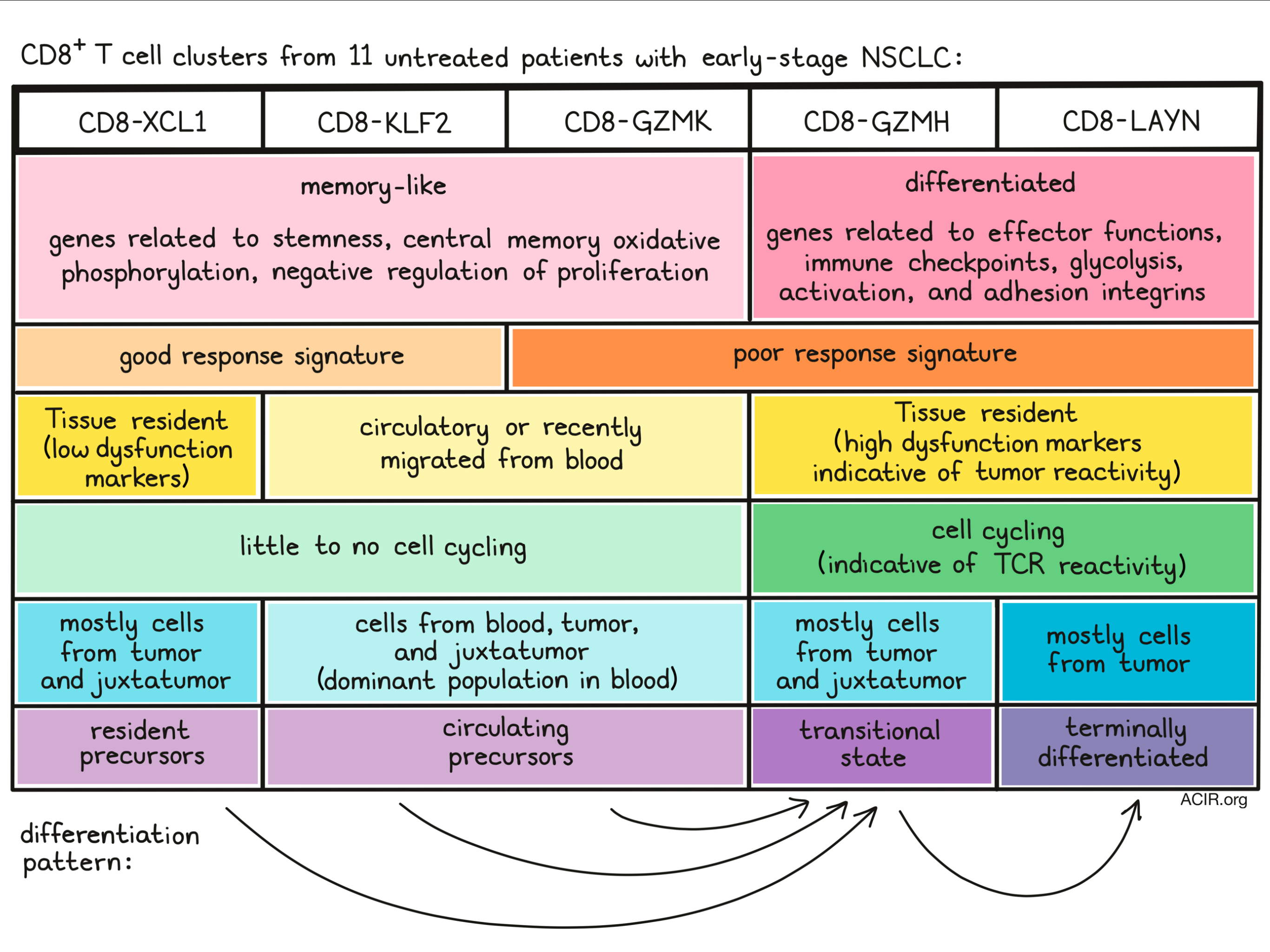
Given their role in antitumor immunity, the researchers then focused on the CD8+ TIL populations. In a UMAP analysis of scRNAseq data, the CD8+ cells segregated into clusters that contained memory-like precursors (CD8-GZMK, CD8-KLF2, and CD8-XCL1) and clusters that were more differentiated, related to exhausted and dysfunctional states (CD8-GZMH and CD8-LAYN). The memory-like precursors expressed stemness, central memory, oxidative phosphorylation, and negative regulation of cell proliferation gene signatures. The more differentiated clusters expressed higher levels of effector-related genes, immune checkpoints, glycolysis signatures, lymphocyte activation-related genes, and adhesion integrins.
The researchers then compared their dataset with a dataset of melanoma patients treated with checkpoint inhibitors, and segregated data based on good and poor therapeutic response signatures. The good response signatures were mainly found in the circulating CD8-KLF2 and the tissue-resident precursor CD8-XCL1 clusters. On the other hand, the CD8-LAYN and CD8-GZMH clusters and part of the CD8-GZMK cluster expressed poor response gene signatures. Therefore, even though the CD8-KLF2 and CD8-GZMK were transcriptionally similar, they were prognostically different, suggesting subtle differences between the subsets. Indeed, a differential gene expression analysis showed that the CD8-GZMK cluster overexpressed T cell activation, effector, and chemokine genes, while the KLF2 cluster overexpressed genes related to control of differentiation.
The differentiated clusters CD8-GZMH and CD8-LAYN and the precursor CD8-XCL1 clusters had high levels of tissue-resident markers, as compared to the CD8-GZMK and CD8-KLF2 clusters, which were characterized by KLRG1 expression. The tissue-resident status of these clusters was confirmed by flow cytometry, in which tissue-resident CD103+CD8+ TIL expressed high levels of the dysfunction markers PD-1, TIM3, and CD39. This is consistent with previous studies suggesting this population has high checkpoint expression and displays features of tumor reactivity. The small subset of CD103+ cells with low levels of dysfunction markers was hypothesized to represent CD8-XCL1 resident precursors, as this cluster also showed low levels of inhibitory receptors.
The researchers hypothesized that the memory-like populations might correspond to tissue-resident cells (CD8-XCL1) and cells that recently migrated from the blood (CD8-KLF2, CD8-GZMK) and that these precursors could differentiate into the differentiated and dysfunctional/exhausted subtypes (CD8-GZMH and CD8-LAYN). They tested this by using unsupervised approaches and pseudotime alignment to deduce the transitions between clusters. The CD8-GZMH cluster was found to be a transitional cluster, suggesting these cells are undergoing differentiation from early memory-like subsets (CD8-XCL1, CD8-KLF2, and CD8-GZMK) to terminally differentiated clusters (CD8-LAYN).
To further assess the clusters' characteristics, the researchers then used scTCRseq data from six patients to determine the clonal expansion in the clusters. More prominent clones were found in the CD8-GZMH and CD8-LAYN differentiated clusters. In most cases, there was a higher probability of finding the same clone in the same cluster. However, there was also some clone sharing between clusters with similar functions, such as between the CD8-GZMK and CD8-KLF2 and the CD8-LAYN and CD8-GZMH populations. The researchers hypothesized this clone sharing represents dynamic transitions between the clusters. Further analysis showed that the two primary circulating precursor memory-like clusters (CD8-GZMK and CD8-KLF2) and the two differentiated clusters (CD8-GZMH and CD8-LAYN) shared multiple TCRs. Overall, the CD8-GZMH cluster exhibited most sharing with the other clusters, further confirming this cluster as a ‘hub’ in the transitional patterns.
Since T cells only cycle upon TCR signaling, actively cycling TIL encountering local antigens in the tumor microenvironment might represent tumor-reactive cells. Using the scRNAseq transcriptome of 11 patients, two cycling populations were found. Labeling transfer, a bioinformatic method to integrate cell classification data from other external datasets, of the CD8+ populations revealed that the cycling clusters were almost exclusively found in the differentiated effector clusters, CD8-GZMH and CD8-LAYN. Flow cytometry from TILs from an additional set of 10 patients confirmed that late differentiated effector cells were cycling, as PD-1+TIM3+CD39+CD8+ cells also expressed Ki67.
Next, the researchers aimed to uncover the origins of the various TIL clusters. To do so, they went back to the initial data and mapped the blood and juxtatumor T cells on the TIL clusters. This revealed that the CD8-LAYN cluster almost entirely consisted of cells found in the tumor. Given that CD8-LAYN was not found in other tissues, it confirmed the data suggesting CD8-GZMH become CD8-LAYN in the TME. The CD8-XCL1 cluster contained mostly tumor and juxtatumor cells, indicating these cells might be tissue precursors. Since the CD8-GZMK and CD8-KLF2 clusters were most common in the blood, these cells might reflect circulating precursors. There was TCR clonal sharing between the CD8-GZMK and CD8-KLF2 clusters in blood and tumor, suggesting that the CD8-KLF2 TIL might be derived from blood CD8-GZMK.
Finally, Gueguen and Metoikidou et al. analyzed the top 20 TCR clones of each cluster in the blood and tumor and compared these. The top 20 of the differentiated TIL clusters were not observed in the blood, suggesting expansion in the tumor. The top 20 TCRs in the KLF2 and GZMK tumoral clusters were found mainly in the blood as well. The most abundant clones in the blood were found in the GZMK cluster, and these were also found in the tumoral KLF2 and GZMH clusters. These data suggest that the blood CD8-GZMK may be the precursor source for tumor infiltration.
These data provide new insights into the origin and differentiation of TILs in NSCLC and may provide new clues for targeting specific populations for cancer immunotherapy and for identifying T cell prognostic markers.
Write-up by Maartje Wouters, image by Lauren Hitchings
Meet the researcher
This week, first co-author Christina Metoikidou answered our questions.
What prompted you to tackle this research question?
Immunotherapies have emerged as a promising treatment option for many types of cancer. Their goal is to re-activate tumor-specific CD8+ T cells through blockade of immune checkpoint (ICB) inhibitors. However, not all T cells are reinvigorated, and thus, there is still a large proportion of patients exhibiting limited or no responses to immunotherapeutic strategies. How this reinvigoration is actually achieved and which CD8+ T cell populations are targeted by ICB therapies, remains not fully understood. Characterization of the CD8+ T cell landscape, as well as CD8+ T cell differentiation in cancer, is crucial for designing enhanced cancer therapy strategies. For these reasons, we wanted to better understand CD8+ TIL heterogeneity in cancer and unveil the mechanisms underlying CD8+ T cell differentiation towards exhaustion. “What are the fates of CD8+ T cells in tumor microenvironment?” and “what are the roles of the different CD8+ T cell subsets in antitumor immunity?” were the questions we had in mind when we started working on this project.
What was the most surprising finding of this study for you?
In our study, we identified two distinct TCF1+ memory-like CD8+ T cell precursor populations: one found only in tissue (both in tumor tissue and normal adjacent-to-tumor tissue), and the other one being present also in blood. These 2 populations seemed quite similar in terms of their transcriptomic profile, since they both expressed memory markers and genes related to CD8+ T cell precursor phenotype, like TCF1, IL7R, etc. What was quite interesting was that despite their similarities, they were quite different when it came to their origin. We gained more insight on that thanks to the single-cell RNA sequencing, which revealed that one of the two populations was enriched in tissue resident marker expression, together with the analysis on matched tumor, juxtatumoral tissue and blood samples, which unveiled the tissue distribution of our populations. Surprisingly, these two precursor subsets were also found to show quite different relevance to clinical outcomes. Comparison of our dataset with a previous single-cell study (Sade-Feldman et al., 2018) showed that the tissue-resident CD8+ T cell precursor subset, rather than the circulating one, was correlated with better response to ICB. Certainly, a lot of work remains to be done on understanding the relevance of the different CD8+ T cell populations described in our study to treatment response and disease prognosis. Future scRNAseq studies on NSCLC patients responding or not to ICB could give more light to this issue.
What was the coolest thing you’ve learned (about) recently outside of work?
Due to the COVID-19 pandemic, I’m spending more and more time inside at home, as everyone is. This gave me the chance to discover new hobbies, and that’s how gardening came into my life! I’ve recently started planting vegetable seeds and they have already started growing! It’s so exciting watching the seeds sprouting! I can’t wait for the first harvesting!