Weekly Digests
‹ Back to February

Found: tumor-reactive regulatory T cells

February 20, 2019

Elevated levels of CD4+ regulatory T cells (Tregs) are often found in human tumors and are associated with poor prognosis. However, the antigen specificity of intratumoral Tregs is not well understood. In a paper recently published in Science Immunology, Ahmadzadeh et al. analyzed the relationships of TCR sequences between intratumoral FOXP3+ Tregs, circulating CD4+ Tregs, and conventional FOXP3-CD4+ cells, as well as the specificity of intratumoral Tregs, with the ultimate goal of improving immunotherapeutic approaches.

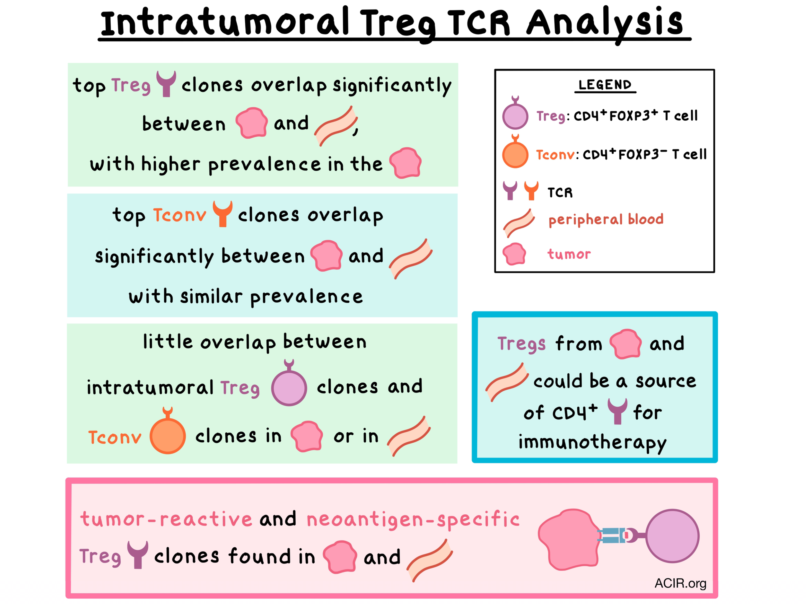
The researchers began by deep sequencing the TCRβ chains of intratumoral Tregs and conventional T cells isolated from six patients with metastatic melanoma, gastrointestinal, or ovarian cancers. Tregs exhibited a diverse TCRβ repertoire that was distinct from conventional T cells, with only a small, variable (<1 to 13%) fraction of shared clonotypes between the two cell populations. These results were consistent with previous findings from peripheral blood of healthy adults showing a small extent of overlap between the populations. The most dominant intratumoral Treg clonotypes overlapped significantly with Tregs from peripheral blood, but not with circulating or intratumoral conventional T cells. Meanwhile, conventional T cells also had a significant overlap in clonotypes between the tumor and circulation.

Intratumoral, but not circulating, Tregs, have previously been shown to be activated. Consistent with these findings, the authors found that the frequency of Tregs was higher in the tumor than in circulation, with almost 20% of intratumoral Tregs in a state of active division. The top 10 most frequent intratumoral Treg clones were much more expanded in the tumor than in peripheral blood, while no significant difference in expansion of the top clones in conventional T cells was observed between the tumor and circulation. These results suggest that the clonal expansion of the intratumoral Tregs may have resulted from tumor antigen stimulation.

With these observations in mind, Ahmadzadeh et al. analyzed the dominant intratumoral Treg clones for antigen specificity. In Tregs from one patient’s metastatic melanoma tumor (patient 3107), 6 of 11 analyzed TCRs specifically recognized the tumor, with 3 of these being among the 10 dominant FOXP3+ clonotypes. Conventional T cells from the same tumor showed 7 of 9 clones reacting to the tumor, with 3 of these being among the 10 dominant clonotypes in the intratumoral conventional T cell subset. Intratumoral Tregs from two more patients with metastatic melanoma were analyzed. In patient 3919, one Treg clone displayed tumor-specific reactivity, while in patient 4066, none of the top 10 Treg clonotypes reacted to the tumor.

Digging deeper, the researchers explored whether the intratumoral Tregs recognized neoantigens arising from patient-specific mutations. Using whole-exome sequencing and RNAseq, the team identified 163 somatic mutations in patient 3107. Utilizing peptide-pulsed autologous dendritic cells and autologous or HLA class II-matched donor peripheral blood cells transduced with Treg-derived TCRs, the authors screened 11 Treg TCR clones and found that one of them exhibited reactivity to mutated annexin A1 (ANXA1). In patient 3919, the screening revealed that one Treg TCR clone reacted to mutated (but not wild-type) CCL-5/RANTES with high functional avidity. With these experiments, the researchers demonstrated that intratumoral Tregs were reactive against patient-specific neoantigens. Interestingly, neither of these TCR clones were reactive to autologous tumor cells, possibly because in both cases RNA expression was low or absent for the gene (CCL5) or mutated allele (ANXA1).

Finally, the researchers asked if tumor-specific Tregs could also be found in peripheral blood. Analyzing the TCRβ deep sequencing data from patient 3107, they found that 6 tumor-reactive Treg TCRs (including the TCR specific for the mutated ANXA1) were found in circulating Tregs. These results suggest that peripheral blood could be a source of tumor-reactive and neoantigen-specific Tregs.

Overall, the results of this study demonstrate that the elevated levels of Tregs in the tumors are likely due to tumor antigen stimulation and the resulting clonal expansion. Intratumoral Tregs displayed a TCR repertoire that was distinct from conventional T cells but overlapped with circulating Tregs. Most importantly, the most dominant intratumoral Treg TCR clones showed reactivity to the tumor and patient-specific neoantigens. Identification of Treg specificity may inform the design of future TCR-based immunotherapies against cancer.

by Anna Scherer

References:

Ahmadzadeh M., Pasetto A., Jia L., Deniger D.C., Stevanović S., Robbins P.F., Rosenberg S.A. Tumor-infiltrating human CD4(+) regulatory T cells display a distinct TCR repertoire and exhibit tumor and neoantigen reactivity. Sci Immunol. 2019 Jan 11.

In the Spotlight...

Distinct Immune Cell Populations Define Response to Anti-PD-1 Monotherapy and Anti-PD-1/Anti-CTLA-4 Combined Therapy

Gide and Quek et al. characterized melanoma samples from patients treated with anti-PD-1 or combination anti-PD-1/anti-CTLA-4 and found that in responders tumor infiltration by activated CD8+ T cells likely drove responses. Non-responders to anti-PD-1 often coexpressed other targetable immune checkpoints. A gene expression profile for EOMES+CD69+CD45RO+ effector memory CD4+ or CD8+ T cells was more abundant in responders and associated with longer PFS in anti-PD-1 therapy and greater tumor reduction in both mono- and combination therapy. The presence of this subset was a better predictor of response than PD-L1 or CD8 alone.

Local delivery of OX40L, CD80, and CD86 mRNA kindles global anti-cancer immunity

Using charge-altering releasable transporters (CARTs), Haabeth and Blake et al. found that the combined intratumoral delivery of OX40L, CD80, and CD86, or OX40L and IL-12 mRNA led to eradication of injected A20 lymphomas in most mice, eradication of both injected and distal tumors in 40% of mice, increased survival, and establishment of immunological memory. OX40L/CD80/CD86 delayed tumor growth in uncured mice better than OX40L/IL-12. The systemic effect was attributed to local activation and mobilization of tumor-specific immune cells, which migrated to the local draining lymph node and to distant tumors.

Ribosomal Proteins Regulate MHC Class I Peptide Generation for Immunosurveillance

Using a lentiviral shRNA panel to knockdown (KD) each of the 80 ribosomal proteins (RPs), Wei et al. identified three RPs modulating MHCI-peptide levels independent of source protein and transcriptome-wide effects. In HEK293(MuH2-Kb) cells infected with a virus containing the SIINFEKL epitope, RPL28 KD enhanced SIINFEKL-Kb presentation. RPL6 KD did the opposite by inhibiting ubiquitin- and proteasome-dependent generation of rapidly degraded nascent polypeptides (DRiPs). RPS28 KD increased HLA-A2 levels by increasing non-canonical translation. Somatic mutations in ribosome subunits may thus affect immune surveillance.

Contributed by Alex Najibi

Mechano-regulation of Peptide-MHC Class I Conformations Determines TCR Antigen Recognition

Building on earlier work showing that separating force applied to the TCR-peptide-MHC I complex counterintuitively strengthened interactions for T cell stimulating agonist peptides, Wu et al. explored the detailed dynamic interactions using simulations and biophysical assays. A stronger peptide-TCR interface with agonist peptides allowed separating forces to induce new interactions between MHC and TCR domains, which were further strengthened as the MHCα domain separated from β2-microglobulin. These dynamic changes may help explain the impact of HLA polymorphisms and cancer-associated mutations on TCR antigen recognition.

PVRIG and PVRL2 Are Induced in Cancer and Inhibit CD8+ T-cell Function

Investigating nectin family receptors, Whelan et al. found that PVRIG and TIGIT (but not CD96) inhibit cytokine production and cytotoxic activity in human CD8+ T cells via non-redundant pathways (PVRIG/PVRL2 and TIGIT/PVR). Single or combination blockade of PVRIG, TIGIT, and PD-1 increased IFNγ production by human TILs, with triple blockade yielding the highest increase. In various human cancer types, PVRIG expression was upregulated on TILs (in line with TIGIT and PD-1), and PVRL2 was upregulated on cancer cells and TAMs. PVRIG was regulated through complex mechanisms, including rapid internalization in the absence of TCR stimulation.

Mouse PVRIG Has CD8+ T Cell-Specific Coinhibitory Functions and Dampens Antitumor Immunity

Murter et al. found that PVRIG acts as a coinhibitory receptor on CD8+ T cells in mice, with the primary ligand being PVRL2. PVRIG was upregulated in CD8+ T cells upon activation, while PVRL2 could be detected on tumor cells and myeloid cells. PVRIG-deficient mice mounted more effective antitumor responses, mediated by CD8+ TILs with increased T cell function and upregulated inflammatory, cytotoxic gene signatures. The efficacy of PD-L1 blockade was enhanced in PVRIG-/- mice or in wild-type mice when combined with a PVRIG blocking antibody, leading to reduced tumor growth and increased survival.

NK cells expressing a chimeric activating receptor eliminate MDSCs and rescue impaired CAR-T cell activity against solid tumors

Parihar et al. developed human NK cells with a chimeric receptor (NKG2D.ζ) containing the extracellular domain of the activating NKG2D receptor fused to the intracellular cytotoxic TCR ζ-chain. NKG2D.ζ receptor expression was maintained upon exposure to TGFβ and soluble NKG2D ligands. NKG2D.ζ-NK cells killed immunosuppressive MDSCs (which express NKG2D ligand), but not other immune cells, in vitro and in a xenograft neuroblastoma model, and shifted the secreted cytokine milieu toward a more immune-stimulatory state. The altered TME allowed GD2.CAR-T cells to effectively localize to the tumor site, potentiating greater antitumor activity.

Durable anticancer immunity from intratumoral administration of IL-23, IL-36γ, and OX40L mRNAs

Hewitt and Bai et al. used lipid nanoparticles to intratumorally deliver IL-23, IL36γ, and OX40L mRNAs in multiple tumor models, inducing strong antitumor responses, control of both treated and distal tumors, and immunological memory. The effects of treatment were attributed to altered cytokine and chemokine expression in the TME, activation of multiple innate and adaptive immune cells, and immune cell mobilization. Efficacy was dependent on Batf3-dependent DCs and CD8+ T cells. The addition of this therapy to checkpoint blockade overcame resistance and improved outcomes. Human PBMCs showed similar cytokine responses in vitro.

Everything New this Week In...

Close Modal

Small change for you. Big change for us!

This Thanksgiving season, show your support for cancer research by donating your change.

In less than a minute, link your credit card with our partner RoundUp App.

Every purchase you make with that card will be rounded up and the change will be donated to ACIR.

All transactions are securely made through Stripe.