Weekly Digests
‹ Back to February

It’s a cytokine! It’s an antibody! It’s a new cancer immunotherapy!

February 28, 2018

Interleukin 12 (IL-12) is a heterodimeric cytokine with potent antitumor efficacy based on its abilities to stimulate the proliferation and cytotoxicity of activated T and NK cells, induce IFNγ and other cytotoxic enzymes and cytokines, and promote differentiation into memory phenotypes. Its actual efficacy as an immunotherapy, however, is limited by its short half-life and dose-related toxicity. In an effort to harness the positive effects of IL-12 and bypass the negatives, Jung et al. constructed an Fc-fused version of monovalent IL-12 and observed remarkably improved effects, which were reported in Oncoimmunology.

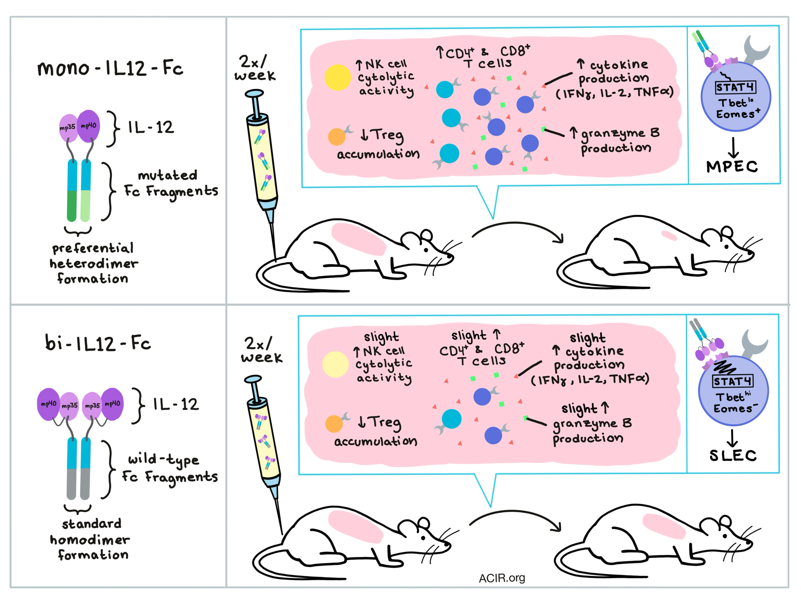
To generate monovalent Ig Fc-fused murine IL-12 (mono-mIL12-Fc), each of the two subunits of IL-12 (p40 and p35) were fused separately to the N-terminus of one of the two respective engineered Fc chains; these Fc chains were mutated such that only heterodimeric Fc would form, ensuring that each dimer would include both subunits of IL-12. As a control, the researchers also generated Fc-fused bivalent IL-12 (bi-mIL12-Fc), which consisted of linked IL-12 subunits attached to a wild-type Fc chain, forming homodimers.

Both mono-mIL12-Fc and bi-mIL12-Fc had longer half-lives compared to recombinant IL-12, and twice-weekly injections of either mono-mIL12-Fc or bi-mIL12-Fc proteins completely cured small CT26-HER2/neu tumors in mice. Jung et al. then went on to test different concentrations of their immunocytokines twice weekly in larger, more challenging tumors (CT26-HER2/neu, CT26, and B16F10), where mono-mIL12-Fc consistently outperformed bi-mIL-12-Fc, eliciting robust tumor regression and durable cures, even at fairly low doses. At the optimal dosing regimen tested, 73% of mice treated with mono-mIL12-Fc experienced tumor-free survival. Even a single dose of mono-mIL12-Fc at a higher concentration substantially impeded tumor growth in almost 50% of tumor-bearing mice. Bi-mIL12-Fc had antitumor efficacy as well, but was consistently inferior. All treatments were well tolerated, and cured mice rejected rechallenge.

To understand the observed antitumor effects, the researchers explored the underlying cellular mechanisms 27-35 days after tumor inoculation and IL-12 treatment, and found that mono-mIL12-Fc significantly increased the numbers of CD4+ and CD8+ T cells in both spleens and tumors and reduced accumulation of Treg cells in the tumor. A closer look at the tumor microenvironment showed that mono-mIL12-Fc treatment increased IFNγ production in tumor-bearing mice, and that tumor-infiltrating T cells (especially CD8+ T cells) expressed elevated levels of cytokines including IFNγ, IL2, and TNFα; in addition, CD8+ T cells showed increased levels of granzyme B. Further, splenic T cells treated with mono-mIL12-Fc were more cytotoxic when tested against tumor target cell lines in vitro. NK cells also showed increased cytotoxicity, suggesting a partial contribution to tumor control.

Jung et al. next explored whether mono-mIL12-Fc and bi-mIL12-Fc differentially affected the generation of memory CD8+ T cells, and thus analyzed T cell subsets in the spleen after therapy, classifying CD8+ T cells as either effector, effector memory (TEM), or central memory (TCM), based on their expression levels of CD62 and IL7R. While low-dose bi-mIL-Fc treatments slightly increased populations of TEM and TCM cells, mono-mIL12-Fc significantly increased both populations in a dose-dependent fashion.

The researchers next analyzed the induction of a memory phenotype by evaluating the proportions of short-lived effector cells (SLECs) versus memory precursor effector cells (MPECs), which can be differentiated by expression levels of the terminal differentiation marker KLRG1 or the memory marker IL-7R. They found that mono-mIL12-Fc moderately transduced IL-12 signaling to cause weak STAT4 activation in CD8+ T cells, yielding low T-bet expression and more incidents of Eomes expression, thus programming effector CD8+ T cells to differentiate into MPECs. Bi-mIL12-Fc, on the other hand, potently transduced IL-12 signaling, triggering strong STAT4 activation, high T-bet expression, and prevention of Eomes expression, directing CD8+ T cells to differentiate into SLECs.

To better understand how these findings might translate to a clinical setting, Jung et al. developed human versions of both mono- and bi-mIL12-Fc. Both fusion proteins potently induced proliferation of activated human PBMCs in vitro. As in mice, mono-hIL12-Fc also led to reduced expression of pSTAT4 and T-bet compared to bi-hIL12-Fc in stimulated human CD8+ T cells.This indicates that, like its murine analog, mono-hIL12-Fc could induce CD8+ T cell differentiation into MPECs for a durable memory T cell response in humans, supporting clinical evaluation.

by Lauren Hitchings

References:

Jung K., Ha J.H., Kim J.E., Kim J.A., Kim Y.J., Kim C.H.& Kim Y.S. Heterodimeric Fc-fused IL12 shows potent antitumor activity by generating memory CD8+ T cells. Journal of OncoImmunology. 2018 February 5.

In the Spotlight...

Efficacy of intracellular immune checkpoint-silenced DC vaccine

Wang et al. designed an adenoviral vector (Ad-siSSF) encoding SOCS1 shRNA (an intracellular checkpoint inhibitor), two tumor-associated antigens, and bacterial flagellin (TLR5 agonist). Ad-siSSF-transduced human PBMC-derived DCs induced an antigen-specific cytotoxic T cell response in vitro and in rhesus monkeys. In a phase I clinical trial in relapsed AML and ALL, Ad-siSSF-DC treatment combined with cytokine-induced killer cells increased survival compared with donor lymphocyte infusion, and exhibited a favorable safety profile.

Rapid activation of tumor-associated macrophages boosts preexisting tumor immunity

The time-dependent depletion of inhibitory tumor-associated macrophages (TAMs) following antibody blockade of CSF-1R signaling provided a window for co-treatment with agonist CD40 antibody to act on the monocytic populations (including antigen-presenting cells) to induce an inflammatory state, resulting in T cell recruitment and activation, effective T cell memory, and improved tumor control in multiple mouse models. Human colorectal and mesothelioma TAMs demonstrated CD40 expression by IHC, although levels varied between patients.

Myeloid-targeted immunotherapies act in synergy to induce inflammation and antitumor immunity

Using a combination of surface marker expression, single-cell RNA sequencing, and single-cell secretion analysis, Perry et al. distinguished changes in multiple tumor-associated macrophage populations (TAMs) in uninflamed autochthonous BRAF/PTEN melanoma tumors treated with agonist CD40 and a CSFR1 inhibitor. They demonstrated a shift to an inflammatory polyfunctional macrophage population, which resulted in synergistic tumor control that was dependent on T cells, TNFα, and IFNγ.

Targeted BiTE expression by an oncolytic vector augments therapeutic efficacy against solid tumors

Speck et al. created an oncolytic measles virus encoding CD20- or CEA-targeted bispecific T cell engagers (MV-BiTEs) and demonstrated that intratumoral administration of MV-BiTEs increased T cell infiltration into the tumor, improved survival, and led to durable remissions without signs of toxicity in immunocompetent mice with B16 melanoma, as well as in immunocompromised mice with xenografts of human colorectal carcinoma. T cell infiltration, CD8/FoxP3 mRNA ratio, chemokine mRNA, and markers of exhaustion were all increased.

Eradication of Triple-Negative Breast Cancer Cells by Targeting Glycosylated PD-L1

Li et al. showed that glycosylation of PD-L1 (enhanced by epidermal growth factor regulation of B3GNT3 glycotransferase) is required for ligation with the PD-1 receptor and identified monoclonal antibodies that specifically recognize N-linked glycosylated PD-L1. One such antibody caused caveolae-dependent endocytosis and subsequent lysosome degradation of PD-L1, enhancing antitumor immunity in a TNBC mouse model. Conjugating the antibody with a cytotoxic drug further improved antitumor efficacy.

CD8 T Cell Exhaustion in Chronic Infection and Cancer: Opportunities for Interventions

Hashimoto et al. thoroughly review properties of exhausted CD8+ T cells, including sustained expression of inhibitory receptors, altered metabolism, unique cytokine expression profile, and a distinct epigenetic signature, with emphasis on the role of PD-1. They discuss the strategies for reinvigorating exhausted CD8+ T cells in cancer and chronic infections, the dependency on CD28, and the recently identified stem cell-like subset of PD-1+CD8+ T cells that localize in lymphoid tissue and proliferate in response to PD-1 blockade in chronic viral infections.

Dendritic cell vaccine induces antigen-specific CD8(+) T cells that are metabolically distinct from those of peptide vaccine and is well-combined with PD-1 checkpoint blockade

Nagaoka et al. compared the effects of a peptide-pulsed dendritic cell vaccine (DC-V) and a short peptide vaccine (SP-V) in a B16 melanoma model and found that the combination of DC-V/anti-PD-1 slowed tumor growth, while SP-V/anti-PD-1 did not. DC-V-primed T cells in the spleen had a higher central/effector memory phenotype ratio, fewer exhaustion markers, and metabolism that was shifted from glycolysis to oxidative phosphorylation compared to SP-V-primed T cells.

T cells specific for a TAP-independent self-peptide remain naive in tumor-bearing mice and are fully exploitable for therapy

Doorduijn et al. evaluated transferred CD8+ T cells specific for the TAP-independent Trh4 epitope, which is associated with impaired peptide processing (TEIPP), in the context of TAP-deficient MHC-Ilow tumors. Although Trh4-specific T cells did not infiltrate and were not activated by such tumors, the T cells were robustly activated by irradiated TAP-proficient tumor cells overexpressing Trh4. Prophylactically, this controlled tumor growth and increased survival in mice, indicating the potential of TEIPP antigen targeting in some immune-escaped tumors.

Everything New this Week In...

Close Modal

Small change for you. Big change for us!

This Thanksgiving season, show your support for cancer research by donating your change.

In less than a minute, link your credit card with our partner RoundUp App.

Every purchase you make with that card will be rounded up and the change will be donated to ACIR.

All transactions are securely made through Stripe.