The helper function of CD4+ T cells in antitumor immune responses has been characterized, but much less is known about the role of cytotoxic CD4+ Th cells (Th-CTX) in tumors. Cachot, Bilous, and Liu et al. investigated the presence, phenotype, and cytolytic function of tumor-specific Th-CTX using single-cell transcriptomics, peptide–MHC-II multimers, and a newly developed high-throughput single-cell cytotoxicity assay. Their exciting results were recently published in Science Advances.
The researchers started by mining publicly available single-cell RNAseq datasets of tumor-infiltrating T cells in advanced melanoma for CD4+ T cell subsets. Unsupervised clustering of more than 2,000 CD3+CD4+ T cells revealed four main clusters, which were annotated as regulatory, memory, cycling, and cytotoxic clusters based on gene expression. The cytolytic cluster was enriched for lytic granule genes and genes previously associated with effector and cytolytic functions in CD4+ T cells, such as CRTAM, NKG7, KLRG1, and CST7. This cluster was also enriched for genes involved in the cellular response to IFNγ and chemotaxis, with upregulation of the chemokines CCL5, CCL4, and CCL4L1/2. No clear single transcription factor appeared to be critical in the cytotoxic cell population, suggesting that multiple factors may be involved in defining this phenotype.
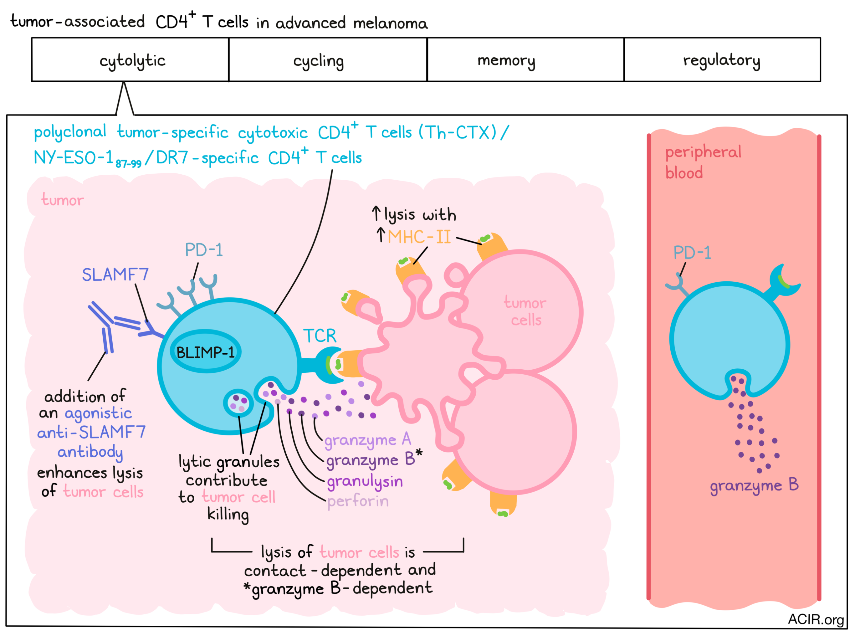
To further investigate the cytolytic features of the Th-CTX population, the researchers used peptide–MHC-II multimers to isolate NY-ESO-187-99/DR7-specific CD4+ T cells from peripheral blood, and from tumors and tumor-infiltrated lymph nodes from patients with melanoma. These multimer-positive T cells were found in 8/9 blood samples and 5/8 tumor samples. The peripheral cells showed higher granzyme B expression than those in the tumor, while tumor-associated cells expressed higher perforin, granzyme A, and BLIMP-1 levels.
Direct cytolytic antitumor activity of Th-CTX would require tumoral MHC-II expression, which was investigated by assessing HLA-DR by fluorescent IHC staining of melanoma tissue sections. Five out of 18 cases had HLA-DR+ tumor cells, and in vitro experiments confirmed that both IFNγ and TNFα could elevate MHC-II expression on melanoma cell lines. These findings demonstrate that interaction between Th-CTX and tumor cells in the tumor microenvironment is possible.
To assess the cytolytic capacity of Th-CTX, the researchers isolated and expanded multimer-positive sorted cells from patient samples and performed a 51chromium release assay. Each clone was tested in the presence of peptide-loaded tumor cells that were either pretreated with IFNγ or stably transduced with CIITA to increase MHC-II expression. Up to 46% lysis occurred in CIITA-transduced tumor cells, but less lysis was observed in IFNγ-treated and wild-type cells, reflecting differences in MHC-II expression. Similar results were found with Mage-A3 epitope-specific T cells.
The researchers then developed a high-throughput single-cell tumor recognition assay to monitor interactions between effector and target cells and evaluate cytotoxicity at the single-cell level. The data were analyzed using deep learning tools, with an algorithm that enabled the quantitation of various effector to target cell ratios and the target cell lysis specificity. Although cytolysis started after a few minutes of coculture, it persisted 15-20 hours, with an average killing time of 5.3 hours; this is slower than the average killing time of 2.4 hours by CD8+ cells in similar assays. There was evidence of serial killing activities and a trend toward a higher lytic capacity when there was an excess of targets, which is in line with previous work on the cytolytic capacities of CD8+ T and NK cells.
To look into the pathways that induced the cytolytic activity of the Th-CTX, the researchers assessed cytokines and other molecules produced by antigen-specific Th-CTX and Th CD4+ T cell clones after coculture with peptide-loaded target cells. Th-CTX and Th cells produced little perforin, granzyme K, and granzyme M, but high concentrations of granzyme A. Additionally, the Th-CTX cells produced significantly more granzyme B and granulysin than Th cells. The importance of lytic granules for these cells' killing capacity was confirmed in a coculture experiment performed in the presence of concanamycin (a compound that degrades perforin present in vacuoles), which resulted in a decrease of lysis. The lysis was found to be contact- and granzyme B-dependent.
Next, the TCR α and β chains of 32 NY-ESO-187-99/DR7-specific CD4+ T cell clones isolated from blood were assessed. The clones showed diverse, clonally distributed TCRs in both Th and Th-CTX, with high variability between patients. Both shared and distinct TCR α and β chains were identified in the two cell populations, with similar membrane expression of TCRαβ in all clones, suggesting the TCR expression had minimal impact on the functional differences between these populations.
To determine specific surface markers of the Th-CTX, the researchers used the scRNAseq data. SLAMF7 was identified as predominantly expressed by cells in the cytolytic cluster, while PD-1 was expressed mainly in the cycling and cytolytic clusters. Assessing the protein levels on blood and tumor-associated NY-ESO-187-99/DR7-specific CD4+ T cells, both SLAMF7 and PD-1 were detected directly ex vivo, with lower expression of PD-1 on peripheral cells and more double-positive cells in the tumor. The expression of SLAMF7 could be induced in vitro by IL-12 or the histone deacetylase inhibitor Entinostat. Although SLAMF7 expression was favorably associated with a good clinical outcome in the TCGA dataset, further studies are needed to link this effect to Th-CTX.
To establish the roles of SLAMF7 and PD-1 in the cytolytic function of the Th-CTX, the 51chromium release assay was repeated with the addition of either an anti-PD-1 or an agonistic anti-SLAMF7 antibody. The percentage of specific lysis was not enhanced by PD-1 signaling blockade, but SLAMF7 stimulation slightly increased lysis. Finally, Th-CTX were found in scRNA-seq datasets from breast, head and neck, and hepatocellular cancers. However, SLAMF7 was only upregulated in melanoma, breast cancer, and hepatocellular carcinoma – not in head and neck cancer.
In summary, the data presented here demonstrate the presence of an interesting tumor-specific CD4+ T cell subpopulation with cytolytic capacity in multiple tumor types and characterize their phenotypic and functional properties, supporting the potential of MHC-I-independent targeting. Further, these studies reveal SLAMF7 agonism on Th-CTX cells as a potential new immunotherapy target.
Write-up by Maartje Wouters, image by Lauren Hitchings
Meet the researcher
This week, lead author Camilla Jandus answered our questions.
What prompted you to tackle this research question?
Immunotherapy, an innovative treatment strategy aimed to boost the immune response against cancer, has significantly changed management of patients with cancer. However, many patients still do not respond to treatment or develop resistance. To date, most studies have focused on boosting and re-invigorating cytotoxic CD8+ T cell functions, while still little is known about the role of CD4+ T cells in antitumor immunity, and particularly in their direct contribution in eliminating tumors. We were really interested in combining innovative approaches down to the single cell level to investigate whether human CD4+ T cells can directly eliminate tumor cells. Further we aimed at identifying the determinants that are linked with their antitumor functions, to be exploited in cancer patients.
What was the most surprising finding of this study for you?
CD4+ T cell cytotoxicity has been reported in the context of viral infections, vaccination, and tumor mouse models, but still little is known regarding the presence and overall frequency of these cells in human tumors. I was surprised to observe very frequent and efficient CD4+ T cell killing of targets in the picowells of the real-time nanobiosensor that we developed together with Professor Altug at the EPFL. Even more surprising was the fact that when a single CD4+ T cell was outnumbered by target cells, serial engagement in multiple killing activities was observed.
What was the coolest thing you’ve learned (about) recently outside of work?
In the recent cold winter days in the Swiss mountains, I discovered the beauty of the Mpemba effect, a phenomenon that refers to the fact that, contrary to natural prediction, in certain circumstances, hot water freezes faster than cold water. Although the theoretical basis of this physical paradox, initially noticed by Aristotle, remains a matter of controversy, the practical result of it is that during icy days such as the ones of last February, you can easily create ice rainbows using a bottle of boiling water. Seeing is believing!